A Hundred Years of Diagnosing Superficial Fungal Infections: Where Do We Come From, Where Are We Now and Where Would We Like To Go? - Acta ...
←
→
Page content transcription
If your browser does not render page correctly, please read the page content below
Centenary theme section: CUTANEOUS AND GENITAL INFECTIONS
REVIEW ARTICLE
ActaDV
A Hundred Years of Diagnosing Superficial Fungal Infections: Where
Do We Come From, Where Are We Now and Where Would We
Like To Go?
Yvonne GRÄSER1 and Ditte Marie Lindhardt SAUNTE2–5
1
Institute of Microbiology and Infectious Immunology, Charité – University Medicine Berlin, corporate member of Freie Universität Berlin,
Humboldt-Universität zu Berlin, and Berlin Institute of Health, Nationale Reference Laboratory of Dermatophytes, Berlin, Germany, 2Department
Acta Dermato-Venereologica
of Dermatology, Zealand University Hospital, Roskilde, 3Department of Clinical Medicine, University of Copenhagen, Copenhagen, 4Unit
of Mycology, Department of Bacteria, Parasites and Fungi, Statens Serum Institut, Copenhagen, Denmark, and 5European Academy of
Dermatology and Venerology Task Force for Mycology Co-chair
Superficial fungal infections have been known for
hundreds of years. During the 20th century new di-
SIGNIFICANCE
agnostic methods were developed and the taxonomy Superficial fungal infections (e.g. ringworm, thrush and
changed several times, which, unfortunately, resulted fungal nail infections) have been known for hundreds of
in many fungi having several names (synonyms). The years. It is crucial to diagnose the fungus correctly, in or-
taxonomy is important, as species-specific identifica- der to choose the correct anti-fungal medication, and to
tion guides clinicians when choosing the most appro- provide information about the source of infection. Traditio-
priate antifungal agent, and provides an indication of nally, diagnosis is based on microscopy, culture and histo-
the source of infection (anthropophilic, zoophilic or pathology of the specimen (hair, skin, nails). More recent
geophilic). Traditional diagnostic tests (direct micros- molecular-based methods have been developed, but there
copy, culture and histopathology) are still widely used, is no standardization as to which fungi they detect. This pa-
but molecular-based methods, such as PCR, have many per presents an update on fungal taxonomy and describes
advantages, and increasingly supplement or replace the diagnostic tools available.
conventional methods. Molecular-based methods pro-
ActaDV
vide detection of different genus/species spectra. This
paper describes recent changes in dermatophyte taxo- teignes”, a monograph based on the standardization of
nomy, and reviews the currently available diagnostics test media and studies on clinical features of skin and
tools, focusing mainly on commercially available PCR hair infections and morphology in cultures (1). At the
test systems. beginning of the 20th century different nomenclatural
Key words: diagnostic; microscopy; PCR; dermatophytes; der-
systems were suggested, based on clinical presentation
matomycoses. and culture characteristics.
Accepted Mar 19, 2020; Epub ahead of print Mar 24, 2020
The taxonomy of superficial fungal infections has
changed several times since then, due to the development
Advances in dermatology and venereology
Acta Derm Venereol 2020; 100: adv00111.
of new diagnostic methods. Unfortunately, this has resul-
Corr: Ditte Marie L. Saunte, Department of Dermatology, Zealand Univer- ted in many fungi having several names (synonyms). An
sity Hospital, Roskilde, Health Sciences Faculty, University of Copenha-
gen, Sygehusvej 5, DK-4000 Roskilde, Denmark. E-mail: disa@regions- attempt to simplify this, by giving “one fungus one name”
jaelland.dk has been initiated, and the development of molecular
diagnostic methods has contributed to this process. This
S uperficial fungal infections have been known since paper describes the latest changes in dermatophyte taxo-
the 5th century BC, when Hippocrates wrote about nomy and reviews currently available diagnostic tools.
thrush in children. It took hundreds of years before the
first scientific proof of infection was made by Agostino TAXONOMY
Bassi in 1835, who showed that the muscardine disease
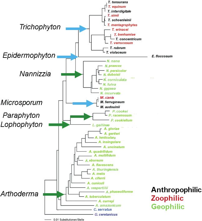
of the silkworm was caused by a fungus (1). In the The taxonomy of dermatophytes changed most recently
following years Audouin from France suggested that at the beginning of 2017 (2). The phylogenetic tree in
some human diseases were caused by the same types Fig. 1, based on molecular data, shows the current valid
of plant parasites (fungi). By the end of the 19th century nomenclature of the family Arthrodermataceae. C. ser-
important microbiological methods, such as obtaining ratus and G. ceretanicus were used as outgroups. Before
pure cultures of the dermatophytes Trichophyton and that, the family of Arthrodermataceae, encompassing
Achorion schoenleinii, were introduced. A morphologi- the dermatophyte fungi, included 3 anamorphic (fungi
cal classification was not established until 1910, when that have no sexual phase in their life cycle, also called
the famous mycologist R. Sabouraud published “Les imperfect fungi), Trichophyton, Microsporum, Epider-
doi: 10.2340/00015555-3467 This is an open access article under the CC BY-NC license. www.medicaljournals.se/acta
Acta Derm Venereol 2020; 100: adv00111 Journal Compilation © 2020 Acta Dermato-Venereologica.Diagnosis of superficial fungal infections 217
ActaDV
Acta Dermato-Venereologica
ActaDV
Fig. 1. Phylogenetic tree of the majority of species of the family Arthrodermataceae, based on the internal transcribed spacer region of
Advances in dermatology and venereology
the ribosomal DNA.
mophyton, and one teleomorphic (fungi that have a sexual aforementioned genera were minor at this time. Most of
phase in their life cycle) genus (Arthroderma). As early as these taxonomic changes were proposed at the begin-
2011, this dual nomenclature of fungi was abolished (3), ning of the 21st century. For example, the previous 50
mainly because the basis of taxonomy moved away from anthropophilic and zoophilic Trichophyton species were
using morphological features towards molecular and reduced to 19 (4), and in 2017 they were reduced by a
phylogenetic data. On this basis, the teleomorphic genus further 3 due to the disappearance of the teleomorphic
in dermatophytes was abolished and 4 additional genera genus. Here, corrections were carried out that mainly af-
(Nannizzia, Lophophyton, Paraphyton and Arthroderma) fected the classification of the dermatophytes into groups
were introduced to account for the former geophilic Mi- that encompassed their natural sources (anthropophilic,
crosporum and Trichophyton spp. according to the rules zoophilic, geophilic). For example, the anthropophilic
of the botanical code. In principle, a separate genus was and zoophilic strains of T. interdigitale, were separated
established at all main clusters (tips of the arrows in Fig. once again, i.e. the zoophilic strains again received their
1) of the phylogenetic multilocus tree. Medical concerns own species name, T. mentagrophytes, whereas the anth-
were also addressed, i.e. the anthropophilic and zoophilic ropophilic strains were called T. interdigitale. Due to this
species names were retained in the well-known genera name change, the previous species T. mentagrophytes,
Microsporum, Trichophyton and Epidermophyton (2). which was phylogenetically closely related to T. scho-
At the species level, the nomenclatural changes that enleinii, had to be renamed. The name T. quinckeanum
affected the medically relevant dermatophytes of the was used, because the originally described strains of T.
Theme issue: Cutaneous and genital infections218 Y. Gräser and D. M. L. Saunte
mentagrophytes var. quinckeanum clustered here (Fig. Direct “non-specific” detection of fungal elements in
1). The skin fungus, Trichophyton sp. of Arthroderma clinical specimens
ActaDV
benhamiae, which was isolated mainly from guinea
Direct microscopy is used for the primary identification of
pigs, was reduced to T. benhamiae. T. soudanense was
fungal elements in specimens after treating with sodium
removed from the T. rubrum complex and is now again
hydroxide or potassium hydroxide (KOH). Conventional
listed as a separate species. New name combinations were
light microscopy, without the benefit of any contrast with
also added, which were mostly geophilic species, due to
the background, is difficult to interpret, and stains, such
the introduction of new genus names, e.g. Microsporum
as lactophenol cotton blue, Parker ink, chlorazol black
gypseum was renamed Nannizzia gypsea. The overall
E or Congo red, are therefore often added. Fluorescence
purpose of these changes was to base the new system on
microscopy after treatment of the specimen with opti-
Acta Dermato-Venereologica
genetically robust determinants and to retain well-known
cal brighteners, such as blankophor or calcoflour, can
dermatophyte names familiar to clinicians (2).
enhance the detection rate after microscopy (12, 13).
Malassezia species show characteristic unipolar bud-
CLINICAL SIGNIFICANCE OF FUNGAL ding blastoconidia, but with the exception of this genus
DIAGNOSTICS it is important to note that direct microscopic findings
are neither genus- nor species-specific, even though it is
Taxonomy may seem remote from everyday clinical possible to distinguish yeasts from hyphae and to detect
practice, but it is important in many ways: first, an ac- pigmented fungal cells. Some very experienced technici-
curate diagnosis is important for choosing the correct ans may be able to suggest a differentiation between other
antifungal treatment (5, 6). Species-specific diagnosis is specific yeasts, dermatophytes and non-dermatophyte
sometimes also necessary, as different species may have moulds, but without absolute certainty (14).
diffierent antifungal susceptibility patterns (7). Secondly, Direct microscopy of hair is important, as the growth
the species name also informs clinicians about the source pattern of the dermatophyte classifies it as either favus,
of the infection. By knowing the source of infection, it endothrix (arthroconidia are present within the hair shaft)
is possible to treat the index patient or animal in order or ectothrix (where the fungus invades the hair shaft at
to reduce the risk of further spread of disease. Thirdly, mid-follicle and the arthrospores then grow out of the
sub-species identification (strain typing) is useful in hair follicle and surround the surface of the hair shaft).
outbreaks as, for example, in India, where a specific
ActaDV
The growth pattern, combined with the conidial size, can
T. mentagrophytes genotype VIII has been uniformly be used as a preliminary indication of the genus of the
isolated as a causative agent of a countrywide spread infecting dermatophyte (15–17). Histology is not used
of a chronic, relapsing dermatophyte epidemic (8). By routinely in skin and hair infections, but is useful when
thoroughly studying this sub-species new knowledge Malassezia folliculitis is suspected, in order to rule out
about virulence and resistance may become available. other causes of folliculitis. Some dermatologists use
Finally, a negative fungal laboratory test is also important histology routinely for fungal identification in nails,
as a diagnosis of exclusion, when other dermatological as it may rule out contamination and is able to confirm
diagnoses have also suspected. Even though identifica- the growth of the fungus directly in the specimen (11,
Advances in dermatology and venereology
tion to genus or species level is important it is not always 18–20). However, the prerequisite for this is an invasive
performed in the clinical setting (9–11). Oral antifungal biopsy.
therapy should not be administered without a confirmed
laboratory diagnosis, because up to 40% of the suspected
diagnoses are wrong (9), and due to the possible side- Genus- and species-specific identification
effects and drug interaction, particularly in older patients Culture is highly dependent on growth media, e.g. some
who often have other underlying diseases and take media are more dermatophyte-specific, while others are
additional medications. A third point is the potentially better for yeasts and non-dermatophyte moulds. Malas-
negative impact that unnecessary treatment may have on sezia is lipid-dependent and, as a consequence, is often
the human microbiome, and the increasing threat of drug difficult to culture on normal laboratory media. Culture of
resistance, which is well recognized with antibacterials, nail material is challenging, as up to 30% of microscopy-
but can be equally applicable to antimycotics. positive nail specimens are culture-negative (21, 22).
This may be due to the presence of non-viable material,
either because of insufficient material from the proximal
TRADITIONAL DIAGNOSTICS: DIRECT
area of infection, or due to previous antifungal treatment.
MICROSCOPY, HISTOLOGY AND CULTURE
Combination of the different techniques is usually
Direct microscopy and culture have been used for the practiced, as it enhances the chances of fungal detec-
purpose of fungal identification over the last 100 years tion and provides more clinically useful information.
and are still used worldwide. The methods will be de All traditional diagnostic methods are dependent on the
scribed in the following section. skills of the laboratory technicians, whereas molecular
Theme issue: Cutaneous and genital infectionsDiagnosis of superficial fungal infections 219
diagnosis does not depend on the acquired skill sets of processed manually. On the other hand, the thermal cy-
the laboratory staff, but may have other limitations, as clers required are less expensive than real-time devices.
ActaDV
described below. However, the advantages of real-time PCR are that both
the amplification and hybridization steps are performed
in the same closed reaction tube without the risk of con-
MOLECULAR-BASED DETECTION OF SUPER tamination. This also eliminates additional bench hand-
FICIAL FUNGAL INFECTIONS ling. However, it must be kept in mind that the number
Development of molecular-based methods for detection of probe hybridizations in conventional techniques is
of dermatomycosis larger (e.g. 78 in the microarray format) than the number
of colour labels (4–6), which are used to label different
With the introduction of molecular tools into the taxo-
Acta Dermato-Venereologica
probes in real-time PCR technology. Thus, melting curve
nomy of dermatophytes approximately 30 years ago, analysis is used to extend the spectrum of species to be
species-specific markers, such as the internal transcribed detected. Nevertheless, these methods are not yet able
spacer (ITS) region of ribosomal DNA, were subsequent- to differentiate, at the same time, more than 20 clinically
ly used for the diagnosis of this fungal group. In the mid- relevant dermatophyte species, including the few non-
1990s, PCR methods were initially applied to cultured dermatophytes that can play a role in onychomycosis
skin material. This included methods such as restriction as infectious agents. Such an all-in-one detection test
fragment length polymorphism (RFLP) and random would replace protracted phenotypic diagnostics based
amplification of polymorphic DNA (RAPD) analyses, on culture, which ultimately requires expert knowledge
but also PCR fingerprinting (23–25). Later, so-called because morphological features in this fungal group are
in-house PCR methods were developed, which were also both polymorphic and partially overlap.
able to identify the fungus directly in clinical specimens.
These methods are generally based on amplification with
Commercial kits for direct detection of fungal
a broad range and/or specific primers and, in a second
infections on skin, hair and nail samples
stage, use hybridization with species-specific probes
with or without a combination of high-resolution mel- Since 2008, commercial systems, that use the above-
ting curve analysis. A distinction can be made between mentioned detection methods and cover different spe-
conventional and real-time PCR techniques. The former cies spectra, have been available. The Dermatophyte
ActaDV
are more personnel-intensive because the hybridization PCR Kit was developed by the Statens Serum Institute
step is performed separately and requires additional (SSI), in Copenhagen Denmark in 2 versions; firstly, as
washing steps (enzyme-linked immunoassay (ELISA), a conventional PCR, and later as a real-time PCR that
blot or microarray technique) and are more susceptible solely detects T. rubrum at species level, as it is the most
to contamination because the amplified DNA is further common pathogen in onychomycosis and tinea pedis
Table I. Species spectra detected by the commercially available test systems
Species/KIT DPK FTD MMD MMD LF DG 1.0 DG 2.0 DD EADM
Advances in dermatology and venereology
T. tonsurans X V
T. equinum X V
T. interdigitale X V
T. mentagrophytes X V
T. schoenleinii X X V
T. quinckeanum X X V
T. simii X nd nd nd nd V
T. erinacei X X X X X V
T. benhamiae X X X X X V
T. verrucosum X X X X V X V
T. bullosum X nd nd nd X X nd V
T. rubrum V V V V V V V
T. violaceum X V V V V V V
E. floccosum X V V V V V V V
M. audouinii X V V V V
M. ferrugineum X V
M. canis X V
N. gypsea X X V V X X V V
N. fulva X X X X X X X V
N. incurvata X X X X X X X V
N. persicolor X X X X X X X V
pan Dermatophyte V X X V X X X V
non Dermatophyte X X V V V V X V
Same coloured boxes refer to the detection of species complexes, but not individual species. V: detects, X: do not detect. The identically coloured boxes mark the species
in the respective kit, which are detected together (as a complex), i.e. not separated from each other.
Nd: no data; DPK: Dermatophyte PCR Kit; FTD: Fast Track Dermatophytes; MMD: Mentype Mycoderm; MMD LF: Mentype220 Y. Gräser and D. M. L. Saunte
(Table I). Otherwise, the kit offers the possibility of is. T. benhamiae is clustered together with T. equinum
detecting dermatophytes as a group (pan-dermatophyte), (27). At the same time, the DERMADYN kit developed
ActaDV
but this will include any non-pathogenic geophilic genera by DYN Diagnostics Ltd in Ha’eshel St., Israel became
present. The conventional test system is based on a PCR available. This test system is also based on a 2-tube
with subsequent size analysis of the amplified DNA multiplex PCR with a melting curve analysis and detects
fragments in an agarose gel, whereas real-time PCR uses a similar spectrum to FTD dermatophytes (Table I). In
hybridization probes instead. Both test systems can be addition N. gypsea is detected, but the T. simii complex
used for screening for dermatophytes, and this may be is not included (28). In 2018, the last of the kits discus-
followed by subsequent species identification of non- sed here, Euroarray Dermatomycosis from Euroimmun,
rubrum species via culture or other molecular techniques, was launched in Lübeck, Germany. This is a multiplex
Acta Dermato-Venereologica
such as sequencing (26). In 2011, the FTD Dermatophyte PCR reaction with subsequent probe hybridization in the
test from Fast Track Diagnostics, was made available form of a “microarray”. This format enables the detection
in Sliema, Malta. This test system, a 2-tube real-time of all relevant (approximately 20) dermatophytes at the
PCR with probe hybridization, but without melting species level, including a pan-dermatophyte probe and 6
curve analysis, is able to detect 3 species (T. rubrum, non-dermatophytes at the species level (Scopulariopsis
T. violaceum, E. floccosum). The remaining detections brevicaulis, Fusarium and Candida spp.). Furthermore,
are performed at species complex level, i.e. more than 1 there is species detection for rare pathogens, such as T.
species is detected here, but not differentiated from each eriothrephon and T. bullosum. Only T. concentricum, a
other. This includes mainly dermatophytes species with pathogen endemic to the Pacific Islands, is not separated
different ecological niches: the T. tonsurans complex from T. benhamiae, and T. soudanense is not differentia-
(no differentiation between T. equinum, zoophilic and T. ted from T. rubrum because the taxonomic change that
tonsurans, anthropophilic), the T. interdigitale complex separated them came after the development of the kit.
(T. interdigitale, T. schoenleinii antropophilic; T. menta- Overall, the clinician should be aware that there is a
grophytes, T. quinckeanum zoophilic) and the M. canis difference between what the commercial tests are able to
complex (M. canis zoophilic, M. audouinii, M. ferrugi- detect (Table I). Most importantly, the majority of tests
neum anthropophilic). The kit does not have a detection do not discriminate between zoophilic and anthropophilic
option for the dermatophytes as a group. Biotype in Dres- species, which is a necessary step in order to find (and
den, Germany launched the first version of the Mentype treat) the sources of infection. Another challenge is that
ActaDV
MycoDerm kit in 2013. These utilize 2 conventional PCR many of the non-dermatophytes involved in the pathoge-
reactions, which can differentiate 2 species (E. floccosum nesis of onychomycosis are not detected in many of the
and N. gypsea) on the basis of fragment size analyses. kits. The broadest species-specific spectrum is offered by
There is no differentiation between the T. tonsurans and the Euroarray. The other test systems do not detect non-
the T. interdigitale complexes. T. rubrum is identified dermatophytes (SSI, FTD), apart from C. albicans (Der-
in a complex together with T. violaceum, as is M. canis maGenius), or they provide detection of Scopulariopsis
complex. The second version of the test system (Mentype and Candida to genus level only (Mentype) (Table I).
MycoDerm Lateral Flow) was available 2 years later
with 3 PCR reactions. Further developments affect, on
Advances in dermatology and venereology
Non-commercial molecular-based tests
the one hand, the procedure, because fragment analysis
was replaced by probe hybridization on a blot strip. On A considerable number of in-house PCR techniques have
the other hand, the species spectrum to be detected has been developed for the diagnosis of dermatophytes and
been enlarged. Now it is possible to detect T. rubrum, T. other skin pathogenic fungi. We do not describe these
violaceum, M. audouinii at species level and M. canis developments in detail here, because they are not stan-
in combination with M. ferrugineum. The T. tonsurans dardized, and in the vast majority of cases are used only
was separated from the T. interdigitale complexes. This by individual, or a few, laboratories involved in routine
was also the first kit that could detect T. benhamiae in diagnostics. Of particular interest are the methods based
a complex together with T. erinacei and T. verrucosum. on real-time PCR. Many of these approaches are able
Another concurrent test system, DermaGenius 1.0, from to identify up to 6 taxa and dermatophytes in general
PathoNostics, produced in Maastrict, the Netherlands, (29, 30). Ohst and colleagues (31) were able to detect
based on a single multiplex real-time PCR with melting 9 dermatophyte taxa by combining up to 10 PCRs in a
curve analysis, had the same identification gaps and a sequential algorithm, and Bergmans and colleagues (32)
similar species spectrum as the FTD Kit. Neither kit could differentiate 11 species in a single-tube assay with
included pan-dermatophyte detection. This changed probes and melting-curve analysis. Walser & Bosshard
with the 2nd version of the kit (DermaGenius 2.0), which (33) report that using sloppy molecular beacons with
became available in 2018. The species detection is the species-specific melting temperature signatures allows
same as for Mentype MycoDerm Kit Lateral Flow, with 2 the identification of 19 dermatophyte species. Until
exceptions. N. gypsea is not included, but T. verrucosum now it has been possible to detect a similar number of
Theme issue: Cutaneous and genital infectionsDiagnosis of superficial fungal infections 221
species only by applying post-PCR techniques, such as They are dependent on the skills of the laboratory
an oligonucleotide array (34). This may be a promising technicians and culture is time-consuming. The culture
ActaDV
approach for a commercial test system, if it is possible to method is still the only diagnostic method that is able to
increase the sensitivity of the 2nd species-differentiating confirm the viability of the fungus, which is important
PCR reaction, which was negative in 76% of cases where for treatment assessment (11). Microbiological laborato-
PCR1 (pan dermatophytes) was positive. ries appreciate the automation possibilities in molecular
Another molecular-based method, matrix-assisted la- diagnostics and often have already established similar
ser desorption/ionization time-of-flight mass spectrome- methods and devices that can also be used for derma-
try (MALDI-TOF), is used to identify micro-organisms tophyte identification. Decisive factors in determining
based on the characteristic protein spectrum of each spe- whether to set up a molecular mycology service are the
Acta Dermato-Venereologica
cies matched with a database. It has been applied to the number of samples, the availability of trained personnel
identification of superficial fungal infection directly on for direct microscopy, culturing and cost-effectiveness,
culture material, both yeast, dermatophytes and moulds which depends strongly on whether and how molecular
(35). This technique is fast and reproducible, but until dermatophyte diagnostics are remunerated. Whether
now not applicable directly to clinical hair, skin or nail conventional diagnostics will still be used after the wider
specimens (36–40). Protein spectra of 7 and 10 dermato introduction of the molecular identification method de-
phytes species (T. tonsurans, T. rubrum, T. interdigitale, pends primarily on the differentiated pathogen spectrum
T. mentagrophytes, T. verrucosum, T. violaceum, M. ca- of the test system used. If not all relevant pathogens are
nis, M. audouinii, E. floccosum, N. gypsea) are included covered, a pan-dermatophyte detection should be used
in the widely used Bruker and Biomerieux reference in order not to miss a possible pathogen. However, even
spectrum databases (41). So far, this method is not able then, the detection of potential non-dermatophytes must
to differentiate the phylogenetically closely related be considered and, if not included, covered by diagnostics
species, e.g. T. rubrum/soudanense, T. interdigitale/ based on culture. It is, therefore, important to be aware
mentagrophytes, T. tonsurans/equinum or T. benhamiae/ of what fungi any locally available molecular test can
concentricum complex (42, 43). Identification can be or cannot detect.
improved by establishing an in-house database (44). It Although molecular diagnostics are up to 30% more
has also been used to detect antifungal resistance (45). sensitive than culture diagnostics, the detection limit is
more than one fungal cell (31). Therefore, the clinical
ActaDV
specimens must be taken from the correct location and in
OTHER DIAGNOSTIC TOOLS sufficient quantity. In this respect, there is no difference
Wood’s light, filtered ultraviolet light, is often used as from culture diagnostics. The detection of pathogenic
a bedside tool for differentiate tinea capitis caused by a dermatophytes, whether in culture or PCR, always
Microsporum species (canis, audouinii and ferrugineum) requires antifungal therapy, because asymptomatic car-
from other dermatophyte infections, as it fluoresces gre- riers also spread the fungi and can become symptomatic.
enish under Wood’s light, and endothrix infections are Disadvantages of the available molecular tests, in ge-
non-fluorescent (17). neral, are that the evolution of fungi can lead to (point)
mutations, especially in species-specific sequences used
Advances in dermatology and venereology
Dermoscopy, reflectance confocal microscopy, opti-
cal coherence tomography and confocal laser scanning in the primers and probe, so that they can no longer bind,
microscopy, all of which are non-invasive methods, can and false-negative results may be generated.
be used as add-on tools to differentiate tinea capitis and/ This can be remedied by sequencing with broad-range
or onychomycosis from other dermatological conditions or only dermatophyte-specific primers, which are more
(17, 20, 46–48). conserved and therefore less susceptible to mutations.
Dermatophyte screening test media, an agar medium Sequencing can then provide accurate species identi-
containing a dermatophyte colour indicator can be used fication. Some laboratories already routinely use these
for dermatophyte screening. The anti-dermatophyte methods for fungal diagnostics. However, the purchase
monoclonal antibody test, an immunochromatographic of a sequencing device is expensive and, like an in-house
detection test, is able to confirm a dermatophyte infec- PCR, the method has to be validated. Furthermore, there
tion, detectable at genus level. Some are known to give must be appropriately validated databases to enable cor-
a false-positive reaction when non-dermatophytes are rect identification.
grown (22, 49–51).
CLINICAL AND LABORATORY INTERACTION
CURRENT ROUTINE DIAGNOSTIC AND THEIR CAN IMPROVE THE DIAGNOSTIC OUTCOME
CHALLENGES
It seems logical that there should be coherence between
The use of phenotypic methods (microscopy and culture) what the clinician suspects and what the mycology labo-
for the detection of pathogens in tinea is still widespread. ratory is able to detect. Nevertheless, in our experience
Theme issue: Cutaneous and genital infections222 Y. Gräser and D. M. L. Saunte
this it often not the case. As described, different fungi already negative at this time, but, microscopically, fungal
have different needs for substrates in order to grow, and elements could still be detected in the KOH preparation.
ActaDV
some molecular-based tests do not detect all relevant The most plausible explanation for this is that resting
fungi. It is therefore important to inform the mycology fungal cells (e.g. in the form of arthroconidia) are still
laboratory, as a minimum, from which anatomical region present and may potentially germinate again after dis-
(hair, skin or nail) the specimen is obtained, which der- continuation of therapy. The survival of dormant fungal
matological disease, and fungal (dermatophyte, Candida, cells inside the nail is supported by follow-up studies,
Malassezia or non-dermatophyte mould) genera is sus- which after 18 months show a complete cure in only 76%
pected (Table II). Furthermore, the attending physician of elderly patients receiving 3-month terbinafine therapy
should note on the referral form whether an animal (54). Dormant cells are missed in the culture. Therefore,
Acta Dermato-Venereologica
contact is probable and whether, for example, a mycosis therapy control with PCR procedures may be suitable in
with a non-dermatophyte is considered in onychomyco- the future, not to mention the short time-span in which
sis. The microbiologist needs this information in order such a finding is available, in order to decide whether to
to interpret the results of the molecular tests correctly, continue the therapy. Only very special PCR procedures
but also to decide whether a culture should be created in are able to discriminate between live and dead cells;
parallel if the kit has gaps in its repertoire. however, it is not known how long dormant fungal cells
survive in the nail, hair or skin of the human body (55).
To date, there has been no significant development of
FUTURE PERSPECTIVES
resistance in dermatophytes to the use of antimycotics.
The advantages of molecular diagnostics for the initial This has suddenly changed with the Indian epidemic,
diagnosis of dermatophytosis are beyond question. A few which has lasted for approximately 6 years, and goes
studies have, so far, shown that the method can also be hand in hand with the use of over-the-counter ointments
used for therapy monitoring (52, 53). Iwanaga et al., in containing antimycotics (e.g. terbinafine), antibiotics and
particular, have demonstrated that the fungal load after steroids (e.g. clobetasol). Terbinafine resistance or partial
16 weeks of terbinafine therapy is significantly reduced resistance in T. mentagrophytes strains with genotype
(from 100% to 36%) (53). The patients’ culture were VIII and T. rubrum reach rates of more than 65% and
ActaDV
Table II. Helpful information for the clinician to differentiate between suspected fungal pathogens, which is needed for the laboratory
for choosing the most appropriate diagnostic methods
Essential information Information helpful for
Help for the clinician to differentiate between suspected fungal pathogens for the microbiologist the microbiologist
Anatomical
region Disease Most common clinical signs Age Suspected pathogen Exposure
Scalp (hair Tinea capitis Broken hairs Children Dermatophyte Animal exposure
region) Kerion Endemic contacts
Favus Woods light results
Alopecia Earlier treatment
Scaling
Seborrhoeic dermatitis/ Greasy skin scales on erythematous skin Newborn Malassezia
Advances in dermatology and venereology
Dandruff Adults
Face Tinea faciei Area with raised erythematous border or All ages, but mostly Dermatophytes Animal exposure
red patch children Signs of tinea capitis
Seborrhoeic dermatitis Greasy skin scales on erythematous skin Adults Malassezia
primary centro-facial and eyebrows
Upper body Pityriasis versicolor Hypo- or hyperpigmented maculae Young and adults Malassezia Immunosuppression
Malassezia folliculitis Monomorphic pustules mainly located at Young and adults Malassezia Immunosuppression
seborrhoeic areas. No comedones
Tinea corporis Area with raised erythematous border or All ages Dermatophyte Animal exposure
red patch. Skin scales Other signs of tinea
Hands Tinea manuum Area with raised erythematous border or All ages Other signs of tinea e.g.
red patches. Skin scales. Hyperkeratosis. tinea pedis
Groin & pubic Cutaneous candidiasis Erythematous skin folds with satellite All ages Candida Immunosuppression
area pustules (and skin scales)
Tinea cruris Area with raised erythematous border or Adults Dermatophytes
red patch. Skin scales
Seborrhoeic dermatitis Greasy skin scales on erythematous skin Adults Malassezia
Feet Cutaneous candidiasis Interdigital maceration All ages Candida Immunosuppression
Tinea pedis Interdigital maceration, skin scales, raised All ages Dermatophytes
erythematous boarder, ’Moccasin foot’,
thickening of the soles
Cutaneous non-D mould Interdigital maceration Mostly adults Non-D moulds Immunosuppression
infection
Nails Candida onychomycosis Paronychia All ages Candida Immunosuppression
Nail dystrophy Moist exposure
Tinea unguium Hyperkeratosis, superficial white All ages, but prevalence Dermatophytes Concomitant tinea
discoloration, yellow streaks. increases with age pedis?
Non-D onychomycosis Hyperkeratosis, discoloration, paronychia/ All ages, but prevalence Non-D moulds
inflammation, nail dystrophy increases with age
Non-D: non-dermatophyte.
Theme issue: Cutaneous and genital infectionsDiagnosis of superficial fungal infections 223
17%, respectively, in India and are spread globally (56). taxonomy for the dermatophytes. Mycopathologia 2017;
182: 5–31.
This means that T. mentagrophytes strains will have to be
ActaDV
3. Hawksworth DL, Crous PW, Redhead SA, Reynolds DR,
fine-typed by molecular genetics in order to determine Samson RA, Seifert KA, et al. The amsterdam declaration
the exact identity, or a susceptibility test has to be per- on fungal nomenclature. IMA Fungus 2011; 2: 105–112.
4. Gräser Y, Scott J, Summerbell R. The new species concept
formed. The advantage of the latter is that breakpoints in dermatophytes-a polyphasic approach. Mycopathologia
can be defined; the disadvantage is that the inoculum, 2008; 166: 239–256.
due to the filamentous growth and the often poor coni- 5. Chen X, Jiang X, Yang M, González U, Lin X, Hua X, et al.
Systemic antifungal therapy for tinea capitis in children.
dia formation of the fungi, is challenging and therefore Cochrane Database Syst Rev 2016; 5: CD004685.
not done routinely. Molecular methods, in particular 6. Fuller LC, Barton RC, Mohd Mustapa MF, Proudfoot LE, Pun-
sequencing with detection of specific genetic mutations jabi SP, Higgins EM. British Association of Dermatologists’
Acta Dermato-Venereologica
guidelines for the management of tinea capitis 2014. Br J
leading to antifungal resistance (e.g. squalene epoxidase Dermatol 2014; 171: 454–463.
gene mutation leading to terbinafine resistance), which 7. Saunte DM, Mrowietz U, Puig L, Zachariae C. Candida infec-
are independent of the fungal growth could overcome tions in patients with psoriasis and psoriatic arthritis treated
with interleukin-17 inhibitors and their practical manage-
this problem (8, 56–59). ment. Br J Dermatol 2017; 177: 47–62.
8. Nenoff P, Verma SB, Vasani R, Burmester A, Hipler U, Wittig
F, et al. The current Indian epidemic of superficial dermatop-
CONCLUSION hytosis due to Trichophyton mentagrophytes —a molecular
study. Mycoses 2019; 62: 336–356.
The diagnosis of superficial fungal infections has evolved 9. Effendy I, Lecha M, Feuilhade de CM, Di CN, Baran R. Epi-
demiology and clinical classification of onychomycosis. J Eur
from the first microscopic description more than 100
Acad Dermatol Venereol 2005; 19: 8–12.
years ago to current techniques that are able to detect a 10. Narang T, Bishnoi A, Dogra S, Singh TD, Mahajan R, Kavita
wide range of clinically relevant fungi using molecular- K. Disease burden and prescription patterns treating der-
based techniques. Worldwide traditional diagnostic matophytosis in North India: salient findings from an online
survey of 1041 dermatologists. J Eur Acad Dermatology
methods, such as direct microscopy and culture, are still Venereol 2019; 33: e391–e393.
used, as they are cheap and the equipment is already 11. Saunte DML, Piraccini BM, Sergeev AY, Prohić A, Sigurgeirs-
available. The development of molecular-based methods son B, Rodríguez-Cerdeira C, et al. A survey among derma-
tologists: diagnostics of superficial fungal infections – what
has already improved a lot during the last years, from is used and what is needed to initiate therapy and assess
only being able to detect fungi in cultures to now being efficacy? J Eur Acad Dermatol Venereol 2018; 33: 421–427;
able to detect fungi directly in clinical samples. The jdv.15361.
ActaDV
12. Monod M, Jaccoud S, Stirnimann R, Anex R, Villa F, Balmer
molecular species-specific fungal detection of clinically S, et al. Economical microscope configuration for direct
relevant fungi is possible, as well as detection of spe- mycological examination with fluorescence in dermatology.
cific mutations causing antifungal resistance. If it were Dermatology 2000; 201: 246–248.
13. Ovrén E, Berglund L, Nordlind K, Rollman O. Dermatophyto-
possible to combine all these tests, it would enable the sis: fluorostaining enhances speed and sensitivity in direct
clinician to obtain the correct species identification, the microscopy of skin, nail and hair specimens from dermatology
possible source of infection and the susceptibility pattern outpatients. Mycoses 2016; 59: 436–441.
14. Pihet M, Le Govic Y. Reappraisal of conventional diagnosis
of the involved pathogen by sending a single sample. The for dermatophytes. Mycopathologia 2017; 182: 169–180.
evolution of the diagnosis of superficial fungal infections 15. Gupta AK, Hofstader SL, Adam P, Summerbell RC. Tinea
Advances in dermatology and venereology
is not far from this goal. capitis: an overview with emphasis on management. Pediatr
Dermatol 1999; 16: 171–189.
16. Morar N, Dlova NC, Gupta AK, Aboobaker J. Tinea capitis in
Kwa-Zulu Natal, South Africa. Pediatr Dermatol 2004; 21:
ACKNOWLEDGEMENT 444–447.
17. Hay RJ. Tinea Capitis: current status. Mycopathologia 2017;
Professor Roderick Hay is acknowledged for his constructive 182: 87–93.
comments and for reviewing the text. 18. Karaman BF, Açıkalın A, Ünal İ, Aksungur VL. Diagnostic
Conflicts of interest: DMLS was paid as a consultant for advisory values of KOH examination, histological examination, and
board meeting by AbbVie, Janssen, Sanofi and received speaker’s culture for onychomycosis: a latent class analysis. Int J
Dermatol 2019; 58: 319–324.
honoraria and/or received grants from the following companies:
19. Velasquez-Agudelo V, Cardona-Arias JA. Meta-analysis of the
Abbvie, Galderma, Astellas, Novartis and Leo Pharma during the utility of culture, biopsy, and direct KOH examination for the
last 3 years. YG received speaker’s honoraria and grants from diagnosis of onychomycosis. BMC Infect Dis 2017; 17: 166.
Euroimmun. 20. Lipner SR, Scher RK. Onychomycosis. J Am Acad Dermatol
2019; 80: 835–851.
21. Monod M, Bontems O, Zaugg C, Lechenne B, Fratti M, Pa-
REFERENCES nizzon R. Fast and reliable PCR/sequencing/RFLP assay for
identification of fungi in onychomycoses. J Med Microbiol
1. Kane J, Summerbell R, Sigler L, Krajden S, Land G. Labo- 2006; 55: 1211–1216.
ratory handbook of dermatophytes: a clinical guide and 22. Monod M, Méhul B. Recent findings in onychomycosis and
laboratory manual of dermatophytes and other filamentous their application for appropriate treatment. J Fungi 2019;
fungi from skin, hair and nails. In: Kane J, editor. Belmont: 5: 20.
Star Publishing Co.; 1997. 23. Kawasaki M, Aoki M, Ishizaki H, Nishimura K, Miyaji M. Phylo-
2. de Hoog GS, Dukik K, Monod M, Packeu A, Stubbe D, geny of Epidermophyton floccosum and other dermatophytes.
Hendrickx M, et al. Toward a novel multilocus phylogenetic Mycopathologia 1996; 134: 121–128.
Theme issue: Cutaneous and genital infections224 Y. Gräser and D. M. L. Saunte
24. Mochizuki T, Sugie N, Uehara M. Random amplification of 42. Su H, Packeu A, Ahmed SA, Al-Hatmi AMS, Blechert O, İlkit
polymorphic DNA is useful for the differentiation of several M, et al. Species distinction in the Trichophyton rubrum
ActaDV
anthropophilic dermatophytes. Mycoses 1997; 40: 405–409. complex. J Clin Microbiol 2019; 57. pii: e00352-19.
25. Graser Y, El Fari M, Presber W, Sterry W, Tietz HJ. Identifica- 43. Suh S-O, Grosso KM, Carrion ME. Multilocus phylogeny of the
tion of common dermatophytes (trichophyton, microsporum, Trichophyton mentagrophytes species complex and the app-
epidermophyton) using polymerase chain reactions. Br J lication of matrix-assisted laser desorption/ionization-time-
Dermatol 1998; 138: 576–582. of-flight (MALDI-TOF) mass spectrometry for the rapid iden-
26. Gräser Y, Czaika V, Ohst T. Diagnostic PCR of dermatophytes tification of dermatophytes. Mycologia 2019; 110: 118–130.
– an overview. JDDG J der Dtsch Dermatologischen Gesell- 44. da Cunha KC, Riat A, Normand A-C, Bosshard PP, de Almeida
schaft 2012; 10: 721–725. MTG, Piarroux R, et al. Fast identification of dermatophytes
27. Uhrlaß S, Wittig F, Koch D, Krüger C, Harder M, Gaajetaan by MALDI-TOF/MS using direct transfer of fungal cells on
G, et al. Halten die neuen molekularen Teste – Microarray ground steel target plates. Mycoses 2018; 61: 691–697.
und Realtime-Polymerasekettenreaktion – zum Dermatop- 45. Delavy M, Dos Santos AR, Heiman CM, Coste AT. Investigating
hytennachweis das, was sie versprechen? Hautarzt 2019; antifungal susceptibility in Candida species with MALDI-TOF
Acta Dermato-Venereologica
70: 618–626. MS-based assays. Front Cell Infect Microbiol 2019; 9: 19.
28. Sherman S, Goshen M, Treigerman O, Ben-Zion K, Carp M-J, 46. Veasey J V., Meneses OMS, da Silva FO. Reflectance confocal
Maisler N, et al. Evaluation of multiplex real-time PCR for microscopy of tinea capitis: comparing images with results
identifying dermatophytes in clinical samples – a multicentre of dermoscopy and mycological exams. Int J Dermatol 2019;
study. Mycoses 2018; 61: 119–126. 58: 849–851.
29. Wisselink GJ, van Zanten E, Kooistra-Smid AMD. Trapped in 47. Pharaon M, Gari-Toussaint M, Khemis A, Zorzi K, Petit L,
keratin; a comparison of dermatophyte detection in nail, skin Martel P, et al. Diagnosis and treatment monitoring of to-
and hair samples directly from clinical samples using culture enail onychomycosis by reflectance confocal microscopy:
and real-time PCR. J Microbiol Methods 2011; 85: 62–66. Prospective cohort study in 58 patients. J Am Acad Dermatol
30. Alexander CL, Shankland GS, Carman W, Williams C. Intro- 2014; 71: 56–61.
duction of a dermatophyte polymerase chain reaction assay 48. Rothmund G, Sattler EC, Kaestle R, Fischer C, Haas CJ, Starz
to the diagnostic mycology service in Scotland. Br J Dermatol H, et al. Confocal laser scanning microscopy as a new valua-
2011; 164: 966–972. ble tool in the diagnosis of onychomycosis – comparison of
31. Ohst T, Kupsch C, Gräser Y. Detection of common derma- six diagnostic methods. Mycoses 2013; 56: 47–55.
tophytes in clinical specimens using a simple quantitative 49. Li X-F, Shen Y-N, Chen W, Chen H, LV G-X, Liu W-D. A new
real-time TaqMan polymerase chain reaction assay. Br J medium for diagnosis of dermatophyte infection. Eur J Der-
Dermatol 2016; 174: 602–609. matology 2009; 19: 34–37.
32. Bergmans AMC, van der Ent M, Klaassen A, Böhm N, An- 50. Rahman MA, Chowdhury OA, Debnath MR, Ahmed SM, Das
driesse GI, Wintermans RGF. Evaluation of a single-tube S, Choudhury R, et al. Comparison among different culture
real-time PCR for detection and identification of 11 der- media for the detection of dermatophytes. Mymensingh Med
matophyte species in clinical material. Clin Microbiol Infect J 2018; 27: 626–630.
2010; 16: 704–710. 51. Tsunemi Y, Hiruma M. Clinical study of Dermatophyte Test
33. Walser M, Bosshard PP. Development and evaluation of a Strip, an immunochromatographic method, to detect tinea
ActaDV
pan-dermatophyte polymerase chain reaction with species- unguium dermatophytes. J Dermatol 2016; 43: 1417–1423.
level identification using sloppy molecular beacon probes. 52. Kupsch C, Czaika V-A, Deutsch C, Gräser Y. Trichophyton
Br J Dermatol 2019; 180: 1489–1497. mentagrophytes – a new genotype of zoophilic dermatophyte
34. Li HC, Bouchara J-P, Hsu MM-L, Barton R, Chang TC. Identi- causes sexually transmitted infections. J Dtsch Dermatol Ges
fication of dermatophytes by an oligonucleotide array. J Clin 2019; 17: 493–501.
Microbiol 2007; 45: 3160–3166. 53. Iwanaga T, Ushigami T, Anzawa K, Mochizuki T. Pathogenic
35. Patel R. A Moldy Application of MALDI: MALDI-ToF mass dermatophytes survive in nail lesions during oral terbinafine
spectrometry for fungal identification. J Fungi 2019; 5: 4. treatment for tinea unguium. Mycopathologia 2017; 182:
36. Denis J, Machouart M, Morio F, Sabou M, Kauffmann-LaCroix 673–679.
C, Contet-Audonneau N, et al. Performance of matrix-assisted 54. Loo DS. Onychomycosis in the elderly: drug treatment op-
laser desorption ionization–time of flight mass spectrometry tions. Drugs Aging 2007; 24: 293–302.
Advances in dermatology and venereology
for identifying clinical Malassezia isolates. J Clin Microbiol 55. Soejima T, Minami J-I, Xiao J-Z, Abe F. Innovative use of
2017; 55: 90–96. platinum compounds to selectively detect live microorga-
37. Cassagne C, Normand A-C, L’Ollivier C, Ranque S, Piarroux nisms by polymerase chain reaction. Biotechnol Bioeng
R. Performance of MALDI-TOF MS platforms for fungal iden- 2016; 113: 301–310.
tification. Mycoses 2016; 59: 678–690. 56. Süß A, Uhrlaß S, Ludes A, Verma SB, Monod M, Krüger C,
38. Karabıçak N, Karatuna O, İlkit M, Akyar I. Evaluation of the et al. Ausgeprägte Tinea corporis durch ein Terbinafin-resi-
Bruker matrix-assisted laser desorption–ionization time-of- stentes Trichophyton-mentagrophytes-Isolat vom indischen
flight mass spectrometry (MALDI-TOF MS) system for the Genotyp bei einem Säugling aus Bahrain in Deutschland.
identification of clinically important dermatophyte species. Hautarzt 2019; 70: 888–896.
Mycopathologia 2015;180: 165–171. 57. Singh A, Masih A, Khurana A, Singh PK, Gupta M, Hagen F,
39. Hedayati MT, Ansari S, Ahmadi B, Taghizadeh Armaki M, et al. High terbinafine resistance in Trichophyton interdigitale
Shokohi T, Abastabar M, et al. Identification of clinical der- isolates in Delhi, India harbouring mutations in the squalene
matophyte isolates obtained from Iran by matrix-assisted epoxidase gene. Mycoses 2018;61: 477–484.
laser desorption/ionization time-offlight mass spectrometry. 58. Khurana A, Masih A, Chowdhary A, Sardana K, Borker S,
Curr Med Mycol 2019; 5: 22–25. Gupta A, et al. Correlation of in vitro susceptibility based
40. Intra J, Sarto C, Tiberti N, Besana S, Savarino C, Brambilla on MICs and squalene epoxidase mutations with clinical re-
P. Genus-level identification of dermatophytes by MALDI-TOF sponse to terbinafine in patients with tinea corporis/cruris.
MS after 2 days of colony growth. Lett Appl Microbiol 2018; Antimicrob Agents Chemother 2018; 62: e01038-18.
67: 136–143. 59. Saunte DML, Hare RK, Jørgensen KM, Jørgensen R, Deleuran
41. Normand A-C, Cassagne C, Gautier M, Becker P, Ranque S, M, Zachariae CO, et al. Emerging terbinafine resistance in
Hendrickx M, et al. Decision criteria for MALDI-TOF MS-based trichophyton: clinical characteristics, squalene epoxidase
identification of filamentous fungi using commercial and gene mutations and a reliable EUCAST method for detection.
in-house reference databases. BMC Microbiol 2017; 17: 25. Antimicrob Agents Chemother 2019; 63. pii: e01126-19.
Theme issue: Cutaneous and genital infectionsYou can also read