A porcupine skeleton of Hystrix (Hystrix) primigenia (Wagner) from the Upper Maeotian (Turolian) of Hadzhidimovo, SW Bulgaria
←
→
Page content transcription
If your browser does not render page correctly, please read the page content below
GEOLOGICA BALCANICA, 41. 1–3, Sofia, Dec. 2012, p. 3–20.
A porcupine skeleton of Hystrix (Hystrix) primigenia (Wagner)
from the Upper Maeotian (Turolian) of Hadzhidimovo,
SW Bulgaria
Dimitar Kovachev
Asenovgrad Palaeontological Branch, National Natural History Museum, Asenovgrad
(Accepted in revised form: November 2011)
Abstract. Relatively well preserved fossil skeleton of Hystrix primigenia (Wagner) is described. The local-
ity near the town of Hadzhidimovo, Blagoevgrad district from which it was collected dated back as Late
Maeotion, that is Turolian faunistic unit, MN12 zone. Comparisons are made with the bones of the modern
species Hystrix cristata Linnaeus. It is concluded that it was a very large and adult animal whose character-
istics strongly correspond to Hystrix primigenia. Some differences have been found which do not contradict
to taxonomical assignment.
Kovachev, D. 2012. A porcupine skeleton of Hystrix (Hystrix) primigenia (Wagner) from the
Upper Maeotian (Turolian) near Hadzhidimovo, SW Bulgaria. Geologica Balcanica 41(1–3),
3–20.
Key words: Fossil porcupine, Hystrix primigenia, Maeotian (Turolian), Hadzhidimovo, SW Bulgaria,
complete skeleton.
INTRODUCTION njanova-Rumenova, 1983; Ivanov, 1995; Yaneva et
al., 2002; Ivanov et al., 2011).
The skeleton of a porcupine Hystrix primigenia here The Nevrokop Formation is represented by conglo
described was excavated in the summer of 1987 in the merates, sands, sandstones, siltstones and clays. It overlays
Girizite ravine, south of the town of Hadzhidimovo, the Baldevo Formation or lies directly on the Precambrian
Blagoevgrad district. It is situated 15 km south-east of basement to the west. For decades, the Nevrokop Formation
the town of Gotse Dlechev in the Mesta Graben that is famous with its rich and well preserved mammal faunas.
is developed along the Mesta River. The paleontologi- Nikolov (1985) was the first to publish a list of fossil
cal site is located east of Sadovo village and north of mammal species supposing a Maeotian age (MN12-13) of
Petrelik village. the Hadzhidimovo 1 locality. The fossil collection of this
The lithostratigraphy of the Miocene and Plio locality was made by D. Kovachev and is stored in the
cene in the Gotse Delchev Basin was presented Paleontological Museum of Asenovgrad.
by Vatsev (1980) and Vatsev and Petkova (1996). The first stratigraphic description of this area was
Four formations were described from bottom to provided by Nenov et al. (1972); Stoyanov et al. (1974).
top: Valevitsa, Baldevo, Nevrokop and Sredna. The Modern data firmly indicated a Maeotian age, i.e. Turo
fossil porcupine here described from Hadzhidimovo lian stage in terms of mammal stratigraphy, base of zone
locality was excavated from the Nevrokop Formation. (Spassov, 2000, 2002). It may appear paradoxal than
The underlaying Baldevo Formation crops out in the Nevrokop Formation which in stratigraphic order
the eastern and north-eastern part of the basin. This is “over” is older in age than the Baldevo Formation. It
formation consists of silts, clays and sands, as well is due to lateral thinning out of the Baldevo Formation
as diatomite levels and coal seams. Its age was westward where the base of Nevrokop Formation is di-
determined as Pontian-Dacian on the basis of diatom rectly on the Valevitsa Formation or on pre-Neogene
and pollen analysis (Temniskova-Topalova and Og rocks (see Vatsev, 1980).
3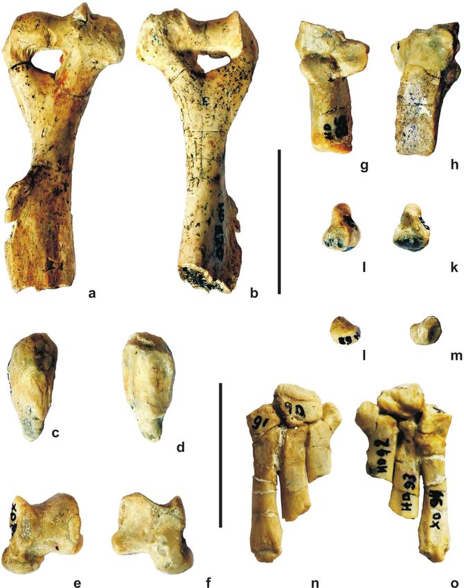
SYSTEMATIC PART parafoseta, mesofoseta and metafoseta, respectively. The
lower molars, on the other hand, possess one labial and a
Order Rodentia Bowdich, 1821 few (normally three) lingual inflexions (synclinias) that
Family Hystricidae Burnett, 1830 are known as hypoflexid, paraflexid and metaflexid.
Subfamily Hystricinae Lyon, 1907 When transformed into islets, the synclinias are called
Genus Hystrix Linnaeus, 1758 fossetidae.
Subgenus Hystrix (Hystrix) Ellerman, 1940
Skull (Figures 1a-d, Table 1)
Hystrix (H.) primigenia (Wagner, 1848)
It is poorly preserved, flattened, the bones are broken.
Locality. Girizite site, Hadzhidimovo Town, Blagoevgrad Some of them are absent. Maxilarae and premaxilarae
district, South-Western Bulgaria. are well preserved, as in some extent also platum durum
Age. Late Miocene, Maeotian; Turolian faunal unit, and condylus occipitalis. The ventral part of the skull is
MN12 vertebrate zone (Spassov, 2002). better preserved.
Material. An almost complete skeleton, hosted in the Due to lateral pressure, the width of the skull is
Asenovgrad Museum of Palaeontology, a branch of the considerably changed. Its length is 189 mm measured
National Museum of Natural History (NMNH) in Sofia, from the labial part of the incisors to the posterior part
Bulgarian Academy of Sciences. of the condyli. This size is here accepted as normal of
Museum No: HD 20 to HD 120. H. primigenia. Corbet and Jones (1965) reported values
in the interval 118–170 mm for H. cristata. The skull of
H. cristata (NMNH) is 165 mm long, i.e. smaller than the
DESCRIPTION AND COMPARISONS fossil skull of H. primigenia.
All teeth are found in situ and are perfectly preserved.
Introduction Premaxilarae are narrow and highly curved downwards.
A small crest, 18 mm long, is seen on their antero-dorsal
The taxonomy of the living species of the genus Hystrix part. The same was previously observed by Niethammer
is based on the differences in spiny covering and cranial (1982). H. cristata also possesses such a crest. A crest
features. Since the spiny covering does not fossilize and that is transversal to the maxiliarae was described by de
the finds of skulls are very rare, the taxonomy of the fos- Bonis et al. (1992) which is not observed in the studied
sil Hystricidae is mainly based on the size of the cheek here skull.
teeth. The latter are the most often found fossils. A small remain of acrus zigomaticus is seen on the
left side of our skull. It is horizontal and starts from the
Terminology middle of P4. The curving upwards is not observable
as acrus zigomaticus is broken. Acrus zigomaticus in
Description of teeth follows the terminology that ac- H. cristata starts much before the beginning of P4.
cording to van Weers (1985) was introduced by Modyer The nasal opening is of isosceles-trainglular form, its
(1946) and Bossma (1968). Nomenclature to describe base being upward. After fossa glenoidalis the braincase
the crown of the upper cheek teeth consists of a lingual narrows and is almost cylindrical. Crista sagitalis is not
concavity, sinus – hypoflexus, and three labial inflex- preserved. Nasal bullae are fairly large. Their anterior
ions, namely paraflexus, mesoflexus and metaflexus. In margins are situated much behind the fossa glenoidalis.
advanced degree of development these inflexions trans- The same position was documented by de Bonis et al.
form into islets (or rings, or lakes) that are known as (1992) in the skull of H. primigenia from Macedonia
Table 1
Measurements of skull (mm)
Hystrix Hystrix
primigenia cristata
1. Length from the anterior end of I through the facial bones up to the posterior end
255 230
of condiles occipitale
2. Length from the anterior end of I through the palate up to the posterior end of condiles
189 165
occipitale
3. Length from the anterior field of the nasal cavities up to the connection between crista
180 155
sagitalis and crista occipitalis
4. Length of the palate from the anterior end of I up to М3 113 91
5. Horizontal diameter of foramen magnum 19 17
6. Vertical diameter of foramen magnum 18 16
7. Distance between the anterior end of I up to the posterior end of M2 177 94
8. Distance between the anterior end of I up to the anterior end of P4 85 56
9. Distance between the anterior end of I up to the posterior end of M3 44 32.4
4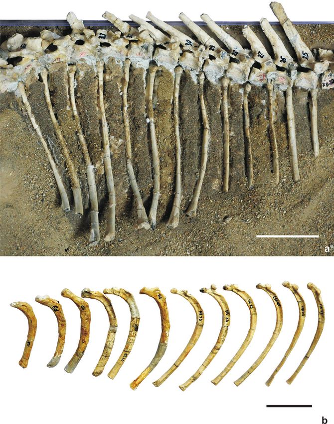
Fig. 1. Skull and mandible. Bar equals 5 cm
        a – skull and mandible, lateral view;
        b – lateral view;
        c – occlusal view;
        d – view from above.

                                                5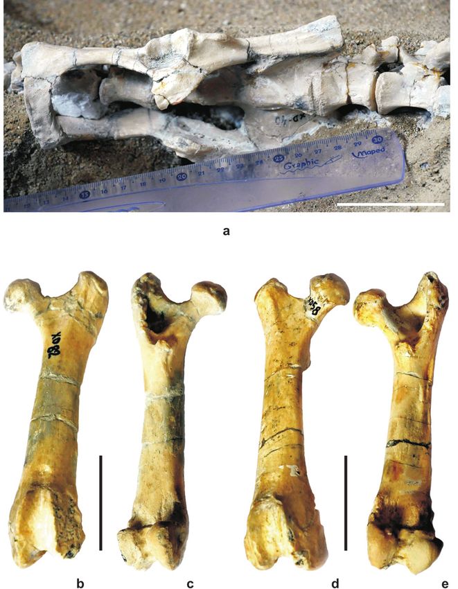
district in Greece. Processus mastoides and processus He emphasized on three main differences. In the genus
paraoccipitalis are absent due to brokening. Hystrix the enamel embraces the anterior side of the inci-
sors reaching up to the mid of the lateral sides; the crest
1 0 1 3 between the anterior and lateral sides is highly rounded;
Teeth formula -----------
the cross section of the incisors of Hystrix is quite smaller
1 0 1 3
than of Castor. J sup. sin et dex here described completely
correspond to the above mentioned features of the genus
Upper jaw teeth (Table 2) Hystrix. Such is the case with H. cristata from NMNH.
A huge diastema separates J sup and P4 (Figs. 1a b). It
The teeth rows are complete and well preserved. Due to is 70.4 mm long. All cheek teeth are excellently preserved.
lateral pressure the two teeth rows became closer. The They are slightly longer and narrower than H. primigenia
primary divergence, however, is still preserved in some from Pikermi described by Gaudry (1862). The material
extent. The upper incisors are shorter, and rather strongly from Hadzhidimovo possesses rounded sides and angles,
curved downwards than the lower incisors are curved up- the lingual surfaces being just slightly curved. Their size
wards. Their roots reach close to the labial side of P4. decreases from M1 to M3. Gaudry (1862) also considered
Neithammer (1982) described the same features in the the cheek teeth of H. primigenia as squarish.
extant H. cristata. They have also been seen in H. crista P4 sin et dex. These are the longest ckeek teeth (11.4 mm).
ta specimen from NMNH. The upper incisors are stouter The occlusial surface shows six enamel islets – fosse-
than the lower ones. The antero-posterior diameters are tae, that originated from inflexions. Six enamel islets are
8.6 mm in the upper incisors, and 8.2 mm in the lower largely reported in H. primigenia from other localities
incisors. Their widths are 7.2 mm and 6.7 mm, respec- (see Sen, 1994). The morphology of islets corresponds to
tively. Thus, the incisors are thicker than wide. The ante- that described in H. bessarabica (=H. primigenia) from
rior surface is covered by completely smooth enamel, as Taraklia. Riabinin (1929), however, mentioned the pres-
is normally in all Hystricidae. The enamel embraces the ence of three exterior grooves (inflexi), whereas in our
incisors also laterally reaching the mid of their external material they are four in number. The third one is the
sides and up to 1/3 of their internal sides. The color of deepest. The lingual side possesses only one groove. The
enamel is yellow-brownish. Such increasing on enamel latter gave rise of another groove (hypoflexus), just op-
is a characterstic feature of the Hystricidae according to posite to the third exterior one, the metaflexus. The hy-
Koliadimou and Koufos (1990). The tipes are grey-black- poflexus and metaflexus divide the tooth into anterior and
ish in color due to manganese compounds. According posterior parts. In the former, the pattern of the enamel
to Malez (1963) the coloration varies depending on the figures (fossetae) is unclear. Four islets are seen. In the
host beds. Gaudry (1862) and Deperet (1890), however, posterior part the islets are only two, one of them being
considered that the yellow-brownish color of the incisors “8”-shaped. A smaller islet is in front of it. The height of
is typical for the porcupines in Pikermi. Deperet (1890) the P4 sin et dex could not be precisely estimated as the
pointed out the triangle form of the upper and lower inci- teeth were “pushed back” into the bone due to pressure.
sors. In our material the form is rather quadrangle, the pos- This does not allow a correct determination of the degree
terior sides being curved and slightly shorter than the an- of hypsodonty. It should be added that these are the least
terior ones. This makes them seemingly triangle. The well worn teeth as they are the latest erupted.
known vertical groove on the anterior side of the incisors M1 sin et dex. A small diastema (2 mm long) separates
that clearly separates their enamel is not deep and is hardly M1 sin et dex from the preceding P4. The both left and
visible in HD-20. Harle (1910) compared his Quaternary right cheek teeth are strongly worn and the two exte-
material of the genus Hystrix with that of the genus Castor. rior enamel grooves (inflexi) are hardly visible. Vekua
Table 2
Measurements of the upper jaw teeth (mm)
Length Width Height Ratio Н/L
Diastema I – P4 H. primigenia 70.4 – – –
H. cristata 50.0 – – –
I H. primigenia 8.6 7.2 – –
H. cristata 6.0 6.8 – –
P4 H. primigenia 11.4 9.3 7.5 0.65
H. cristata 8.7 6.0 2.0 –
M1 H. primigenia 10.0 9.0 4.0 0.40
H. cristata 9.4 8.0 2.0 –
M2 H. primigenia 10.4 9.6 4.5 0.43
H. cristata 9.5 7.5 2.0 –
M3 H. primigenia 9.0 8.5 5.3 0.58
H. cristata 8.2 6.2 2.0
6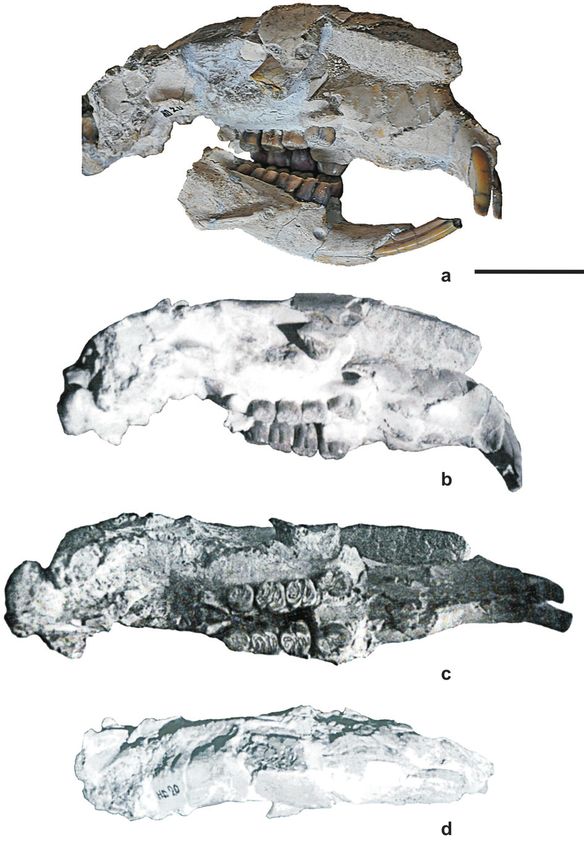
(1972) described a first molar with two inflexi, whereas                The length of the upper teeth rows is 44 mm. This sug-
that from Taraklia has only one inflexus (Riabinin, 1929).          gests that the described porcupine is a large-sized speci-
One hypoflexus is obvious on the lingual side. It reaches           men. Greenwood (1955) considered that Xenohystrix de-
the base of the tooth crown, thus suggesting the pres-              scribed by him (teeth rows 40 mm long) is a large-sized
ence of a hypoflexid. Each of the molars possesses five             Hystricidae. Our specimen is even greater. Actually,
enamel islets. Only the most posterior one is rounded, the          Xenohystrix is now considered as a junior synonym of
rest are worm-like. The material figured by Vekua (1972)            Hystrix (van Weers, 1994).
shows a cheek tooth with seven fossetae, as the tooth was
slightly worn. The roots are hidden in the bone and their           Lower jaw (Table 3)
number is not known. The tooth shape is slightly rounded
rectangular, almost squarish. The labial sides are almost           Only the two rami horizontali are preserved. Their lower
flat, and the lingual sides are more rounded. M1 in HD-             surfaces are smoothly arc-shaped. This form is a result of
20 shares same morphological features with the Pliocene             the fact that the roots of the two lower incisors pass through
Hystricidae. The width of M1 (HD-20) is similar to that             the whole lower surfaces of the rami. Curving upwards, the
of Weze, Poland, and Traklia, Moldova (Sulimski, 1960;              crowns of the incisors form an angle of 40° with the alveo-
Riabinin, 1929), and is smaller than that of Perpignan,             lar row. The material described from Dytiko 3 in Greece
France. Actually, Sen (2001a) described the Perpignan               (de Bonis et al., 1992) shows the same angle. Those au-
material as a new species, H. depereti. It should be noted          thors concluded that, therefore, their Upper Miocene fossil
that the described teeth are higher than all Quaternary             is brachycephalous. Our specimen has a longer skull and is
and extant Hystrix from Africa and Asia.                            a large animal as a whole. Fossa masseterica reaches up to
M2 sin et dex. These cheek teeth share similar form and             the labial sinus of m1. It has a well pronounced crista mas-
size with M1. Their extero-labial sides are more rounded            seterica in its lower portion. Crista masseterica reaches to
and the lingual grooves are less expressed. These molars            the posterior portion of p4 where it gradually disappears.
are slightly longer than M1. Their width is equal with H.           The slightly inclined and less pronounced anterior margin
parvae from Hohfidish, Austria (van Weers and Montoya,              of the coronoid appendice also goes to the posterior part of
1996). Two specimens of H. aryanensis from Molagan,                 p4. Thus, fossa masseterica is embraced by another crista
Afganistan, described by Sen (2001 b) are as long as the            in its upper part. The height of the jaw is 23.5 mm at p4
material from Hadzhidimovo. Sen (1996) pointed out that             and 27.0 mm at m3, i. e. it is slightly lower anteriorly. The
in all specimens of H. primigenia except that from Weze,            thickness is 18.0 mm throughout its length. The length of
M1 is shorter than M2. Many authors considered this fea-            the symphysis is 41 mm. Mandibular index is calculated as
ture more expressed in the living porcupines H. cristata            the height of the lower jaw between m3 and p4 multiplied
and H. indica (e.g. Sen and Kovatchev, 1987; de Bonis et            ×100 and divided to the alveolar length between p4 and
al., 1992; Sen, 1996). Garevski (1956) mentioned that the           m3. According to Sen (1996) this index is quite indica-
first molars are the largest, and the last one – the smallest.      tive of the degree of teeth hypsodonty. The values of this
Two well visible grooves could be seen on the labial side,          index are 53.4 in three specimens of H. primigenia from
and one groove – on the lingual side. The islets are four           Kalimantsi (Sen and Kovatchev, 1987), 56.3 in specimens
in number. All these features are same in the molar from            from Pikermi and 78.5 in extant H. cristata. The described
Taraklia described by Riabinin (1929).                              lower jaw has a mandibular index 54.4 that is rather close
M3 sin et dex. These are the smallest upper cheek teeth.            to the type material of Pikermi. As a whole, the degree of
All angles are rounded. This gives an almost circular               hypsodonty of fossil H. primigenia in Bulgaria and Greece
shape already mentioned in Pikermi, Kalimantsi and                  is almost equal and significantly lower than living porcu-
Weze (Gaudry, 1862; van Weers and Montoya, 1996;                    pines. The foramen mentale is large. It is open just in front
Sen and Kovatchev, 1987; Sulimski, 1960). The external              of p4. A peculiar morphological feature of HD-21 is the
and internal inflexi are hardly visible only on M3 dex.             presence of a second foramen, one centimeter behind the
The porcupine H. cf. primigenia from Kvabebe, Georgia,              first one, right in the middle of p4.
has circle-shaped M3 (Vekua, 1972). The M3 have four                    When compared with H. cristata (NHNM, Sofia), the
roots. The lingual roots are fused, and the two labial roots        lower jaw (HD-21) of H. primigenia exhibits the follow-
are well shaped and separated.                                      ing differences: 1) a steeper symphysis; 2) pars incisiva

Table 3
Measurements of the lower jaw (mm); * – broken bone

                                                                                          H. primigenia          H. cristata
 1. Length from the anterior part of I up to the posterior part of procesus angularis         122.0*                123.0
 2. Total length of the teeth raw I – m3                                                       98.0                  86.0
 3. Length of p4 – m3                                                                          44.0                  36.0
 4. Height of ramus mandibuli through p4                                                       23.5                  26.0
 5. Height of ramus mandibuli behind m3                                                        27.0                  29.0
 6. Thickness of ramus mandibuli through p4                                                    18.0                  15.0
 7. Thickness of ramus mandibuli behind m3                                                     18.0                  12.0

                                                                                                                                7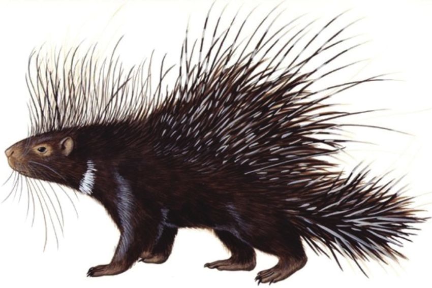
is not higher than the incisor alveolar margin; 3) fossa       worn-out, almost invisible. This sinus is equivalent to the
masseterica is shallower; a slight convergence anteriorly      sinclinid that divides the teeth into two lobes – anterior
is hardly visible; 4) teeth rows are almost parallel; and      and posterior. The former is longer and narrower than the
5) all teeth are shorter and thinner, arranged in arc rather   latter. In the anterior lobe five enamel islands (fossetidae)
than in a straight line.                                       are seen touching each other. Due to their complicated
                                                               pattern, it is hard to determine which are the sinclinids
Lower jaw teeth (Table 4)                                      giving rise to each islets. Four sinclinids are observed on
                                                               the lingual faces of the incisors. The sinclinid IV occurs
All mandible teeth are excellently preserved. Due to           in the anterior lobe and is island-shaped (flexid).
lateral pressure the distance between them in the poste-       m1 sin et dex. The occulusial surface is rectangular,
rior end is obviously decreased. The pressure increased        11.2 mm × 8.5 mm in size, and is slightly wider than p4.
the divergence in anterior direction. This divergence is       Koliadimou and Koufos (1990) stated that the margins,
slighter in the upper jaw teeth.                               especially the lingual ones, are rounded. Vekua (1972)
    The lower incisors have somewhat broken cut-               described a specimen of m1 which has rounded margins,
ting edges. The cutting anterior faces are completely          too. In HD-21 they are less rounded. Vekua’s (1972)
smooth. Grooves of varying depth, described by Sen             materisl is wider than long. This is a rare exception.
(1994), are absent. This feature makes the material from       HD-21 is quite different. m1 teeth from Hadzhidimovo
Hadzhidimovo closer to extant porcupines. The lower            are characterised by seven enamel islands (fossetidae),
incisors are much longer than the upper ones. The roots        whereas the Kvabebian fauna has only four. According
of the former pass under the roots of all cheek teeth thus     to Koliadimou and Koufos (1990) the sinclinid I is much
forming an enlarged arc-shaped impression of the man-          complcated and consists of a pair of small fossetidae
dible, terminating right in its posterior end, immediately     and one „s“-shaped on the lingual edge. HD-21 has also
in front of the processus angularae. The latter is broken.     a pair of enamel islets but one of them is considerably
Fortunately, this break reveals the interior of the two        elongated and angular. The third one is „s“-formed, and
rami horisontali with the incisor roots inside. Koliadimou     enlarged in its end. The sinclinid II consists of two al-
and Koufos (1990) mentioned that in H. major Gervais           most equal islets, one of them being minute and rounded.
(= H. refossa Gervais) the incisor roots are shorter reach-    Sinclinid III is narrrow and long. The axis of sinclinid IV
ing up to m3. Compared to the upper incisors the lower         is parallel to the lingual and labial enamel borders. Some
incisors are seemingly gentle and elegant.                     authors stressed on the pyramide-like form of sinclinid
    The diastema I – p4 is 56 mm long, i.e. rather short-      IV which is not clear in our material. The teeth have
er than the upper diastema I – P4. The cutting edges of        four roots like the materail from Weze (Sulimski, 1960)
the lower incisors do not reach those of the upper ones.       and not three roots like H. cf. primigenia from Kvabebe
Cutting tips of the lower incisors do not reach those of       (Vekua, 1972)
the upper incisors and remained significantly behind.          m2 sin et dex. The anterior faces are straight, the rest are
Thus, they do not match.                                       rounded. Angles are also rounded. The anterior margins
p4 sin et dex. Sulimski (1960) showed a schematic draw-        are less rounded. The anterior lobe is slightly greater than
ing of the roots of all mandible teeth of H. primigenia.       the posterior one. The buccal sinuses are deep and orient-
In that scheme, p4 has three sockets. Some authors sug-        ed backwards. Among the lingual sinuses those related
gest the presence of four alveoli. Our specimen of p4 sin      to the sinclinid III are almost transversal. The sinclinid
has three short roots with shallow sockets. The occlusial      III consists of three eliptical islets in the meso-lingual
outline is rounded rectangular. The buccal face has a          portion of the occlusial surface. The sinclinid II is similar
deep sinus. It starts from the occlusial surface and reachs    in shape to m1. Sinclinid III, along with the buccal sinus,
the roots (hypoflexidae). The sinus on the lingual side is     divide the occlusial surface into anterior and posterior

         Table 4
         Measurements of the lower jaw teeth (mm)

                                                     Length          Width            Height        Ratio Н/L
          Diastema I – p4     H. primigenia           56.0              –               –                 –
                              H. cristata             46.0              –               –                 –
          I                   H. primigenia            7.2             6.7              –                 –
                              H. cristata              6.0             5.4              –                 –
          p4                  H. primigenia           10.6             8.5             9.2              0.86
                              H. cristata              7.5             6.7             2.0                –
          m1                  H. primigenia           11.2             8.5             6.2              0.55
                              H. cristata              8.0             7.0             2.0                –
          m2                  H. primigenia           10.5             9.6             7.0              0.66
                              H. cristata              9.6             7.2             2.6                –
          m3                  H. primigenia            9.1             8.2             8.0              0.82
                              H. cristata              8.2             5.0             2.0                –

8
lobes. The anterior one is greater. The sinclinid IV is an (1993) were so deeply convinced in the brachyodon-
enlarged islet. ty of their Mondecian material that they assigned the
m3 sin et dex. Compared to the other molars, m3 have teeth to H. primigenia mainly based on brachyodonty.
more expressed triangle occlusial surfaces. This is due Hadzhidimovo material is here also idenfied as H. primi
to width decreasing in the posterior part. m3 teeth from genia considering the brachyodonty.
Weze have same smaller width but the form is rounded. Following Greenwood (1955) concerning the number
In 1890, describing his Pliocene Hystrix from Perpignan, and development of the teeth roots, one diagnostic fea-
Deperet wrote that m3 possesses better outlined triangle ture of Hystrix is that the upper molars are rootless. They
form than other lower cheek teeth but this difference is only have small nodular roots and shallow alveoli. M3,
within the intraspecific variability. Thus, the porcupine m3 and p4 have aslo underdeveloped roots. Sulimski
from Perpignan was attributed to H. primigenia. Sen (1960) regarded the clear root differentiation of all teeth
(2001 a), however, reexamined that material and atrrib- as a primitive characteristic.
uted the bulk of it to the new species Hystrix depereti.
One deep sinus passes through the labial portion of m3 Columna vertebralis (Table 5)
(HD-21) from crown to roots. This sinus is oriented back-
wards. On the lingual face three deeply eroded sinclinids Thirty five vertebrae are relatively well preserved. Their
are seen. Riabinin (1929) wrote that m3 has two external distribution in groups is given below.
grooves and three internal subdivisions of the enamel.
There is a difference between our material and that from
Taraklia. m3 has three relatively short roots.
To summarise, in Hystrix the order of eruption
Hystrix Hystrix
of lower and upper teeth is from front to back, like in primigenia cristata
all mammals: the incisors erupt first, followed by the
premolars, and then the cheek teeth m1, m2 and m3 one Number of vertebrae
after another. After the change of milk teeth P4, when the Vertebrae cervicales 2 7
animal is one and a half years old, permanent P4 teeth Vertebrae thoracales 14 14
erupt (Frenkel, 1970). For this reason, when all teeth are Vertebrae lumbales 5 5
available (the case with HD-21), M1 is always the most Vertebrae sacrales 5 5
worn. The attrition ot the occlusial surface is slightest in Vertebrae caudales 9 7
P4. The latter are preserved as the highest teeth. This fact
Total 35 38
was described in 1890 by Deperet and is confirmed by
our material. The cited author pointed out that the upper
cheek teeth are more rounded than the lower ones. Again,
HD-21 obviously reveals the same features. According
to Frenkel’s (1970) scale, the above described teeth from The table shows differences in the number of cer-
HD-20 and HD-21 correspond to class „D“ of high de- vical and caudal vertebrae between H. primigenia and
gree of attrition. All described teeth are yellow-brownish H. cristata. Five cervical vertebrae of H. primigenia were
in color. Their sizes are similar to previously reported H. not found. The number of the caudal vertebrae indicates
primigenia. Their slight hypsodonty, rather brachyodon- that the tail of H. cristata has two vertebrae less. This
ty, together with other morphological features are also corresponds to Neithammer’s (1982) description demon-
comparable to H. primigenia from other localities. If we strating that the tail of H. cristata is shorter, just 1/5 of
accept Greenwood’s (1955) concept that in the brachyo- the total lenght of thorax. This is due to the significantly
dont teeth of the lower jaw in Hystrix the lower enamel shorter bodies (corpus vertebrae) of the caudal vertebrae
boundary almost reaches the contact point between the (Table 5). The two species differ in the shape of spine. In
crown and the roots, then the cheek teeth of HD-21 are H. primigenia the spine is convex upward in the area of
brachyodont, indeed. the lumbar spine. Curve starts immediately after cervical
All Miocene porcupines of Hystrix are relatively low- spine, cimbs quite steeply, and in the area of the lum-
er-crowned, the ratio H/L of the molars being normally bar spine slopes down to the sacrum. The latter is almost
slightly greater than 1.0. These ratios of the mollars from vertical. Seen from the side, the back of the porcupine
HD-20 and HD-21 are presented in Tables 2 and 4. All has a hemispherical shape. In H. cristata the curve starts
values are under 1.0 which means brachyodonty. One from the middle of the thoracic spine and is not as steep
should remain, however, that all teeth are strongly worn as in H. primigenia. The highest part of the arc is a little
and P4 are pushed back into the sockets. Thus, these re- further back, near the sacrum which is shaped slightly
sults should be interpreted very cautiously. Van Weers concave arc.
and Montoya (1996) recognised that cheek teeth dimen-
sions can not be taken to clear due to varying degrees of Vertebrae cervicales(C1 – C7)
wear of teeth.
The onset of high-crowned porcupines like Hystrix Atlas. This first cervical vertebra has preserved acrus
occurred in the latest Miocene or Lower Pliocene (van anterior and arcus posterior. Its body was attibuted to
Weers, 1996). Our material is in agreement with that the second cervical vertebra, epistrofey, which was not
as teeth material of Hadzhidimovo is older (MN12), found. All processes of the atlas are significantly dam-
of late but not latest Miocene age. Massini and Rook aged. There are only traces of the processus transver-
9Table 5
Measurements of vertebrae (mm)
Vertebrae Thoracales Lumbales Sacrales Caudales
H. primigenia 1 13.0 25 29 20
H. cristata 1 14.5 19 22 17.5
H. primigenia 2 16.0 27 28 23
H. cristata 2 15.0 23 22 17
H. primigenia 3 16.0 32 25 19
H. cristata 3 14.0 23 21 15.5
H. primigenia 4 16.0 29 24 20
H. cristata 4 15.0 24 19 13.5
H. primigenia 5 17.0 29 22 21
H. cristata 5 15.0 22 19 12.5
H. primigenia 6 17.5 23
H. cristata 6 15.5 11.5
H. primigenia 7 17.5 24
H. cristata 7 14.5 70
H. primigenia 8 17.5 20
H. cristata 8 15.0
H. primigenia 9 19.0 19
H. cristata 9 14.0
H. primigenia 10 19.0
H. cristata 10 14.0
H. primigenia 11 21.0
H. cristata 11 15.5
H. primigenia 12 22.0
H. cristata 12 17.5
H. primigenia 13 24.0
H. cristata 13 17.0
H. primigenia 14 24.0
H. cristata 14 16.0
sus. Only the right foramen transversarium is preserved. processes are less developed and turned backward, op-
Foramen vertebrale has a very round shape. Its transver- posite to the same processes in thoracic spine.
sal diameter is 12 mm and the lateral diameter is 12 mm,
too. Articular surfaces of the head and second vertebrae Vertebrae sacrales (S1 – S5)
are well preserved. Only a fragment of the acrus verte-
brae of the last cervical vertebra C7 has remained. These vertabrae are completely fused with each other
to form a sacrum (os sacrum). Tops of the spinal proc-
Vertebrae thoracales (Th1 – Th14)(Fig. 2a) esses retain their individuality but their bases are fused to
form sacral crest (crista sacralis). This bone has a trian-
All 14 thoracic vertebrae are preserved. They have well- gular shape. On the upper and lower surfaces, four paired
developed spinal processes. Spinal process of Th2 is openings (foramina sacralia pelvina) are merged to form
longest, and that of Th1 is broken. All spinal processes highly elongated holes of the dorsal and ventral sides.
are inclined backward. Forward to the posterior end this This is certainly a result of deformation. Both sides of the
inclination increases and the lenght of spinal processes sacrum, the right and left, are connected with the respec-
decreases. The last cervical vertebra Th14 is much like tive bones of the pelvis. Canalis sacralis passes along its
the lumbar spine. It differs from lumbar vertebrae in the entire lenght.
presence of fovea costalis which serves as an articular
connection with the corresponding rib bones. Each tho- Vertebrae caudales (Cau1 – Cau 9)
racic vertebra has two such fovea costalis except the first
and last that only have one. Measurements of thoracic Caudal vertebrae are two more in number than H. crista
vertebrae are shown in Table 5. ta. Although their sizes decrease towards the end of the
tail, they remain much more massive than H. cristata. In
Vertebrae lumbales (L1 – L5) the latter species, last caudal vertebra is strongly reduced,
the lenght being 7 mm, and width – only 3 mm. It should
These are the largest vertebrae. They have developed cor- be stressed that even the last caudal vertebrae are almost
pus and transverse processes (processus laterales). These as long as the preceding ones in H. primegenia. Their
features clearly distinguish them from H. cristata. Spinal processes are, however, atrophied, especially the lateral
10ones. Same is true of their spinal processes, and the thick- sizes. After the tenth pair of ribs their sizes decrease, but
ness of their bodies. still remain larger than the first pair. Each rib has a head,
caput costae, which has two articular surfaces intented
Rib bones (ossa costae)(Figs 2a, b; Table 6) to be connected to vertebrae. Exceptions are the first and
last ribs which have only one such surface.
25 single rib bones are collected, 13 left and 12 right.
The total number should be 14 pairs, as the number of Skeletal elements of the fore limb (Figs. 3a, b)
the thoracic spines. The first pair of ribs are the short-
est. The length of the following pairs of ribs increases Only the distal part of the left humerus was found. This
to the sixth pair. Then, up to ninth pair they retain their bone is broken in the area of diaphysis. 35 mm of the
Fig. 2. Columna vertebralis and ribs. Bar equals 5 cm.
a – left ribs with thoracic vertebrae;
b – right ribs.
11Table 6
Measurements of ribs (mm)
Costae dextra Costae sinistra
Length along the Width at the curve Length along the Width at the curve
outher margin of costae outher margin of costae
H. primigenia 1 110 11 80 –
H. cristata 1 40 14 37 14
H. primigenia 2 120 11 90 –
H. cristata 2 32 14 32 14
H. primigenia 3 125 12 110 –
H. cristata 3 75 17 74 17
H. primigenia 4 140 12 110 –
H. cristata 4 98 8.5 95 9.5
H. primigenia 5 154 10.5 115 –
H. cristata 5 115 9.0 115 9.0
H. primigenia 6 165 8.5 140 –
H. cristata 6 120 10.0 121 10.0
H. primigenia 7 168 11.3 150 –
H. cristata 7 135 7.9 138 7.5
H. primigenia 8 170 12.5 170 –
H. cristata 8 140 7.5 140 7.5
H. primigenia 9 175 9.5 170 –
H. cristata 9 142 7.0 142 7.0
H. primigenia 10 180 9.0 165 –
H. cristata 10 135 8.5 135 9.5
H. primigenia 11 170 9.2 165 –
H. cristata 11 135 8.0 135 8.0
H. primigenia 12 155 9.2 130 –
H. cristata 12 110 6.0 110 6.0
H. primigenia 13 – – 115 –
H. cristata 13 – – 110 6.0
H. primigenia 14 – – – –
H. cristata 14 – – 108 6.0
lower portion of tuberositas deltoidea are preserved. Skeletal elements of the hind limb
It shows that this process reaches up to 57 mm above
the lower end of the trohlea. Condilus medialis and Pelvis (Fig. 4a; Table 7)
condilus lateralis are perfectly preserved. The same is
true for the two crests which derive from them. Medial It consists of three bones – sacrum (os cacrum), and two
crest is sharper. hip bones (ossa coxae). All three bones are present al-
Measurements (mm) of this femoral fragment as though slightly damaged. Pelvis is larger and more up-
compared with the same bone in H. cristata are given in right than H. cristata. All three parts of ossa coxae are
the table below. Diameters of diaphysis are measured at preserved. Flank bone (os ilium) builds broader anterior
the bottom of tuberositas deltoidea. part of the bone. It is slightly concave compared with that
in H. cristata. Seatbone (os ischii) builds the back of the
pelvis. Tuber ischiadicum which is located on the cur-
vature of the backbone is much more developed than H.
H. primigenia H. cristata cristata. Pubic bone (os pubis) builds frontal part. It is
broken in both nameless bones.
1. Length of the preserved
97 –
part
Femur (Fig. 4b; Table 8)
2. Antero-posterior
27 21
diameter of diaphysis Both left and right hipbones are preserved. These are the
3. Lateral diameter of longest skeletal elements. They are much more solid,
12 11
diaphysis longer and more flattened than H. cristata. In both H.
4. Antero-posterior primigenia and H. cristata hipbones are quite right. In
23 12
diameter of distal epiphysis many mammals they are more or less curved. Proximal
5. Lateral diameter of distal
40 35 epiphysis has a head (caput femori) with underdeveloped
epiphysis fovea capitis femori. Neck (collum femori) is not very
12long and makes an angle of 130o with the hipbone body All these skeletal details form fossa trochanterica, a
(corpus femori). Trochanter major has a lateral position, pit of rounded triangle outline. Trochanter major rises 11
and trochanter minor is placed in medial position and mm over caput femori. Diaphysis is completely smooth
beneath the neck; between the two trochanters is crista and third trochanter, trohanter tercius, is observed as in
trochanterica. H. cristata. Fossa suprapatelaris is barely noticable.
Fig. 3. Skeletal elements of the limb. Bar equals 5 cm except stated otherwise.
a, b – humerus, distal part; a – frontal view; b – view from behind;
c, d – patella; c – frontal view; d – view from behind;
e – m – bones of the metacarpus; e, g, i, l – frontal view; f, h, k, m – view from behind; bar equals 1 cm in i, k, l, m.
n, o – carpus and metacarpus (I, II and III); n – dorsal view; o – planar view.
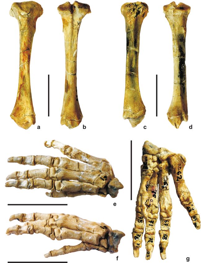
13Fig. 4. Skeletal elements of the hind limb. Bar equals 5 cm.
a – pelvis, view from below;
b, c – left femur; b – frontal view; c – view from behind;
d, e – right femur; d – frontal view; c – view from behind.
Table 7
Measurements of the pelvis (mm)
H. primigenia H. cristata
1. Length of the preserved part 165 154
2. Antero-posterior diameter of the acetabulum 28 21
3. Vertical diameter of the acetabulum 26 19
4. Ratio length of pelvis/length of skull 1/1.1 1/0.67
14Table 8
Measurements of the long bones of the hind limb; * – slightly damaged
Femur Tibia
H. primigenia H. cristata H. primigenia H. cristata
sin dex sin dex sin dex sin dex
1. Total length of the bone 157 157 128 129 152* 154 116 115
2. Proximal epiphysis
a) transversal diameter 51 50 39 39 39 39 30 30
b) antero-posterior diameter 20 21 19 19 30 30 20 20
3. Diaphysis
a) transversal diameter 22 22 15 15 14 14 10 10
b) antero-posterior diameter 15 15 15 14 17.5 17.5 12 11.8
4. Distal epiphysis
a) transversal diameter 36 36 31 31 28 28 22 22
b) antero-posterior diameter 40 41 34 37 21 20 14 12
5. Diameter of caput femori 21 21 18 18 – – – –
Tibia (Figs 5a, b) the preserved bones of the right limb, a “complete” ossa
pedis is formed consisting of tarsus, metatarsus and
As with the femur, at the left and right tibia the site of phalanges.
the cartilage (cartilago epiphysialis) is between the meta- Tarsus (Table 9). It includes calcaneus, astragalus, na-
physis and epiphysis. Increase in bone length is achieved viculare, cuboid and three cuneiformes, namely ecto-,
through this cartilage. Proximal epiphysis is formed of meso-, and endocuneiforme. Measurements of all these
condylus medialis and condylus radialis. Each of these skeletal elements are given in Table 9.
has a joint surface with the femur. In their midst is seen Calcaneus sin. It lies in the lower part of the foot. Its
outlined eminentia intercordilaris. Diaphysis, corpus tib- back part, tuber calcanei, is 28 mm long. It associated
iae, is triangular. The distal epiphysis ends with a well- with the Achilles tendon without a visible groove of this
preserved joint surface for connection to plantar bones, coupling. Sustentaculum is in medial position support-
i.e. the tarsus. Measurements of tibia are shown in Table ing the astragalus and bearing one of its three articulating
8. It indicates equal lengths of the femur and tibia. By surfaces. Only one of them is slightly concave. The three
contrast, in H. cristata tibia is shorther than femur. surfaces are divided by sulcus calcanei. The plantar sur-
face of tuber calcanei is damaged. The big processus of
Ossa pedis (bones of the foot) calcaneus is truncate in a manner that the head of astra-
galus is well seen at the distal end of calcaneus. A large
Bones of the foot of the left limb are better preserved. fasseta for the cuboid occurs at that end. The fasseta is
A part of them are missing. If one combines them with slightly concave.
Table 9
Measurements of the tarsus (mm)
Antero- Transv. Transv. Length of susten-
Transv. Vertical
posterior diameter at diameter at taculum up to the
diameter diameter
diameter trohlea head free end
calcaneus H. primigenia 48.0 27.0 – – 40.0 –
H. cristata 36.0 19.0 – – 31.0 –
astragalus H. primigenia 27.5 – 21.0 11.0 11.0 –
H. cristata 20.0 – 18.0 8.0 8.0 –
naviculare H. primigenia 19.5 12.5 12.5 – – 12.5
H. cristata 11.5 11.5 12.0 – – 12.0
endo-cuneiform H. primigenia 19.0 12.5 – – – 12.5
H. cristata 15.5 12.5 – – – 12.0
mеso-cuneiform H. primigenia 10.0 7.0 5.0 – – 5.0
H. cristata – – – – – –
ecto-cuneiform H. primigenia – – 9.0 – – 9.0
H. cristata 9.0 11.5 – – – –
cuboid H. primigenia 15.5 17.2 10.0 – – 16.0
H. cristata 12.0 12.5 10.0 – – 10.0
15Fig. 5. Skeletal elements of the hind limb. Bar equals 5 cm.
a, b – right tibia; a – view from the front; b – view from behind;
c, d – left tibia; c – view from the front; d – view from behind;
e, f, g – tarsus and metatarsus; e – dorsal view; f – lateral view; g – planar view
16Astragalus sin. All its components are well preserved Two fassetae are seen on the fibular surface – one for the
– caput, colum and corpus. Head is oval. Its transversal mesocuneiform, and other, distal for Mt – II.
diameter is 18 mm and the vertical diameter is 11 mm. Cuboid sin et dex. Cuboid is situated on the lateral side
At the distal surface there is a large convexe fasseta for of foot, just opposite to the anterior surface of the calca-
coupling with naviculare. At the upper surface of the cor- neus. The dorsal surfaces are slightly concave in proxi-
pus trochlea occurs with three shallow surfaces for ar- mal direction. The proximal surface connects the cuboid
ticulation with the tibia. The inner condylus is slightly with the calcaneus; it is flat and highly tilted forward.
higher than the outer. In plantar view, there are also three This slope rapidly disappears laterally. At the distal end
articulation surfaces of calcaneus which are divided by of this bone, a large fasseta connects it with the proximal
sulci. The two bones, calcaneus and astragalus, have ends of Mt – IV and Mt – V.
been found connected. Metatarsus (Figs 5e, f, g; Table 10). Metatarsus dex is
Naviculare sin. It is boat-shaped, flat and relatively thin. completely preserved. Metatarsus sin is represented by its
It becomes higher in medial direction. The upper end is Mt – III, and the proximal parts of Mt – IV and MT – V.
boat-shaped concave and forms a fasseta for the astra- Metatarsus – I dex. It is situated at medial position and
galus. The postero-external apophysis is signifigantly represents the greatly reduced metatarsus. Its head is
elevated. On the internal side there is a fasseta surving highly enlarged compared to both antero-posterior and
for connection with the cuboid. It is connected with the transversal dimensions. For this reason, the fasseta on
cuneiform bone through its plantar part. endocuneiform is also large. It is sloped and thus the first
Endocuneiform dex. This bone is long and highly flat- finger points inward. The form is arc-shaped, slightly
tened laterally. Its dorsal diameter is smaller than the convex forward. The distal trohlea is hemisphaerical.
plantar one. The bone is positioned over Mt – I by a Metatarsus – II dex. The connecting surface with me-
highly concave fasseta. Such a fasseta also exists on the socuneiforme is short and slightly concave. The head is
proximal surface making connection with the naviculare, elevated over the heads of Mt – I and Mt – III. In dorsal
however, this fasseta is not concave. On the fibular sur- view, the body of bone is quite straight, whereas in platar
face there are two fassetae – one proximal for the meso- view it looks arc-shaped, convex upwards. This impres-
cunieform, and other, smaller distal fasseta for Mt – II. sion is reinforced by the larger antero-posterior diameters
Mesocuneiform dex. Its antero-posterior diameter is lar- at its both ends. The corp has a form of highly flattened
ger than the transversal diameter, especially on the distal cylindre, widened at its ends. In distal direction, the bone
surface. The fasseta for connection with naviculare is til- widens transversally. The dorsal surface of trohlea is
ted back and remains flat throughout its length. In the sphaeroidal; the plantar surface is divided into two parts
distal part this bone is eleveted by Mt – II. by a crest, a structure which is seen is all metatarsalia.
Ectocuneiform sin et dex. This is the largest of the three Metatarsus – III sin et dex. It is slightly longer than Mt –
cuneiforms. It lies entirely over Mt – III and is apparen- II. The antero-posterior diameter of its head is longer than
tly wider than tall. Its distal fasseta for connection with Mt – II. The fasseta to connect with the ectocuneiforme is
Mt – III is strongly sloped. In dorsal view, it just lies on strongly sloped. To connect with Mt – IV the bone head
its fibular top creating an impression of diamond-shaped. is deeply concave and overthrusts the head of Mt – IV.
This impression is reinforced by the fact that the lateral The diaphysis is slightly more massive than Mt – II. In
surfaces are shorter than the anterior and posterior ones. dorsal view it is quite straight, whereas in plantar view
Table 10
Measurements of the metatarsus (mm)
Length of Antero-posterior Transversal diameter Transversal diameter
bone diameter of head of head of the distal end
Mt-I H. primigenia sin 20 9.0 8.5 6.8
dex – – – –
H. cristata – – – –
Mt-II H. primigenia sin 36.5 – 9.0 9.0
dex – – – –
H. cristata 27.5 14.0 – 5.5
Mt-III H. primigenia sin 41.5 – 12.0 10.5
dex 45.0 12.5 10.5 11.5
H. cristata 31.0 10.0 8.0 8.0
Mt-IV H. primigenia sin 39.5 14.0 12.0 10.5
dex – 12.0 10.0 –
H. cristata 31.0 9.0 6.0 7.0
Mt-V H. primigenia sin 38.0 11.0 17.0 10.0
dex – 9.0 17.0 –
H. cristata 29.0 10.0 7.0 8.0
17the diameter is smaller than the lateral diameter. Thus,
the bone looks oval and flattened. The distal epiphysis is
similar to Mt – II, but is clearly more symmetrical.
Metatarsus – IV sin et dex. Its shape and dimensions are
equal to MT – III. The only difference is that the connect-
ing fasseta to the cuboid is less tilted. Mt – IV is repre-
sented by only its head and a part of diaphysis.
Metatarsus – V sin et dex. It is slightly longer than Mt – II.
This bone is more flattened than the other metatarsalia. In
transeversal sections its diaphysis looks oval and flattened.
The head is strongly tilted outward and upward. The fas-
seta over the head curves in the same direction in order to
articulate with the cuboid. Mt – V dex is only represented
by its proximal part (head and part of diaphysis) which
show no differences with the well preserved Mt – V sin.
Sesamoidal bones. There is only one preserved sesa- Fig. 6. Image of living Hystrix cristata Linn
moidal bone from this group, beside the pattela of the
left limb which is a part of the knee. This single bone
was found lying between the distal trohlea of MT II and
the proximal fasseta of the first bone of second finger. cartilage plates (cartilago epiphysalis) passed before
Patella has a characteristic crescent-shaped outline and is completely ossified.
very small in size. The description, measurement and comparison of the
Phalanges dex (Table 11) fossil material suggest that the skeleton of Hadzhidimovo
Only fingers of the right hind leg are found. These are locality was a quite big animal which univocally belongs
five in number. Each finger has three ossicles, namely to Hystrix primigenia.
proximalis, medialis and distalis. The first finger only has
two ossicles. Proximal ossicles are the longest and distal
ossicles are the shorter. Their arched shape is more obvi- STRATIGRAPHIC AND GEOGRAPHIC
ous than metatrsalia. Proximalia of all fingers are flat- DISTRIBUTION
tened dorso-ventrally. They have a concave pit to articu-
late with the corresponding metatarsus. The distal part of The stratigraphic range of Hystrix (H.) primigenia was
this pit is the trohlea which surves for connection with summarised by Alcala and Montoya (1998), van Weers
the medial ossicle of the finger. These second phalanges and Rook (2003) and Lopatin et al. (2003). The strati-
resemble the proximal ossicles, however, being slightly graphic range of the species is from the early-middle
shorter. The third phalanges (distalis, the nails) are highly Turolian (transition MN 11/12) to the late Ruscinian
thickened in proximal direction. The presence of a big tu- (MN 15). H. primigenia was reported so far from the fol-
ber on their plantar surface contributes to this impression. lowing localities in Europe:
The third phalanges are arc-shaped upwards. They do not Greece: Pikermi, Samos, Chomateras, Palmyopotamus,
sharpen to the distal end as might be expected. Their dis- all four localities middle Turolian in age (MN 12) and
tal ends are as thick as the beginninig of diaphysis. These Dytico – late Turolian (MN 13) according to de Bonis et
nails have had likely a reactive character. They came al. (1992); Koufos (2006); Koufos et al. (2011)
back to the distal ossicles when not used. This assump- Albania: late Miocene to early Pliocene (Festani et al.,
tion is based on the presence of a deep pit on the nails 1997);
dorsal surface. Such a device, much more sophisticated, FYR Macedonia: Umen Dol, (Garevski, 1956);
of course, is seen in cats. Pits similar but much shallower Turkey: Kemiklitepe, Turolian (MN 11, MN 12) (de
can be seen in the extant species H. cristata. Bonis et al., 1994), also Bayirköy, Gülpınar, Şerefköy;
As a whole, in the majority of metatarsus and phalan Moldovia: Taraklia (Riabinin, 1929) as H. bessarabica,
ges bones grooves are visible (in places clearly) where a junior synonym of H. primigenia;
Table 11
Measurements of the phalanges (mm); H. pr. = Hystrix primigenia, H. cr. = Hystrix cristata
1 2 3 4 5
H. pr. H. cr. H. pr. H. cr. H. pr. H. cr. H. pr. H. cr. H. pr. H. cr.
Length of the
– 17.0 32.0 13.0 42.0 15.0 40.0 15.0 30.0 15.0
I phalange
Length of the
14.0 8.5 10.0 7.0 12.0 8.5 19.0 9.0 16.0 9.0
II phalange
Length of the
9.0 7.5 14.0 12.0 13.0 10.0 17.0 12.0 15.6 11.0
III phalange
18European Russia: Morskaya 2, late Turolian (MN 13) Hystrix aryannesis Sen, 2001b
(Lopatin et al., 2003); Hystrix caucasica (Argyropulo, 1939)
Poland: Weze, early Plocene, Ruscinian (MN 14) H. (H.) parvae is a small porcupine which appeared
(Sulimski, 1960); before H. primigenia. It is known from the latest Vallesian
Italy: Brisighella, latest Miocene (Masini and Rook, (MN 10) and Early Turolian (MN 11) of Europe. The lo-
1993); cality Kohfidish in Austria which was thought to be Late
Spain: Las Casiones (Alcala and Montoya, 1998); Vallesian (MN 10) in age was recently dated as Early
Algeria: Menacer, late Miocene (see van Weers and Turolian (MN 11) by Daxner-Höck and Höch (2009).
Rook, 2003); H. (H.) suevica Schlosser, 1884 is a junior synonym.
India: As H. sivalinsis Lydekker, a junior synonym of H. (H.) depereti was first described from the classi-
H. primigenia (Sen, 2001 b); cal material of Perpignan, France. This is the closest fos-
Bulgaria: Kalimantsi, Struma River Basin, SW sil species to H. primigenia. The stratigraphic range of
Bulgaria, MN12 (Sen and Kovachev, 1987); Strumyani, H. depereti is slightly younger than H. primigenia. Van
Struma River Basin, SW Bulgaria, MN 12 (Geraads et Weers and Rook (2003) re-examined fossil collections
al., 2011). The first description of H. primigenia from and considered that the material previously described
Hadzhidimovo locality was by Kovachev and Atanasova as H. primigenia from Samos Island in Greece, Çoban
(2008). Spassov (2000) mentioned this species in the fau- Pınar in Turkey, Kalimantsi in Bulgaria, Weze in Poland,
nal list of Hadzhidimovo I locality. Brisighella in Italy, Las Casiones in Spain, and Menacer in
Vekua et al. (2010) argued that the specimen report- Algeria, should actually be allocated to H. depereti. Lopatin
ed earlier as H. cf. primigenia by Vekua (1972) from et al. (2003) did not accept that and maintained the name
Kvabeba locality in Georgia resembles H. primigenia but H. depereti only for the material of Perpignan, France.
is not well preserved for reliable identification. The age H. (H.) aryanensis Sen is known from its type lo-
of Kvabeba locality is, however, lower Villafranckian cality Molayan, Afganistan, in the Middle Turolian (MN
(MN 16). All other localities of H. primigenia in Europe, 12) (Sen, 2001b). Later it was also documented in the
Asia Minor and North Africa are up to the late Ruscinian Ivand locality, Iran, by Sen and Purabnishemi (2010)
in age (MN 15). again from the Middle Turolian (MN 12).
Van Weers and Rook (2003) made a taxonomical re- Hystrix caucasica (Argyropulo) from Kolyakino lo-
vision of the late Miocene (Turolian) and early Pliocene cality in Northern Caucasus is from the early Ruscinian
(Ruscinian) large-sized fossil porcupines and allocated (MN 14) (Lopatin et al., 2003).
them in the subgenus Hystrix (Hystrix). Those authors
considered Hystrix syvalinensis Lydekker and Hystrix Acknowledgments
besarrabica Riabinin as junior synonyms of H. primi
genia. H. syvalinensis was reported from Siwalkis, India, The author expresses his gratitude to Prof. Nikolay
by Sen 2001b, and H. bessarabica – from Moldovia by Spassov (NMNH, Sofia) for providing him with a skel-
Riabinin (1929), both from the Turolian. eton of extant Hystrix cristata, as well as with literature
Other Miocene and Early Pliocene fossil species of on the topic of this paper. Dr Radostina Mateeva (Institu
large porcupines (Hystrix) of Europe, Asia Minor and für Transfusion Medizin, Berlin) also helped in provid-
Middle East are: ing with articles on Hystricidae. Editorial Board of the
Hystrix parvae (Kretzoi, 1951) journal Geologica Balcanica kindly translated and edited
Hystrix depereti Sen, 2001a this paper.
REFERENCES
Alcala, L., Montoya, P. 1998. Hystrix primigenia (Wagner, Daxner-Höck, G., Höch, E. 2009. New data on Eomidae and
1848) (Rodentia, Mammalia) del Mioceno superior MN13, Gliridae (Rodentia, Mammalia) from the late Miocene of
de las Casiones (Fosa de Teruel, España). Revista Española Austria. Annalen des Naturhistorischen Museums in Wien
de Paleontologia 13, 139–147. 111A, 375–444.
de Bonis, L., Bouvrain, G., Geraads, D., Koufos, D. 1992. Depéret, C. 1890. Les animaux pliocènes de Roussillon.
A skull of Hystrix primigenia from the late Miocene of Mémoires de la Société Géologique de France, Paléonto
Macedonia (Greece). Neues Jahrbuch für Geologie und logie 3, 43–48.
Paläontologie, Monatshefte 2, 75–87. Garevski, R. 1956. Neue Fundstellen der Pikermifauna in
de Bonis, L., Bouvain, G., Geraads, D., Koufos, G., Sen, S., Mazedonien. Acta Musei Macedonici Scientiarum Natu
Tassy, P. 1994. Les gisements de mammiferès du Miocène ralium 4, 69–96.
supérieur de Kemiklitepe, Turquie. 11. Biochronologie, Gaudry, A. 1862. Animaux fossils et geologie de l’Attique.
paléoecologie et relations paléobiogeographiques. Bulletin Savy, F. (ed.). Société Géologique de France, 474 pp.
du Musée National d’Histoire Naturelle Paris, 4e série (C) Geraads, D., Spassov, N., Hristova, L., Markov, G.N., Tzankov,
16, 225–240. T. 2011. Upper Miocene mammals from Strumyani, South-
Corbet, G.B., Jones, L.A. 1965. The specific characters of Western Bulgaria. Geodiversitas 33(3), 451–484.
the crested porcupines, subgenus Hystrix. Proceedings Greenwood, M. 1955. Fossil Hystricidae from the Makapan
Zoological Society London 144(2), 285–300. Valley, Transvaal. Paleontologia Africana 3, 77–85.
19Festani, A. B., Pavlakis, P. P., Symeonides, N. 1997. First
����������
dis- Sen, S. 2001b. Rodents and insectivores from the Upper
covery of Hystrix primigenia Wagner from the Late Miocene Miocene of Molayan, Afganistan. Palaeontology 44(5),
to Early Pliocene deposits of Shahinova, Berat, South-West 913–922.
Albania. Annalen des Naturhistorischen Museums in Wien Sen, S., Kovatchev, D. 1987. The porcupine Hystrix primigenia
98 A, 155–172. (Wagner) from Late Miocene of Bulgaria. Proceedings of
Frenkel, H. 1970. Hystrix angressi sp.nov., a large fossil por- the Koninklijke Nederlandse Akademie van Wetenschappen
cupine from the Levalloiso-Mousterian of the Geula Cave. Series B, 90(4), 317–323.
Israel Journal of Zoology 19(1), 51–82. Sen, S., Purabnishemi, Z. 2010. First porcupine fossils (Mam
Harlé, E. 1910. Porc-épic quaternaire des environs de Montréjeau malia, Rodentia) from the Late Miocene of NW Iran, with
(Haute-Garonne). Bulletin de la Sociètè Géologique de notes on the Late Miocene – Pliocene dispersal of porcu-
France 4(10), 740–744. pines. Paläontologische Zeitschrift 84, 239–248.
Ivanov, D. 1995. Palynological data on the fossil flora from the Spassov, N. 2000. The Turolian Hipparion fauna and the
village of Ognjanovo, Southwestern Bulgaria. Phytologia character of the environment in the Late Miocene of West
Balcanica 1(2), 3–14. Bulgaria. Review Bulgarian Geological Society 61(1–3),
Ivanov, D., Utescher, J., Ashraf, R., Mossbruger, V., Bozukov, 47–59.
V., Djorgova, N., Slavomirova, E. 2011. Late Miocene Spassov, N. 2002. The Turolian megafauna of West Bulgaria
palaeoclimate and ecosystem dynamics in Southwestern and the character of the Late Miocene “Pikermian biome”.
Bulgaria − a study based on pollen data from the Gotse- Bollettino della Società Paleontologica Italiana 41(1), 69–81.
Delchev Basin. Turkish Journal of Earth Sciences 21, Spassov, N., Tzankov, Tz., Geraads, D. 2006. Late Neogene
187–211. stratigraphy, biochronology, faunal diversity and envi-
Koliadimou, K., Koufos, G.D. 1990. The Hystricidae of the ronments of South-West Bulgaria (Struma river valley).
Pleistocene of Macedonia (Greece) and a review of the Geodiversitas 28(3), 417–441.
European representatives of the family. Bulletin of the Stoyanov, I., Nenov, T., Stoykov, S. 1974. Geological structure
Geological Society of Greece 25(2), 453–471. and tectonic development of the Mesta graben. Annual of
Koufos, G.D. 2006. The Neogene mammal localities of Greece: Sofia University, Faculty of Geology and Geography 66,
Faunas, chronology and biostratigraphy. Hellenic Journal Series 1, Geology, 85–100 (in Bulgarian).
of Geosciences 41, 183-214. Sulimski, A. 1960. Hystrix primigenia (Wagner) in the Plio
Koufos, G.D., Kostopoulos, D.S., Vlachou, T.D., Konidaris, cene fauna from Weze. Acta Palaeontologica Polonica 3,
G.E. 2011. A synopsis of the late Miocene mammal fauna of 319–335.
Samos Island, Aegean Sea, Greece. Geobios 44, 237–251. Temniskova-Topalova, D., Ognjanova-Rumenova, N. 1983.
Kovatchev, D., Atanasova, N. 2008. On the presence of genus Diatom fossils from fresh-water Neogene diatomites in the
Hystrix (Rodentia) in the Upper Maeotian from the region Gotse Delchev region. Fitologia 22, 29–45.
south of Hadzhidimovo town, Blagoevgrad district. Review Vatsev, M. 1980. Lithostratigraphy of the Neogene sedimentary
of the Bulgarian Geological Society 69, 1–3, 21–26 (in rocks of the Gotse Delchev Basin. Annual of the University
Bulgarian). of Mining and Geology 25(2), 103–115 (in Bulgarian).
Lopatin, A.V., Tesakov, A.S., Titov, V.V. 2003. Late Miocene Vatsev, M., Petkova, A. 1996. New data about the stratigraphy
– early Pliocene porcupines (Rodentia, Hystricidae) from of the Neogene in Gotse Delchev Basin. Annual of the
South European Russia. Russian Journal of Theriology University of Mining and Geology 41(1), 13–20 (in Bul
2(1), 26–32. garian with English abstract).
Malez, M. 1963. Kvartarna fauna Pecine veternice u medvedni- Vekua, A.K. 1972. The Kwabeb vertebrate fauna of Akchag.
ci. Jugoslavska Akademia Znanosti i Umjetnosti, Odjel za Gruzinskaya Akademiya Nauk, Moskva, 359 pp.
prirodne nauke,133. Vekua, A., Bendukidze, O., Bukhsianidze, M., Vanishvili, N.,
Massini, F., Rook, L. 1993. Hystrix primigenia (Mammalia, Augusti, J., Martinez-Navaro, B., Rook, L. 2010. Porcupine
Rodentia) from the Late Messinian of the Monticino of the Late Neogene and Quaternary of Georgia. Bulletin of
gypsum quarry (Faenza, Italy). Bolletino della Società the Georgian National Academy of Sciences 4(3), 140–149.
Paleontologica Italiana 32(1), 79–87. van Weers, D.J. 1985. Hystrix gigantea, a new fossil porcu-
Nenov, T., Stoyanov, I., Stoykov, S. 1972. The Pliocene pine species from Yava (Rodentia, Hystricidae). Sencken
and Quaternary in the Gotze Delchev Valley. Review bergiana Lethaea 66(1/2), 111–119.
of the Bulgarian Geological Society 32(2), 195–204 (in van Weers, D.J. 1994. The porcupine Hystrix refossa Gervais,
Bulgarian). 1982 from the Plio-Pleistocene of Europe, with notes on
Niethammer, J. 1982. Hystrix cristata Linnaeus, 1758. Hand other fossil and extant species of the genus Hystrix. Scripta
buch der Saugetiere Europas, Band 2/1, 588–603. Geologica 106, 35–52.
Nikolov, I. 1985. Catalogue of the localities of Tertiary mammals van Weers, D.J. 2005. A taxonomy of the Pleistocene Hystrix
in Bulgaria. Paleontologia, Stratigrafia i Litologia 21, 43–62. from Eurasia with notes on the evolution of the family.
Riabinin, A. 1929. Fauna de mammifères de Taraklia. Carnivora Contributions to Zoology 74 (3/4), 301–312.
vere, Rodentia, Subungrilata. Ttudy Geologicheskogo Mu van Weers, D.J., Montoya, P. 1996. Taxonomy and stratigraph-
zeya Akademii nauk SSSR, v. 5, 130–131. ic record of the oldest European porcupine Hystrix parvae
Sen, S. 1994. Les gisements de mammiferès du Miocène (Kretzoi, 1991). Proceedings of the Koninklijke Nederlandse
supérieur de Kemiklitepe, Turquie. 5. Rongeurs, Tubuli Akademie van Wetenschappen 99B(1–2), 131–141.
dentès et Chalicothéres. Bulletin du Musée National van Weers, D.J., Rook, L. 2003. Turolian and Ruscinian por-
d’Histoire Naturelle Paris, 4e serie (C) 16, 97–111. cupines (genus Hystrix, Rodentia) from Europe, Asia and
Sen, S. 1996. Late Miocene Hystricidae in Europe and Anatolia. North Africa. Paläontologische Zeitschrift 77(1), 95–113.
The evolution of western Euroasian Neogene Mammal fau Yaneva, M., Ognjanova, N., Nikolov, G. 2002. Palaeoecological
nas. Columbia University Press, New York, 263–265. development of the Gotse Delchev Basin during the
Sen, S. 2001a. Early Pliocene porcupine (Mammalia, Roden Neogene, South-west Bulgaria. Proceedings International
tia) from Perpignan, France: a new systematic study. Scientific Conference in Memory of Prof. Dimitar Jaranov,
Geodiversitas 23(2), 303–312. Varna 2002, 36–47 (in Bulgarian with English abstract).
20You can also read