Acetaldehyde exposure underlies functional defects in monocytes induced by excessive alcohol consumption - Nature
←
→
Page content transcription
If your browser does not render page correctly, please read the page content below
www.nature.com/scientificreports
OPEN Acetaldehyde exposure underlies
functional defects in monocytes
induced by excessive alcohol
consumption
Shunsuke Shiba1, Nobuhiro Nakamoto1*, Po‑Sung Chu1, Keisuke Ojiro1, Nobuhito Taniki1,
Akihiro Yamaguchi1, Rei Morikawa1, Tadashi Katayama1, Aya Yoshida1, Ryo Aoki1,
Toshiaki Teratani1, Takahiro Suzuki1, Takeshi Miyamoto2, Sachiko Hara3, Akira Yokoyama3 &
Takanori Kanai1*
Increased intestinal permeability and hepatic macrophage activation by endotoxins are involved in
alcohol-induced liver injury pathogenesis. Long-term alcohol exposure conversely induces endotoxin
immune tolerance; however, the precise mechanism and reversibility are unclear. Seventy-two
alcohol-dependent patients with alcohol dehydrogenase-1B (ADH1B, rs1229984) and aldehyde
dehydrogenase-2 (ALDH2, rs671) gene polymorphisms admitted for alcohol abstinence were enrolled.
Blood and fecal samples were collected on admission and 4 weeks after alcohol cessation and were
sequentially analyzed. Wild-type and ALDH2*2 transgenic mice were used to examine the effect
of acetaldehyde exposure on liver immune responses. The productivity of inflammatory cytokines
of peripheral CD14+ monocytes in response to LPS stimulation was significantly suppressed in
alcohol dependent patients on admission relative to that in healthy controls, which was partially
restored by alcohol abstinence with little impact on the gut microbiota composition. Notably,
immune suppression was associated with ALDH2/ADH1B gene polymorphisms, and patients with
a combination of ALDH2*1/*2 and ADH1B*2 genotypes, the most acetaldehyde-exposed group,
demonstrated a deeply suppressed phenotype, suggesting a direct role of acetaldehyde. In vitro LPS
and malondialdehyde-acetaldehyde adducted protein stimulation induced direct cytotoxicity on
monocytes derived from healthy controls, and a second LPS stimulation suppressed the inflammatory
cytokines production. Consistently, hepatic macrophages of ethanol-administered ALDH2*2
transgenic mice exhibited suppressed inflammatory cytokines production in response to LPS
compared to that in wild-type mice, reinforcing the contribution of acetaldehyde to liver macrophage
function. These results collectively provide new perspectives on the systemic influence of excessive
alcohol consumption based on alcohol-metabolizing enzyme genetic polymorphisms.
Alcohol and its metabolites induce systemic damage such as oxidative stress, hepatocyte injury, and mitochon-
drial damage. Habitual and excessive alcohol consumption gives rise to alcohol-related liver diseases (ALDs)
including steatohepatitis, liver fibrosis, and liver cirrhosis. ALD remains the leading cause of death due to alcohol
worldwide.
To date, many studies have tried to elucidate the pathogenesis of ALD from the viewpoint of the gut-liver
axis1,2. Alcohol consumption results in high intestinal permeability, the overgrowth of gram-negative bacteria in
the proximal small intestine and increased circulating endotoxin following bacterial translocation3. Endotoxins
are pathogen-associated molecular patterns (PAMPs) recognized by Toll-like receptors (TLRs). TLR4 and CD14
are essential components for macrophages/monocytes activation by circulating lipopolysaccharides (LPS)4, the
major components of the outer membrane of gram-negative bacteria. A major innate immune response of
TLR4 binding is NF-kB-mediated activation of pro-inflammatory cytokines such as tumor necrosis factor-alfa
(TNF-α) and IL-65–8.
1
Division of Gastroenterology and Hepatology, Department of Internal Medicine, Keio University School of
Medicine, Tokyo 1608582, Japan. 2Department of Orthopedic Surgery, Kumamoto University, Kumamoto,
Japan. 3National Hospital Organization Kurihama Medical and Addiction Center, Kanagawa, Japan. *email:
nobuhiro@z2.keio.jp; takagast@z2.keio.jp
Scientific Reports | (2021) 11:13690 | https://doi.org/10.1038/s41598-021-93086-y 1
Vol.:(0123456789)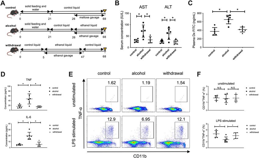
www.nature.com/scientificreports/
Long-term alcohol consumption has negative effects on host immune functions, which might contribute
to suppressed immunity and potential susceptibility to systemic infection9. The negative effects of alcohol and
its metabolites on monocytes have been studied both in humans and in murine models. These studies have
demonstrated the evidence of reduced pro-inflammatory cytokine production in response to LPS stimulation
and dysregulated antigen presentation on monocytes owing to chronic alcohol consumption10–12; however, the
precise mechanism and reversibility with alcohol withdrawal still remain unclarified. Human peripheral blood
monocytes have been classified according to their surface expression patterns of CD14 and CD16 into the
following three major subsets: classical ( CD14highCD16−), intermediate ( CD14highCD16+), and non-classical
(CD14lowCD16+)13. These subsets are functionally distinct and play different roles in various diseases14,15, however,
the contribution of each cell subset to ALD pathogenesis has not been well studied.
Acetaldehyde is a well-known highly toxic metabolite of alcohol. Consumed alcohol is mainly metabolized
by alcohol dehydrogenases (ADHs) or cytochrome P450 2E1 (CYP2E1) to acetaldehyde, which is then metabo-
lized to acetate by aldehyde dehydrogenases (ALDHs). In East Asians, the fast-metabolizing form of ADH1B is
encoded by ADH1B*2 (rs1229984), affecting more than 90% of the Japanese population16. ALDH2 is a major
enzyme in acetaldehyde oxidation in humans and its genetic polymorphism (rs671) determines blood or tissue
acetaldehyde concentration after alcohol consumption. Individuals with ALDH2 *1/*2 or ALDH2 *2/*2 polymor-
phisms have much lower activity than those with homozygous ALDH2 *1/*1. Approximately half of the popula-
tion in East Asia possesses the ALDH2 *2 allele, in contrast to non-Asians, who rarely possess t his17. ALDH2*2
allele carriers show high acetaldehyde concentrations after alcohol c onsumption18, and extensive DNA damage
is induced by chronic excessive d rinking19,20. Acetaldehyde accumulated in the intestine disrupts the barrier
function, which potentially involves the gut–liver axis21,22. In addition to the promotion of ethanol-induced gut
barrier dysfunction in mice with ALDH2 deficiency21, acetaldehyde impairs microtubule-dependent protein traf-
ficking pathways leading to hepatocyte b allooning23, results in the formation of immunogenic protein a dducts24,
and increases hepatic stellate cell activation and production of fibrillar collagen25. In contrast, a meta-analysis
of Asian studies has shown a strong protective effect of ALDH2 deficiency against alcoholic liver cirrhosis as
well as alcohol dependence26. Furthermore, the results of large surveys of Japanese alcohol-dependent (AD)
patients have demonstrated that the inactive ALDH2*1/*2 genotype is associated with a lower risk of alcoholic
liver cirrhosis27,28. These conflicting findings suggest that acetaldehyde is associated with both fascinating and
protective aspects with respect to the development of alcoholic cirrhosis, which prompted us to examine the role
of acetaldehyde as an inflammatory mediator, especially in regulating immune response during alcohol exposure.
In the current study, we examined changes in immunological impairment of monocytes in AD subjects follow-
ing alcohol withdrawal. We unexpectedly noticed that functional impairment of monocytes was closely associ-
ated with genetic polymorphisms in alcohol-metabolizing enzymes and thus explored the potential mechanism
involved using in vitro assays and murine models.
Material and methods
Patients and samples. This study included 72 Japanese male AD patients (average age 52.4 years) who
were admitted to the National Hospital Organization Kurihama Medical and Addiction Center for the treatment
of alcoholism (Table 1). They fulfilled the following criteria: continued to drink more than 60 g/day of ethanol,
did not use alcohol-aversive drugs, were not related to another etiology of liver disease (hepatitis B, C, PBC, and
AIH), and had never been admitted for abstinence previously. Blood samples were collected early in the morn-
ing, after overnight fasting, on the next day of admission, and at the end of the 4-week of hospital stay. A BD
vacutainer cell preparation tube™ was used to isolate peripheral blood monocytes (PBMCs), which were stored
at − 80 °C. Thirteen PBMC samples as controls were collected from healthy male donors without drinking habits
and were processed. The institutional review board of Keio University School of Medicine and National Hospital
Organization Kurihama Medical and Addiction Center approved all human studies (No. 20140211) according
to the guidelines of the 1975 Declaration of Helsinki (2008 revision). The study subjects were prospectively
recruited, and each subject provided prior written informed consent for blood sampling, study participation,
and analysis of clinical data.
ADH1B and ALDH2 genotypes. The DNA of each subject was extracted from their blood samples using a
QIAamp DNA Blood Mini Kit (Qiagen, USA). Polymerase chain reaction–restriction fragment length polymor-
phism methods were used to analyze lymphocyte DNA samples from all subjects, without knowledge of their
status, to determine their ADH1B and ALDH2 genotypes29.
Isolation of human PBMCs. Human PBMCs were isolated by density centrifugation. Human blood was
collected into BD Vacutainer CPT (BD, USA) tubes and centrifuged at 470× g for 20 min at room temperature.
PBMCs were collected at the interphase, washed, and resuspended in FACS buffer.
Flow cytometric analysis. We performed cell surface staining to characterize the cell populations in
each sample. Briefly, cells were incubated with specific fluorescence-labeled monoclonal antibodies at 4 °C for
30 min. CD14 and CD16 were used to identify the monocyte subpopulation. CD3, CD56, BDCA2, and CD123
were used for T cells, NK cells, NKT cells, and plasmacytoid dendritic cells. Events were acquired with a FACS
Canto II (Becton Dickinson, USA) and analyzed with FlowJo software v.10.3 (Tree Star Inc., USA) (https://
www.flowjo.com/solutions/flowjo). The following antibodies were used for cell surface staining: PE-Cy7 Mouse
Anti-Human CD14 (BD Pharmingen), APC-Cy7 Mouse Anti-Human CD16 (BD Pharmingen), FITC Mouse
Anti-Human CD3 (BD Pharmingen), PE-Cy7 Mouse Anti-Human CD56 (BD Pharmingen), APC anti-human
CD303 (BDCA-2) Antibody (BioLegend), and PerCP/Cyanine5.5 anti-human CD123 Antibody (BioLegend).
Scientific Reports | (2021) 11:13690 | https://doi.org/10.1038/s41598-021-93086-y 2
Vol:.(1234567890)
www.nature.com/scientificreports/
Background characteristics Mean ± SD
Age, yrs 52 ± 4
Cirrhosis (Y/N) 8/64
Child Pugh score (A/B/C) 5/3/0
AST, IU/L 86 ± 16
ALT, IU/L 51 ± 8
γ-GTP, IU/L 370 ± 61
Alb, g/dL 4.1 ± 0.6
T-bil, mg/dL 1.0 ± 0.4
PT-INR 1.02 ± 0.22
Type IV collagen, ng/mL 240 ± 20
FBS, mg/dL 92 ± 10
WBC, × 103/μL 6.0 ± 0.3
Hb, g/dL 13.8 ± 0.2
Plt, × 104/μL 20.3 ± 1.1
TC, mg/dL 182 ± 28
HDL-C, mg/dL 58 ± 9
TG, mg/dL 132 ± 14
Ethanol (g/day) n
0–49 2
50–99 20
100–149 32
150–199 8
200–249 3
250–299 3
300–349 4
ADH1B (His47Arg)
ALDH2 (Glu487Lys) *1/*1 *1/*2 *2/*2 Total
*1/*1 14 13 17 44
*1/*2 7 11 10 28
Total 21 24 27 72
Table 1. Characteristics of patients with alcohol dependence.
Isolation of CD14+ monocytes and cell stimulation. CD14+ monocytes were isolated by immuno-
magnetic positive selection using the EasySep™ Human CD14 Positive Selection Kit (STEMCELL technologies,
Canada). Acquired C D14+ monocytes were seeded in 24-well plates at 5 × 10⁴ cells/well and stimulated with
5 ng/mL LPS for 24 h at 37 °C.
RT‑qPCR. Total RNA was extracted from cells using TRIzol reagent (Invitrogen, USA), as per the manu-
facturer’s protocol. Complementary DNA was synthesized by reverse transcription using the iScript™ cDNA
Synthesis Kit (Bio-Rad, Hercules, USA). To measure the quantity, real-time PCR was performed using the SYBR
green RT-qPCR kit with the predesigned primers. The level of target gene expression was normalized against
glyceraldehyde-3-phosphate dehydrogenase (GAPDH) expression in each sample. The following primers were
used for SYBR green assays (BIO-RAD, Japan): TNF (qHsaCEP0040184), IRAK-1 (qHsaCEP0057865), IRAK-3
(IRAK-M; qHsaCIP0031947), CD274 (PD-L1;qHsaCIP0039192), and PDCD1LG2 (PD-L2; qHsaCID0015625).
Quantification and analysis of cytokines. Cytokine production in cell supernatants was measured
using the BD™ Cytometric Bead Array (CBA) Human Th1/Th2/Th17 cytokine kit (BD, USA) and were acquired
on the BD FACS Canto™ II flow cytometer. Acquired data were analyzed using FCAP Array™ software v.3.0 (BD,
USA) (https://www.bdbiosciences.com/jp/applications/research/bead-based-immunoassays/analysis-software/
fcap-array-software-v30/p/652099).
Fecal sample collection and DNA extraction. Fresh fecal samples were collected using stool collection
tubes and an anaerobiosis generator was added to the samples to favor the preservation of anaerobic bacteria
at the outpatient clinic of Keio University Hospital. We selected four AD patients who achieved the recovery
from diminished cytokines production following alcohol abstinence. The samples were processed immediately
and frozen at − 80 °C for bacterial preservation. Bacterial DNA was isolated as described previously30. In brief,
bacterial DNA was isolated by the enzymatic lysis method using lysozyme (Sigma-Aldrich, USA) and achromo-
peptidase (Wako). DNA samples were then purified by treating with ribonuclease A (Wako, Japan), followed by
Scientific Reports | (2021) 11:13690 | https://doi.org/10.1038/s41598-021-93086-y 3
Vol.:(0123456789)www.nature.com/scientificreports/
precipitation with 20% polyethylene glycol solution (PEG6000 in 2.5 M sodium chloride). DNA was then pel-
leted by centrifugation, rinsed with 75% ethanol, and dissolved in tris–EDTA buffer.
16S rRNA metagenomic analysis. The hypervariable V3–V4 region of the 16S gene was amplified using
Ex Taq Hot Start (TAKARA Bio Inc., Japan) and subsequently purified using AMPure XP (Beckman Coulter,
USA). Mixed samples were prepared by pooling approximately equal amounts of each amplified DNA sample
and sequenced using the Miseq Reagent Kit V3 (600 Cycle) and Miseq sequencer (Illumina, USA), according to
the manufacturer’s instructions. Sequences were analyzed using the QIIME2 software package v.2019.10 (https://
qiime2.org) 31,32. Paired-end sequences were joined using a fastq-join tool in the ea-utils software package
(https://doi.org/10.2174/1875036201307010001). High-quality sequences per sample (15,000) were randomly
chosen from quality filter-passed sequences. After trimming both primer sequences using cutadapt (https://doi.
org/10.14806/ej.17.1.200) followed by chimera detection by the USEARCH de novo m ethod33, the sequences
were assigned to operational taxonomic units (OTUs) using the UCLUST a lgorithm34 with a sequence identity
threshold of 96%. Taxonomic assignments of each OTU were made by similarity searching against the publicly
available 16S (RDP version. 10.27 and CORE update 2 September 2012) and NCBI genome database using
the GLSEARCH program. Data were rarefied to 10,000 sequences per sample, as determined by the rarefac-
tion curves. Relative abundances of the community members were determined using the rarefied data. UniFrac
analysis was performed as described previously35. To determine bacterial taxonomy that explained differences
between conditions, the linear discriminant analysis effect size method was u sed36.
Preparation of MAA‑Alb and in vitro stimulation. As previous reported, MAA-Alb was prepared by
reacting 1.0 mM acetaldehyde and 1.0 mM malondialdehyde with 2 g/L of bovine serum albumin in 0.1 M phos-
phate buffer containing 2 mM diethylenetriaminepentaacetic acid and 2 mM phytic acid at 37 °C for 3 days and
ultrafiltration of the phosphate buffer37. CD14+ monocytes collected from healthy donors were stimulated with
LPS (1 ng/mL), MAA-Alb (10–25 µg/mL), or the combination for 18 h in vitro. In some experiments, CD14+
monocytes following the first treatment were further stimulated with 5 ng/mL LPS for 24 h in vitro, followed by
cytokine quantification in supernatants.
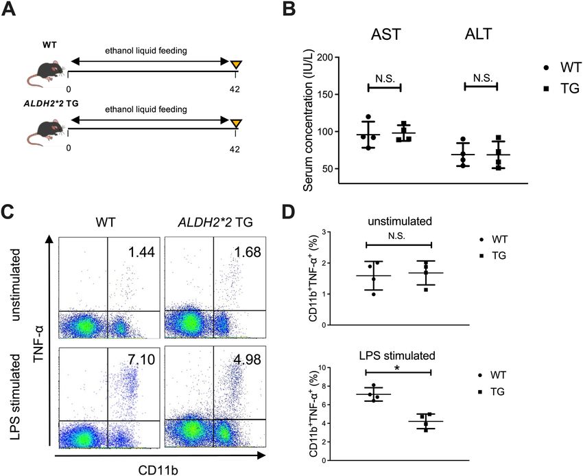
Animal experiments. Animals and chronic‑plus‑binge ethanol feeding. We used 6-week-old male
C57BL/6 J (wild-type; WT) mice weighing between 17 and 19 g. Mice were obtained from CLEA Japan Inc. The
mice were randomly divided into three groups, the control, alcohol group, and withdrawal groups (Fig. 5A),
treated differently as follows:
• The control group received the control Lieber-DeCarli d iet38 ad libitum for 6 weeks. Maltose solution was
administered via gavage twice per week.
• The alcohol group received the control Lieber-DeCarli diet ad libitum for the first week and the ethanol
Lieber-DeCarli diet containing 5% (v/v) ethanol for the following 6 weeks. Ethanol solution was administered
via gavage twice per week.
• The withdrawal group was treated the same as that in the alcohol group for 6 weeks and received the control
Lieber-DeCarli diet for the following 3 weeks.
To ensure the same amounts of ethanol intake, each mouse was housed individually and a consistent ethanol
Lieber-DeCarli diet containing 5% (v/v) ethanol was provided every day. We prepared a 31.5% (v/v) ethanol
solution equivalent to 0.25 g/mL ethanol and 45.0% (w/v) maltose dextrin solution for the gavage. The gavage
dose was 20 µL/g body weight, and each mouse received 5 g ethanol/kg body weight or isocaloric maltose. We
performed gavage between 7:00 and 9:00, and mice were sacrificed 24 h after the last g avage38.
ALDH2*2 transgenic (TG) mice generated as described in prior studies were kindly provided by the Depart-
ment of Orthopedic Surgery, Keio University School of Medicine39. Four WT mice and four TG mice were
included in the alcohol group for 6 weeks and their serum and liver were collected and analyzed. All experiments
were approved by the regional animal study committees (Keio University, Tokyo, Japan) and were performed in
accordance with the ARRIVE guidelines and institutional guidelines and Home Office regulations.
Serum transaminase. As a marker of liver injury, serum aspartate aminotransferase and alanine aminotrans-
ferase were measured by a simple colorimetric method using the Fuji Dri-Chem 3500™.
Intestinal permeabilization in vivo. We prepared 125 mg/mL fluorescein isothiocyanate-dextran (FITC-Dx)
solution. After 4 h fasting, mice were gavaged with the FITC-Dx solution at 4 µL/g body weight (500 mg FITC-
Dx/kg body weight). Blood samples were collected 4 h after gavage, and serum FITC-Dx levels were measured
at an excitation wavelength of 485 nm and emission wavelength of 528 nm.
Serum cytokine concentrations. The serum cytokine concentrations were measured using a BD™ Cytometric
Bead Array (CBA) Mouse Th1/Th2/Th17 cytokine kit. Data acquisition and analysis were performed as that used
for human samples, described previously herein.
Intracellular cytokine production and cell population. Mononuclear cells were isolated from the liver and spleen
by Percoll density gradient centrifugation. They were seeded in 24-well plates at 5 × 10⁵ cells/well and stimulated
with 100 ng/mL LPS for 4 h at 37 °C, in the dark. Before permeabilization, the cells were labeled with CD11b,
Scientific Reports | (2021) 11:13690 | https://doi.org/10.1038/s41598-021-93086-y 4
Vol:.(1234567890)www.nature.com/scientificreports/
CD11c, Ly6C, Ly6G, and F4/80. Labeled cells were permeabilized with the BD Cytofix/Cytoperm™ Fixation/Per-
meabilization Solution Kit, and intracellular staining was performed using Fixable Viability Dye (FVD) eFluor™
780 for TNF-α. To evaluate the subpopulations of T cells, TCR-β, CD4, CD8, and CD1d-tetramer were separately
used for cell surface staining. Prepared samples were acquired with the flow cytometer, BD FACS Canto™ II flow
cytometer.
Statistical analysis. For comparison of two groups, Student’s paired or unpaired t-test was used. For com-
parison of three or more groups, one-way analysis of variance (ANOVA) was used. For categorical variables, the
χ2 test was used. Data are presented as the mean ± SD with p < 0.05 considered significant.
Results
The number of peripheral CD14+CD16− monocytes is decreased in alcohol‑dependent patients
and partially recovered following alcohol abstinence for 4 weeks. We obtained blood samples
from AD patients both at admission and at 4 weeks after abstinence and compared them with those of healthy
controls. The clinical characteristics, alcohol intake, and genetic polymorphisms in the alcohol-metabolizing
enzymes of patients and controls are shown in Table 1. PBMCs were isolated and the surface expression of CD14
and CD16 was analyzed by flow cytometry (Supplementary Fig. 1A). The frequency of CD14+CD16− monocytes
in the PBMCs of AD patients at the time of admission was significantly lower than that in healthy donors and
was recovered following alcohol abstinence for 4 weeks (Fig. 1A). However, the difference between AD patients
and healthy controls was not observed for other immune cells including CD14int CD16+ monocytes (Fig. 1B),
CD14−CD16+ monocytes (Fig. 1C), CD123+BDCA2+ plasmacytoid DCs, CD3+CD56− T cells, CD3−CD56+ NK
cells, and CD3+CD56+ NKT cells (Supplementary Fig. 1B–G).
Peripheral CD14+CD16− monocytes from alcohol‑dependent patients produce less TNF‑α and
IL‑6 after in vitro stimulation with LPS, and this defect is partially recovered following alcohol
abstinence for 4 weeks.. Given that CD14+CD16− monocytes were the main cell subset for which num-
D14+
bers were affected by alcohol abuse, we examined the functional profile of these cells. Sorted peripheral C
monocytes from ADs and healthy controls produced low amounts of the inflammatory cytokines, TNF (Fig. 2A)
and IL-6 (Fig. 2B) without stimulation. In contrast, after in vitro stimulation with LPS for 24 h, which induced
robust inflammatory cytokines production, C D14+ monocytes from AD patients produced significantly lower
amounts of these inflammatory cytokines compared to those from healthy controls (Fig. 2A,B). Importantly, the
potential to produce inflammatory cytokines after LPS stimulation was partially recovered following alcohol
abstinence for 4 weeks (Fig. 2A,B). CD14+ monocytes from AD patients expressed lower IRAK-1 and higher
IRAK-M levels at the time of admission, but their levels were recovered to a similar level as those of healthy
donors (Fig. 2C,D), whereas the expression of PD-L1 and PD-L2 was not different between healthy controls and
ADs (Fig. 2E,F). Of note, both cytokine production in response to LPS stimulation and the recovery following
alcohol abstinence were not affected by any clinical parameter such as age, average ethanol intake before admis-
sion, serum transaminase, and type IV collagen level on admission (Supplementary Fig. 2). We initially hypoth-
esized that the altered composition of gut microbiota mediated by alcohol abstinence affected the recovery of
monocyte function; however, metagenomics analysis of fecal samples from four AD patients whose monocytes
demonstrated a substantial recovery from diminished cytokine production suggested that short-term alcoholic
abstinence did not result in a remarkable change in the gut microbiota composition (Supplementary Fig. 3).
CD14+CD16− monocytes in AD patients with ADH1B*2 and ALDH2*1/*2 genotypes demon‑
strate a deeply suppressed phenotype. As both ethanol and its metabolite acetaldehyde have been
reported to alter the intestinal permeability, in addition to their direct cytotoxic e ffect21, we hypothesized that
genetic factors of alcoholic metabolism might affect the function of these monocytes. For this, patients were clas-
sified according to ADH1B or ALDH2 genotypes and reanalyzed. The ability of CD14+ monocytes to produce
inflammatory cytokines in response to LPS stimulation did not differ by ADH1B polymorphism; however, we
D14+ monocytes from AD patients with the ALDH2*1/*2 genotype produced significantly lower
noticed that C
amounts of TNF-α compared to those in AD patients with ALDH2*1/*1 on admission (Fig. 3A). Furthermore,
CD14+ monocytes from AD patients who carried the combination of ALDH2*1/*2 and ADH1B*2 genotypes,
the most acetaldehyde-exposed group, produced the lowest amount of TNF-α in response to LPS stimulation
compared to that in patients with the three other genotypic patterns on admission (Fig. 3A). The difference was
abolished following alcohol abstinence for 4 weeks (Fig. 3B). We also confirmed that the frequency of CD14+
monocytes was also affected by the combination of the alcohol-metabolizing genes (Fig. 3C). Of note, the daily
ethanol consumption of ADs before admission did not differ according to the genetic polymorphisms (Supple-
mentary Fig. 4A). These results collectively suggest that functional impairment in the monocytes of AD patients
might be regulated by acetaldehyde and not by the simple amount of alcohol consumption. Regardless of the
functional difference in monocytes according to the combination of alcohol-metabolizing genes, clinical param-
eters including serum transaminase levels, and fibrosis markers were comparable between the groups (Table 2
and Supplementary Fig. 4B–G).
MAA‑Alb enhances LPS stimulation of CD14+ monocytes, and cells exposed to MAA‑Alb pro‑
duce less TNF‑α upon secondary LPS stimulation. CD14+ monocytes from the blood of healthy
donors were obtained and stimulated with LPS in the presence of acetaldehyde in vitro to examine the direct
D14+ monocytes demonstrated lower viability
effect of acetaldehyde on the function of peripheral monocytes. C
after incubation with LPS and MAA-Alb in a dose-dependent manner compared to that in cells without MAA-
Scientific Reports | (2021) 11:13690 | https://doi.org/10.1038/s41598-021-93086-y 5
Vol.:(0123456789)www.nature.com/scientificreports/
A
B
C
Figure 1. The number of peripheral CD14+CD16− monocytes is decreased in alcohol-dependent patients,
and partially recovered following alcohol abstinence for 4 weeks. Blood samples were collected from healthy
male donors (n = 13) and alcohol dependent patients (n = 72). Left; frequency of CD14+CD16− monocytes (A),
CD14intCD16+ monocytes (B), and CD14−CD16+ monocytes (C) in the indicated groups. Data represent the
mean ± SD. #p < 0.05 by Student’s unpaired t-test. Right; individual change in the frequency of C
D14highCD16−
monocytes (A), CD14highCD16+ monocytes (B), and CD14lowCD16+ monocytes (C) following abstinence.
*
p < 0.05 Student’s paired t-test (admission vs 4 weeks).
Alb (Fig. 4A). Monocytes incubated with LPS and MAA-Alb showed higher expression of TNF-α and IRAK-M
than those stimulated with LPS alone (Fig. 4B,C). Conversely, monocytes incubated with MAA-Alb and LPS
produced lower amounts of TNF-α following secondary LPS stimulation (Fig. 4D). These results collectively
reinforce the direct contribution of acetaldehyde to the immune response of monocytes upon continuous LPS
stimulation.
Scientific Reports | (2021) 11:13690 | https://doi.org/10.1038/s41598-021-93086-y 6
Vol:.(1234567890)www.nature.com/scientificreports/
A B C
D E F
Figure 2. Peripheral CD14+CD16− monocytes from alcohol-dependent patients produce less TNF-α and IL-6
after in vitro stimulation with LPS, and the defect is partially recovered following alcohol abstinence for 4 weeks.
(A,B) Concentration of TNF-α (A) and IL-6 (B) in the culture supernatants of CD14+ monocytes isolated
from each individual in the indicated groups stimulated with medium or LPS for 24 h in vitro. Data represent
the mean ± SD. (C–F) Gene expression of (C) IRAK-1, (D) IRAK-M, (E) PD-L1, and (F) PD-L2 in peripheral
blood mononuclear cells (PBMCs) isolated from each individual in the indicated groups. Data represent the
mean ± SD. #p < 0.05 by Student’s unpaired t-test. N.S.: not significant.
Continuous alcohol administration to mice results in suppressed production of inflammatory
cytokines from CD11b+ macrophages in the liver. We finally used murine models to examine the
effect of continuous alcohol administration and abstinence on the function of macrophages in the liver. Mice
were divided into the following three groups: alcohol group, orally administered ethanol gavage for 42 days;
withdrawal group, orally administered ethanol gavage for 42 days followed by control liquid feeding for 21 days;
control group (Fig. 5A). As expected, alcohol-group mice developed mild liver injury both serologically and
histologically, and alcohol abstinence for 3 weeks recovered the damage to the control level (Fig. 5B). Impor-
tantly, intestinal permeability, assessed by FITC dextran administration and the level of inflammatory cytokines
in the serum, was significantly increased by continuous alcohol administration and was recovered by alcohol
abstinence (Fig. 5C,D). Conversely, the production of TNF-α by hepatic C D11b+ macrophages after in vitro
LPS stimulation was significantly decreased in the alcohol group and was partially recovered in the withdrawal
group, consistent with the results shown in human subjects (Fig. 5E,F). Finally, we used ALDH2*2 transgenic
mice to examine the direct contribution of alcohol-metabolizing enzymes to the function of liver macrophages
in response to LPS stimulation (Fig. 6A). Although serum transaminase levels were comparable to those of
WT mice (Fig. 6B), hepatic C D11b+ macrophages of ALDH2*2 transgenic mice chronically fed ethanol showed
significantly lower production of TNF-α in response to in vitro LPS stimulation than those from WT mice
(Fig. 6C,D).
Discussion
In this study, we showed that chronic alcohol consumption induces hypo-reactivity of peripheral monocytes to
LPS in Japanese male AD patients, and short-term abstinence partially restored this reactivity. Furthermore, we
obtained similar results with a murine model of chronic alcohol feeding and alcohol withdrawal. Notably, we
demonstrated that the immunological defects in the peripheral monocytes of AD patients are partially regulated
by genetic polymorphisms in alcohol-metabolizing enzymes and that the most acetaldehyde-exposed patients
carrying the combination of ALDH2*1/*2 and ADH1B*2 showed the most impaired function of peripheral
monocytes regardless the similar amounts of alcohol intake.
Clinical observations and experimental data have revealed that excessive alcohol use has significant inhibitory
effects on the immune system. From the viewpoint of the gut–liver axis, habitual ethanol consumption induces
altered composition of the gut microbiota, termed dysbiosis, and subsequent increased intestinal p ermeability40.
This results in elevated LPS levels in circulation and subsequent activation of NF-κβ-mediated transcription
of proinflammatory cytokines as the first line of defense against foreign bacteria or metabolites in the liver41.
In contrast, continuous exposure to bacteria and the subsequent immune active state give rise to an immune
LD9. In the current study, the
paralytic state with a higher susceptibility to infection in a specific condition of A
number of C D14+ CD16– monocytes was significantly decreased in the PBMCs of AD patients, whereas that of
Scientific Reports | (2021) 11:13690 | https://doi.org/10.1038/s41598-021-93086-y 7
Vol.:(0123456789)www.nature.com/scientificreports/
Figure 3. CD14+CD16− monocytes in alcohol-dependent (AD) patients with ADH1B*2 and ALDH2*1/*2
genotypes demonstrate a deeply suppressed phenotype. (A–C) Concentration of TNF-α in the culture
supernatants of CD14+ monocytes isolated from AD patients on admission (A) and at 4 weeks post-abstinence
(B) according to the genetic polymorphism of ADH1B (upper), ALDH2 (middle), and the combination
(lower). (C) Frequency of CD14+CD16− monocytes isolated from AD patients on admission according to the
genetic polymorphism in ADH1B (upper), ALDH2 (middle), and the combination (lower). Data represent the
mean ± SD. *p < 0.05 by Student’s paired t-test for two groups or by one-way ANOVA for three or four groups.
N.S.: not significant.
other subsets was not affected. Furthermore, these cells were functionally impaired as the production of TNF-α
and IL-6 in response to LPS was significantly decreased compared to that in cells of healthy controls. As a poten-
tial mechanism of immunological impairment, previous studies have shown that the IL-1-receptor-associated
kinase (IRAK) family plays important roles in ALD pathogenesis and the compromised status of alcoholism.
In the immune response via TLR signaling, IRAK-M, also known as IRAK-3, inhibits inflammatory cytokine
production to prevent excessive inflammation and tissue damage42. At the early stage of LPS stimulation to
monocytes, the expression of IRAK-1 is predominant and the production of inflammatory cytokines is promoted;
however, the expression of IRAK-M gradually becomes dominant and the cytokine productivity per monocyte
is decreased, termed LPS tolerance43,44. A previous study has demonstrated that IRAK-M-deficient mice show
enhanced intestinal permeability, higher serum endotoxin levels, and worse liver injury after ethanol adminis-
tration, suggesting that IRAK-M negatively regulates the innate immune response with alcoholic liver injuries.
In our study, C D14+ monocytes derived from AD patients expressed higher levels of IRAK-M and produced
fewer inflammatory cytokines in response to LPS than those from healthy controls. These results suggest that
macrophages and monocytes in AD individuals are in a state of LPS tolerance, and this condition is related to
the insufficient production of inflammatory cytokines, leading to the relief of excessive inflammation in the liver,
while contributing to an infectious state. Recent papers have reported the contribution of immune-inhibitory
receptors, such as PD-1 and TIM-3, to the immunological defects in patients with acute alcoholic h epatitis45;
however, the gene expression of PD-L1 was not affected in this study, possibly due to the different state of ALD.
Scientific Reports | (2021) 11:13690 | https://doi.org/10.1038/s41598-021-93086-y 8
Vol:.(1234567890)www.nature.com/scientificreports/
Mean ± SD Mean ± SD Mean ± SD Mean ± SD
Background ADH1B*1/*1 ADH1B*2 ADH1B*1/*1
characteristics ADH1B*2 ALDH2*1/*1 ALDH2*1/*1 ALDH2*1/*2 ALDH2*1/*2 P-valuea
Number of subjects 30 14 21 7 –
Age, yrs 49 ± 5 53 ± 5 53 ± 4 51 ± 7 0.292
Cirrhosis (Y/N) 2/28 2/12 3/18 1/6 –
Child Pugh score
2/0/0 1/1/0 1/2/0 1/0/0 –
(A/B/C)
AST, IU/L 90 ± 35 87 ± 26 79 ± 16 85 ± 24 0.278
ALT, IU/L 56 ± 17 47 ± 10 46 ± 8 50 ± 11 0.450
γ-GTP, IU/L 381 ± 79 369 ± 61 357 ± 59 403 ± 98 0.502
Alb, g/dL 4.2 ± 0.5 3.9 ± 1.1 3.9 ± 0.9 4.0 ± 1.4 0.523
T-bil, mg/dL 0.8 ± 0.2 0.7 ± 0.4 1.1 ± 0.6 1.3 ± 0.8 0.413
PT-INR 1.03 ± 0.36 0.91 ± 0.42 1.15 ± 0.30 0.96 ± 0.37 0.324
Type IV collagen, ng/
237 ± 39 234 ± 33 250 ± 41 232 ± 47 0.354
mL
FBS, mg/dL 100 ± 6 90 ± 10 84 ± 7 96 ± 9 0.372
WBC, × 103/μL 6.1 ± 0.4 6.6 ± 0.6 5.5 ± 0.5 5.7 ± 0.8 0.405
Hb, g/dL 14.2 ± 0.4 13.7 ± 0.5 13.2 ± 0.5 13.6 ± 0.7 0.561
Plt, × 104/μL 20.8 ± 1.7 21.3 ± 2.9 19.1 ± 2.2 20.4 ± 3.2 0.412
TC, mg/dL 177 ± 30 187 ± 38 180 ± 29 191 ± 39 0.333
HDL-C, mg/dL 61 ± 9 56 ± 11 59 ± 13 56 ± 14 0.420
TG, mg/dL 134 ± 19 127 ± 21 135 ± 17 131 ± 19 0.303
Table 2. Characteristics according to the genetic polymorphisms of alcohol metabolizing enzymes.
a
Homogeneity among the four groups based on one-way analysis of variance (ANOVA).
Since alcohol abstinence is still one of the major therapeutic options for ALD, it is critical to determine
whether alcohol withdrawal could restore the immunological impairment. We confirmed that the impaired
function of inflammatory cytokine production in response to LPS stimulation of circulating monocytes was
reversible following short-term abstinence for 4 weeks. As mentioned, gut microbiota affects the intestinal bar-
rier and subsequent immune responses both during the initiation and progression of A LD46. Thus, we initially
hypothesized that the altered composition of gut microbiota induced by long-term alcohol consumption could
be recovered by alcohol withdrawal, leading to improvements in the impaired immune responses. However, the
individual changes following short-term abstinence were not evident in our small cohort.
Rather, it is possible that ethanol or its metabolites directly affect the immunological function of monocytes
via TLR signaling. Regarding this point, we noticed that the production of inflammatory cytokines by peripheral
monocytes in response to LPS stimulation differed according to the combination of ALDH2/ADH1B gene poly-
morphisms in AD patients. Interestingly, we confirmed that AD patients with the ALDH2 *1/*2 and ADH1B*2
genotype combination, affecting approximately 30% of the Japanese population16, showed the lowest number
of and most impaired cytokine production by peripheral CD14+ monocytes. A recent report demonstrated
that the number of circulating monocytes is elevated in AD individuals compared to that in healthy c ontrols47.
Considering that the majority of patients included in this study were Caucasian, patient characteristics based on
different alcohol metabolism-related genes and the extent of alcohol consumption might explain the conflicting
result. The ALDH2*1/*2 genotype is a major determinant of high blood acetaldehyde exposure after alcohol
intake. Although an ADH1B*2 genotype has little effect on blood acetaldehyde levels after alcohol challenge tests
using moderate doses of ethanol in non-AD p atients48, previous studies have suggested that blood acetaldehyde
exposure is the highest in drinkers with the ALDH2*1/*2 and ADH1B*2 genotype combination for the follow-
ing reasons: the slope of the increase in blood acetaldehyde levels according to the increase in blood ethanol
levels were found to be steepest for intoxicated AD individuals belonging to this group49, the highest levels of
N2-ethylidene-dG, an acetaldehyde-DNA adduct, were detected in the leukocytes of AD individuals belonging
to this g roup19, and the most severe macrocytic anemia and l eukocytopenia50 and the slowest recoveries after the
cessation of drinking51 were observed among AD individuals belonging to this group, suggesting the strongest
bone marrow suppression as a result of the highest blood acetaldehyde exposure. In contrast, the direct effect
of acetaldehyde on the function of immune cells has not been elucidated to data. We confirmed that peripheral
monocytes of healthy controls stimulated with LPS and MAA produced lower amounts of TNF-α in response
to the second LPS stimulation in parallel with the upregulation of IRAK-M gene expression. Although we did
not examine the immunological aspect in human livers, we demonstrated that hepatic C D11b+ macrophages of
ALDH2*2 transgenic mice chronically fed ethanol were functionally impaired in response to LPS stimulation,
Scientific Reports | (2021) 11:13690 | https://doi.org/10.1038/s41598-021-93086-y 9
Vol.:(0123456789)www.nature.com/scientificreports/
Figure 4. MAA-Alb enhances LPS stimulation of CD14+ monocytes, and cells exposed to MAA-Alb produce
less TNF-α upon secondary LPS stimulation. CD14+ monocytes collected from healthy donors were stimulated
in the presence of LPS (1 ng/mL) and Alb (10–25 µg/mL) or MAA-Alb (10–25 µg/mL) for 18 h in vitro. (A–C)
Viability of (A) and gene expression of TNF-α (B) and IRAK-M (C) in monocytes following stimulation. Cells
were stimulated with 5 ng/mL LPS for 24 h. (D) Concentration of TNF-α in the culture supernatants of the
monocytes following the second LPS stimulation for an additional 24 h in vitro. Data represent the mean ± SD.
*p < 0.05, as determined by one-way ANOVA, N.S.: not significant.
similar to the results in humans. Of note, another study demonstrated that patients with ALDH2*1/*2 or Aldh2-
deficient mice had much higher levels of blood acetaldehyde and glucocorticoids, leading to immune suppression
and the attenuated liver injury by inhibiting T-cell glucose metabolism52. These findings together might explain
the regulated liver injuries regardless of continuous exposure to a high concentration of acetaldehyde following
alcohol consumption in this specific population, as reported in large cross-sectional studies of Japanese AD
patients27,28.
Scientific Reports | (2021) 11:13690 | https://doi.org/10.1038/s41598-021-93086-y 10
Vol:.(1234567890)www.nature.com/scientificreports/
Figure 5. Increased intestinal permeability and the subsequent influx of PAMPs mediated by continuous
alcohol administration to mice result in suppressed production of inflammatory cytokines from C D11b+
macrophages in the liver. (A) Study design. Control mice (n = 6) received a control liquid diet for 26 days. They
were gavaged with isocaloric maltose dextrin twice per week for the last 6 weeks. Alcohol-treated mice (n = 6)
received the control liquid diet for 5 days and the 5% ethanol liquid diet for 6 weeks. Withdrawal-group mice
(n = 6) received the control liquid diet for 5 days and 5% ethanol liquid diet for 6 weeks, followed by feeding
with the control liquid diet for 3 weeks as a withdrawal period. (B) Serum aspartate aminotransferase (AST)
and alanine aminotransferase (ALT) levels. (C) Intestinal permeability evaluated by measuring the amount
of fluorescence from dextran-FITC (Dx FITC) at 4 h after gavage. (D) Serum levels of TNF-α and IL-6. (E)
Representative surface CD11b and intracellular TNF-α staining of liver mononuclear cells derived from the
indicated mice, either unstimulated (upper) or stimulated with LPS for 4 h (lower). (F) Frequency of hepatic
TNF-α producing CD11b+ macrophages from the indicated mice either unstimulated (upper) or stimulated with
LPS for 4 h (lower). Data represent the mean ± SD. *p value < 0.05 by one-way ANOVA. N.S.: not significant.
Data are representative from three independent experiments.
Collectively, our findings suggest a previously unrevealed mechanism of how chronic excessive alcohol intake
induces immunological tolerance of monocytes. Strikingly, acetaldehyde directly affects the immune response,
which is reversible, following alcohol abstinence. Although further investigations with a large cohort validation
are needed, the results of this study provide a new perspective on the systemic influence of excessive alcohol
consumption according to the genetic polymorphisms of alcohol-metabolizing enzymes in Japanese AD patients.
Scientific Reports | (2021) 11:13690 | https://doi.org/10.1038/s41598-021-93086-y 11
Vol.:(0123456789)www.nature.com/scientificreports/
Figure 6. Productivity of inflammatory cytokines from hepatic macrophages in response to LPS stimulation
is significantly suppressed in ALDH2*2 transgenic (TG) mice chronically fed ethanol. (A) Study design. WT
mice (n = 4) or ALDH2*2 TG mice (n = 4) received a control liquid diet for 5 days and 5% ethanol liquid diet
for 6 weeks. (B) Serum aspartate aminotransferase (AST) and alanine aminotransferase (ALT) levels. (C)
Representative surface CD11b and intracellular TNF-α staining of liver mononuclear cells derived from the
indicated mice either unstimulated (upper) or stimulated with LPS for 4 h (lower). (D) Frequency of hepatic
TNF-α producing CD11b+ macrophages from the indicated mice either unstimulated (upper) or stimulated with
LPS for 4 h (lower). Data represent the mean ± SD. *p value < 0.05 by Student’s paired t-test. N.S.: not significant.
Data are representative of two independent experiments.
Received: 25 February 2021; Accepted: 18 June 2021
References
1. Lieber, C. S. Alcoholic fatty liver: Its pathogenesis and mechanism of progression to inflammation and fibrosis. Alcohol 34(1), 9–19
(2004).
2. Szabo, G. Gut-liver axis in alcoholic liver disease. Gastroenterology 148(1), 30–36 (2015).
3. Chen, P. & Schnabl, B. Host-microbiome interactions in alcoholic liver disease. Gut Liver. 8(3), 237–241 (2014).
4. Enomoto, N. et al. Long-term alcohol exposure changes sensitivity of rat Kupffer cells to lipopolysaccharide. Alcohol Clin. Exp.
Res. 25(9), 1360–1367 (2001).
5. Donnadieu-Rigole, H. et al. Effects of alcohol withdrawal on monocyte subset defects in chronic alcohol users. J. Leukoc. Biol.
100(5), 1191–1199 (2016).
6. Yin, M. et al. Reduced early alcohol-induced liver injury in CD14-deficient mice. J. Immunol. 166(7), 4737–4742 (2001).
7. Crews, F. T. et al. Cytokines and alcohol. Alcohol Clin. Exp. Res. 30(4), 720–730 (2006).
8. Gao, B. Hepatoprotective and anti-inflammatory cytokines in alcoholic liver disease. J. Gastroenterol. Hepatol. 27(Suppl 2), 89–93
(2012).
9. Barros, F. R. et al. Effects of chronic ethanol consumption in experimental sepsis. Alcohol Alcohol. 47(6), 677–682 (2012).
10. Kawaratani, H. et al. The effect of inflammatory cytokines in alcoholic liver disease. Mediators Inflamm. 2013, 495156 (2013).
Scientific Reports | (2021) 11:13690 | https://doi.org/10.1038/s41598-021-93086-y 12
Vol:.(1234567890)www.nature.com/scientificreports/
11. Thakur, V., McMullen, M. R., Pritchard, M. T. & Nagy, L. E. Regulation of macrophage activation in alcoholic liver disease. J.
Gastroenterol. Hepatol. 22(Suppl 1), S53–S56 (2007).
12. Nakamura, Y. et al. Acetaldehyde accumulation suppresses Kupffer cell release of TNF-Alpha and modifies acute hepatic inflam-
mation in rats. J. Gastroenterol. 39(2), 140–147 (2004).
13. Mandl, M., Schmitz, S., Weber, C. & Hristov, M. Characterization of the CD14++CD16+ monocyte population in human bone
marrow. PLoS ONE 9(11), e112140 (2014).
14. Abeles, R. D. et al. CD14, CD16 and HLA-DR reliably identifies human monocytes and their subsets in the context of pathologi-
cally reduced HLA-DR expression by CD14(hi) /CD16(neg) monocytes: Expansion of CD14(hi) /CD16(pos) and contraction of
CD14(lo) /CD16(pos) monocytes in acute liver failure. Cytometry A. 81(10), 823–834 (2012).
15. Ziegler-Heitbrock, L. The CD14+ CD16+ blood monocytes: Their role in infection and inflammation. J. Leukoc. Biol. 81(3), 584–592
(2007).
16. Matsuo, K. et al. Alcohol dehydrogenase 2 His47Arg polymorphism influences drinking habit independently of aldehyde dehy-
drogenase 2 Glu487Lys polymorphism: Analysis of 2,299 Japanese subjects. Cancer Epidemiol. Biomarkers Prev. 15(5), 1009–1013
(2006).
17. Goedde, H. et al. Distribution of ADH2 and ALDH2 genotypes in different populations. Hum. Genet. 88(3), 344–346 (1992).
18. Mizoi, Y., Yamamoto, K., Ueno, Y., Fukunaga, T. & Harada, S. Involvement of genetic polymorphism of alcohol and aldehyde
dehydrogenases in individual variation of alcohol metabolism. Alcohol. Alcohol. 29(6), 707–710 (1994).
19. Yukawa, Y. et al. Combination of ADH1B*2/ALDH2*2 polymorphisms alters acetaldehyde-derived DNA damage in the blood of
Japanese alcoholics. Cancer Sci. 103(9), 1651–1655 (2012).
20. Matsuda, T., Yabushita, H., Kanaly, R., Shibutani, S. & Yokoyama, A. Increased DNA damage in ALDH2-deficient alcoholics. Chem.
Res. Toxicol. 19(10), 1374–1378 (2006).
21. Chaudhry, K. K. et al. ALDH2 deficiency promotes ethanol-induced gut barrier dysfunction and fatty liver in mice. Alcohol. Clin.
Exp. Res. 39(8), 1465–1475 (2015).
22. Kwon, H. J. et al. Aldehyde dehydrogenase 2 deficiency ameliorates alcoholic fatty liver but worsens liver inflammation and fibrosis
in mice. Hepatology 60(1), 146–157 (2014).
23. Tuma, D., Smith, S. & Sorrell, M. Acetaldehyde and microtubules. Ann. NY Acad. Sci. 625(Suppl 1), 786–792 (1991).
24. Rolla, R. et al. Detection of circulating antibodies against malondialdehyde-acetaldehyde adducts in patients with alcohol-induced
liver disease. Hepatology 31(4), 878–884 (2000).
25. Greenwel, P. Acetaldehyde-mediated collagen regulation in hepatic stellate cells. Alcohol. Clin. Exp. Res. 23(5), 930–933 (1999).
26. Li, D., Zhao, H. & Gelernter, J. Strong protective effect of the aldehyde dehydrogenase gene (ALDH2) 504lys (*2) allele against
alcoholism and alcohol-induced medical diseases in Asians. Hum. Genet. 131(5), 725–737 (2012).
27. Yokoyama, A. et al. Genetic polymorphisms of alcohol dehydrogenase-1B and aldehyde dehydrogenase-2 and liver cirrhosis,
chronic calcific pancreatitis, diabetes mellitus, and hypertension among Japanese alcoholic men. Alcohol. Clin. Exp. Res. 37(8),
1391–1401 (2013).
28. Yokoyama, A. et al. Associations among liver disease, serum lipid profile, body mass index, ketonuria, meal skipping, and the
ADH1B and ALDH2 genotypes in Japanese men with alcohol dependence. Hepatol. Res. 50, 565 (2019).
29. Yokoyama, A. et al. Alcohol and aldehyde dehydrogenase gene polymorphisms and oropharyngolaryngeal, esophageal and stomach
cancers in Japanese alcoholics. Carcinogenesis 22(3), 433–439 (2001).
30. Nishijima, S. et al. The gut microbiome of healthy Japanese and its microbial and functional uniqueness. DNA Res. 23(2), 125–133
(2016).
31. Caporaso, J. G. et al. QIIME allows analysis of high-throughput community sequencing data. Nat. Methods. 7(5), 335–336 (2010).
32. Kuczynski, J. et al. Using QIIME to analyze 16S rRNA gene sequences from microbial communities. Curr. Protoc. Bioinform. 36,
107 (2011).
33. Edgar, R. C., Haas, B. J., Clemente, J. C., Quince, C. & Knight, R. UCHIME improves sensitivity and speed of chimera detection.
Bioinformatics 27(16), 2194–2200 (2011).
34. Edgar, R. C. Search and clustering orders of magnitude faster than BLAST. Bioinformatics 26(19), 2460–2461 (2010).
35. Tsuda, A. et al. Influence of proton-pump inhibitors on the luminal microbiota in the gastrointestinal tract. Clin. Transl. Gastro-
enterol. 6, e89 (2015).
36. Segata, N. et al. Metagenomic biomarker discovery and explanation. Genome Biol. 12(6), R60 (2011).
37. Thiele, G. M. D., Willis, M. S., Sorrell, M. F., Tuma, D. J. & Klassen, L. W. Malondialdehyde-acetaldehyde (MAA) modified proteins
induce pro-inflammatory and pro-fibrotic responses by liver endothelial cells. Comp. Hepatol. 14(3), 1 (2004).
38. Bertola, A., Mathews, S., Ki, S. H., Wang, H. & Gao, B. Mouse model of chronic and binge ethanol feeding (the NIAAA model).
Nat. Protoc. 8(3), 627–637 (2013).
39. Endo, J. et al. Metabolic remodeling induced by mitochondrial aldehyde stress stimulates tolerance to oxidative stress in the heart.
Circ. Res. 105(11), 1118–1127 (2009).
40. Yan, A. W. et al. Enteric dysbiosis associated with a mouse model of alcoholic liver disease. Hepatology 53(1), 96–105 (2011).
41. Sung, H., Kim, S. W., Hong, M. & Suk, K. T. Microbiota-based treatments in alcoholic liver disease. World J. Gastroenterol. 22(29),
6673–6682 (2016).
42. Kobayashi, K. H. L., Galán, J. E., Janeway, C. A. Jr. & Medzhitov, R. Flavell RA IRAK-M is a negative regulator of Toll-like receptor
signaling. Cell 110(2), 191–202 (2002).
43. van Veer, C. et al. Induction of IRAK-M is associated with lipopolysaccharide tolerance in a human endotoxemia model. J. Immunol.
179(10), 7110–7120 (2007).
44. Mandrekar, P., Bala, S., Catalano, D., Kodys, K. & Szabo, G. The opposite effects of acute and chronic alcohol on lipopolysaccharide-
induced inflammation are linked to IRAK-M in human monocytes. J. Immunol. 183(2), 1320–1327 (2009).
45. Markwick, L. J. et al. Blockade of PD1 and TIM3 restores innate and adaptive immunity in patients with acute alcoholic hepatitis.
Gastroenterology 148(3), 590–602 (2015).
46. Leclercq, S. et al. Role of intestinal permeability and inflammation in the biological and behavioral control of alcohol-dependent
subjects. Brain Behav. Immun. 26(6), 911–918 (2012).
47. Li, M. et al. MicroRNA-223 ameliorates alcoholic liver injury by inhibiting the IL-6-p47(phox)-oxidative stress pathway in neu-
trophils. Gut 66(4), 705–715 (2017).
48. Mizoi, Y., Yamamoto, K., Ueno, Y. & Fukunaga, T. Involvement of genetic polymorphism of alcohol and aldehyde dehydrogenase
in individual variation of alcohol metabolism. Alcohol. Alcohol. 29(6), 707–710 (1994).
49. Yokoyama, A. et al. Polymorphisms of alcohol dehydrogenase-1B and aldehyde dehydrogenase-2 and the blood and salivary ethanol
and acetaldehyde concentrations of Japanese alcoholic men. Alcohol. Clin. Exp. Res. 34(7), 1246–1256 (2010).
50. Yokoyama, A. et al. Blood leukocyte counts and genetic polymorphisms of alcohol dehydrogenase-1B and aldehyde dehydroge-
nase-2 in japanese alcoholic men. Alcohol. Clin. Exp. Res. 40(3), 507–517 (2016).
51. Yokoyama, A. et al. Recovery from anemia and leukocytopenia after abstinence in Japanese alcoholic men and their genetic poly-
morphisms of alcohol dehydrogenase-1B and aldehyde dehydrogenase-2. Jpn. J. Clin. Oncol. 47(4), 306–312 (2017).
52. Gao, Y. et al. Alcohol inhibits T-cell glucose metabolism and hepatitis in ALDH2-deficient mice and humans: Roles of acetaldehyde
and glucocorticoids. Gut 68(7), 1311–1322 (2019).
Scientific Reports | (2021) 11:13690 | https://doi.org/10.1038/s41598-021-93086-y 13
Vol.:(0123456789)www.nature.com/scientificreports/
Acknowledgements
We thank S. Chiba for technical assistance. We would like to thank Editage (www.editage.jp) for English lan-
guage editing. This study was supported in part by Keio University Academic Development Funds for Individual
Research from Keio University Medical Fund.
Author contributions
S.S. helped design the study, performed experiments, analyzed the data, and wrote the paper; N.N. conceived
and designed the study, analyzed the data, and wrote the paper; P-S.C., K.O., N.T., A.Y., R.M., T. Katayama, A.Y.,
R.A., T.T., T.S., and S.H. performed experiments;T.M. provided transgenic mice; A.Y. designed the study and
wrote the paper; T.Kanai helped conceive and supervise the study.
Competing interests
The authors declare no competing interests.
Additional information
Supplementary Information The online version contains supplementary material available at https://doi.org/
10.1038/s41598-021-93086-y.
Correspondence and requests for materials should be addressed to N.N. or T.K.
Reprints and permissions information is available at www.nature.com/reprints.
Publisher’s note Springer Nature remains neutral with regard to jurisdictional claims in published maps and
institutional affiliations.
Open Access This article is licensed under a Creative Commons Attribution 4.0 International
License, which permits use, sharing, adaptation, distribution and reproduction in any medium or
format, as long as you give appropriate credit to the original author(s) and the source, provide a link to the
Creative Commons licence, and indicate if changes were made. The images or other third party material in this
article are included in the article’s Creative Commons licence, unless indicated otherwise in a credit line to the
material. If material is not included in the article’s Creative Commons licence and your intended use is not
permitted by statutory regulation or exceeds the permitted use, you will need to obtain permission directly from
the copyright holder. To view a copy of this licence, visit http://creativecommons.org/licenses/by/4.0/.
© The Author(s) 2021
Scientific Reports | (2021) 11:13690 | https://doi.org/10.1038/s41598-021-93086-y 14
Vol:.(1234567890)You can also read