Algorithm for the pulmonary metastasectomy based on number of metastases and histology
←
→
Page content transcription
If your browser does not render page correctly, please read the page content below
Review Article
                                                                                                                                          Page 1 of 9

Algorithm for the pulmonary metastasectomy based on number of
metastases and histology
Stefan Welter, Varun Gupta

Department of Thoracic Surgery, Lung Clinic Hemer, Hemer, Germany
Contributions: (I) Conception and design: S Welter; (II) Administrative support: All authors; (III) Provision of study materials or patients: S Welter;
(IV) Collection and assembly of data: All authors; (V) Data analysis and interpretation: S Welter; (VI) Manuscript writing: All authors; (VII) Final
approval of manuscript: All authors.
Correspondence to: PD Dr. Stefan Welter. Department of Thoracic Surgery, Lung Clinic Hemer, Theo-Funccius-Str. 1, 58675 Hemer, Germany.
Email: Stefan.welter@lkhemer.de.

                Abstract: The role of video-assisted thoracic surgery (VATS) in metastasis lung surgery is debatable.
                Randomized trials from early-stage lung cancer show that VATS access is superior to open lung surgery
                in respect to local pain, complication rate, length of hospital stay, recovery time and even long-term
                survival. There is no reasonable doubt that anatomical resection of single lung metastases by VATS can be
                offered with the same oncologic results as open surgery. But there is a considerable risk to miss previously
                undetected nodules when using VATS for the removal of multiple metastases, because through this approach
                manual palpation of the whole lung is impossible. To overcome this dilemma and increase identification of
                smaller nodules, maximum intensity projection (MIP) technique and computer-aided detection (CAD) may
                be used during preoperative computed tomography (CT) scan analysis. The sensitivity of preoperative CT
                scan is decreased in non-epithelial tumors, smaller main nodules (1). Removing deeper lesions by wedge resection is a risk factor for local recurrence at the staple
                margin. Therefore, VATS should be used for superficial lesions or for anatomical resections that guarantee
                adequate safety margins. Radical lymphadenectomy is essential for all anatomical resections as well as
                metastasis of renal cell carcinoma (RCC) and colorectal cancer (CRC). In case of non-anatomical resections,
                a lymph node sampling should suffice. The use of laser during VATS has not been adequately explored yet,
                but may provide an alternative for deeper lesions in the future.

                Keywords: Pulmonary metastasectomy; video-assisted thoracic surgery (VATS); wedge resection; safety margin

                Received: 10 September 2019; Accepted: 17 December 2020.
                doi: 10.21037/vats-2020-lm-09
                View this article at: http://dx.doi.org/10.21037/vats-2020-lm-09

Introduction                                                                  inability to perform a systematic manual palpation of the
                                                                              lung and therefore small nodules may be missed leading
Video-assisted thoracic surgery (VATS) is being increasingly
                                                                              to an incomplete resection (R2) (1). Completeness of
used for treatment of lung metastases. In this paper, we                      resection after metastasectomy is the most important
try to define the practicability and feasibility of the VATS                  prognostic factor described in the largest ever published
and open approaches for lung metastasectomy. Thereby                          metastasectomy collective (2). The lung specimens
comparing the two approaches and indicating when which                        resected with staplers through the VATS approach are
one is more advisable. From an academic point of view, the                    predominantly linear with greater safety margins at the
differences between VATS and conventional thoracotomy                         periphery and lesser margins deeper in the tissue usually
have been listed below (Table 1).                                             directly under the nodule. This results in a greater tissue
   A clear disadvantage of the VATS approach is the                           loss, as compared to that after a precision laser or cautery

© Video-Assisted Thoracic Surgery. All rights reserved.                   Video-assist Thorac Surg 2021 | http://dx.doi.org/10.21037/vats-2020-lm-09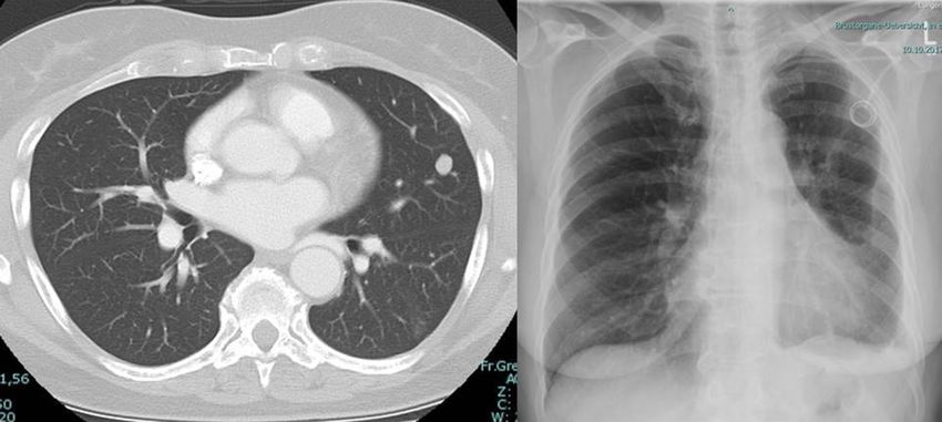
Page 2 of 9                                                                                           Video-Assisted Thoracic Surgery, 2021

 Table 1 Differences between VATS and open lung surgery concerning metastasectomy
 Characteristics                      Conventional thoracotomy                             VATS

 Operative access                     Large incision, rib spreading                        Small incision, muscle spreading

 Pain                                 More, longer periods                                 Less, shorter periods

 Post-thoracotomy pain syndrome High risk                                                  Rare, low risk

 Length of hospital stay              Longer                                               Shorter, enhanced recovery protocols

 Postoperative complications          Increased incidence                                  Less

 Metastasis detection                 Better, manual palpation whole lung                  Inadequate palpation

 Resection technique                  Laser, cautery, staples                              Staples (laser, cautery)

 Resected tissue                      Precision resection possible                         Various margin lengths

 Repeat operations                    Excessive scaring and adhesions                      Rather easy, lesser adhesions

 Oncologic outcome                    Many retrospective large series                      Few reports, small series

 Metastasis recurrences               Depending on histology and number of metastasis “Overseen lesions”? Recurrence at the staple line?
 VATS, video-assisted thoracic surgery.

enucleation (Video 1) and might influence local recurrence                  of stay, fewer complications and a more rapid return to
rates in certain tumor types. These may in turn impact the                  function are the parameters of VATS lobectomy that
functional and oncologic outcome (3). Hence, the number                     are mentioned in the National Comprehensive Cancer
of nodules, the size and location in the lung and the risk for              Network (NCCN) 2020 guidelines (4-7). It can be stated
local recurrence in case of small safety margins impacts the                that a smaller incision guaranteed by VATS is superior as
decision for the surgical approach.                                         compared to open thoracotomy for the patient receiving
                                                                            lobectomy. To our belief, this is also true for lobectomy or
                                                                            segmentectomy in metastasectomy patients and lymph node
Methods
                                                                            dissection can also be easily performed by VATS (Figure 1).
A selective literature review has been undertaken by the
authors to create a clinical guideline, and to shed light upon              VATS and number of metastases
the aforementioned issues. When available, systematic
reviews were preferred to clarify the facts. Finally, our                   As the benefits of VATS rest upon small incisions and no rib
personal experiences with metastasis surgery have been                      spreading, superficial lung palpation is possible “up till the
added to this review.                                                       reaches of the index finger”. Bimanual palpation or at least
                                                                            bidigital palpation of intermediate or central is not possible
                                                                            without rib spreading. Therefore, three questions present
Results                                                                     themselves in concern to metastasectomy by VATS approach:
Open thoracotomy versus VATS                                                is there a greater probability of undetected additional lesions,
                                                                            localization of the nodules detected preoperatively on the
Every pulmonary metastasectomy needs surgical access                        computed tomography (CT) in the lung parenchyma and if
through the thoracic wall, this usually being an incision                   detected, is a resection using staples possible?
through the intercostal space. It is easy to understand
that the VATS approach being associated with smaller
                                                                            Undetected additional lesions: preoperative imaging
incisions, no retraction of the ribs and less scar formation
has some clear advantages over conventional thoracotomies.                  A retrospective study including 521 patients with colorectal
This difference has been studied thoroughly in regard to                    cancer (CRC) lung metastases demonstrated only a
the treatment of non-small cell lung cancer (NSCLC).                        moderate concordance between both CT scan (kappa index:
Improved early outcome, decreased pain, shorter length                      0.42) and fluorodeoxyglucose (FDG)-positron emission

© Video-Assisted Thoracic Surgery. All rights reserved.                 Video-assist Thorac Surg 2021 | http://dx.doi.org/10.21037/vats-2020-lm-09
Video-Assisted Thoracic Surgery, 2021 Page 3 of 9
Table 2 Sensitivity of CT scan depending on number of resected metastases (13)
No. of metastasis Sensitivity of CT scan (%)
N (%) Additional benign nodules, n (%) Additional malignant nodules, n (%)
on CT scan (detection of malignant nodules)
1 65 (54.2) 18 (27.7) 6 (9.2) 90.80%
2 34 (28.3) 6 (17.6) 10 (29.4) 70.60%
3 9 (7.5) 0 (0) 4 (44.4) 55.60%
≥4 12 (10.0) 0 (0) 12 (100.0) 0%
CT, computed tomography.
tomography (PET) (kappa index: 0.42) findings and the 101 nodules were pathologically confirmed to be metastatic
histologically proven number of metastases. Only 61.7% nodules. The sensitivity, specificity, positive predictive value,
and 61.8% of histologically diagnosed metastases were and negative predictive value were lowest in the osteosarcoma
correctly identified with the CT and FDG-PET scans, group. Thin slice CT scan achieved 100% sensitivity when
respectively (8). In a prospective trial, identification of lung nodule size was over 5 mm and less than five nodules were
metastases was compared between radiologists and a cloud detected in the non-osteosarcoma group (12).
cased computer-aided detection (CAD) system validated by Macherey et al. (1) analyzed 18 studies on 1,472
experienced radiologists. From 225 patients, 75 had a total patients with lung metastases having 1,630 pulmonary
of 215 nodules. The sensitivity to detect lesions ≥3 mm was metastasectomies between 1990 and 2014 with 30% of
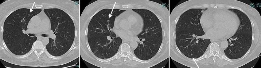
significantly higher using CAD (65% vs. 88%, PPage 4 of 9 Video-Assisted Thoracic Surgery, 2021 Figure 1 Ideal candidate for VATS metastasectomy: single lesion, anatomical resection. A 61-year-old female 1 year after radio- chemotherapy for an adenocarcinoma of the distal esophagus. Uniportal left sided VATS with S4 wedge resection was performed. Frozen section analysis could not differentiate between primary NSCLC and metastasis, so complete lingulectomy with radical lymphadenectomy was performed. Later comparison of both histologies revealed single pulmonary metastasis from esophageal cancer. The postoperative chest X-ray was performed at follow up after 4 weeks. VATS, video-assisted thoracic surgery; NSCLC, non-small cell lung cancer. Figure 2 Unsuitable situation for VATS metastasectomy: three small lesions in the same patient, planned for wedge resection and difficult to identify. A 62-year-old male with two lung metastases from rectal cancer. Manual palpation was necessary to identify the deeper lesions. Laser enucleation of the middle lobe nodule did not provide adequate safety margin alongside the middle lobe artery, so middle lobe lobectomy was performed. The other nodules, in segment 3 and segment 9 were found to be benign. White arrows point at the suspicious nodules. VATS, video-assisted thoracic surgery. NSCLC and metastasis, so complete lingulectomy with radical lymphadenectomy was performed. Later comparison of both histologies revealed single pulmonary metastasis from esophageal cancer. The postoperative chest X-ray was performed at follow up after 4 weeks. VATS, video-assisted thoracic surgery; NSCLC, non-small cell lung cancer. detection of CRC metastases ranged from 35.5% to 95.5%. to look for relationship between chest CT and pathologic Unilateral solitary lesions were found with additional nodule counts in in 404 patients having open pulmonary malignant lesions in less than 5% (P=0.023) (Figure 1) and metastasectomy for CRC lung metastases. Unilateral increasing numbers of metastases were associated with involvement was found in 345 (85%) and single nodule increasing numbers of additional undetected metastases in 253 (63%) patients. The radiologic and malignant (Figure 2), always (100%) when 4 or more lesions were pathologic findings were concordant in 316 (78%) patients. present (13). Independent predictors of discordance were bilateral Marron et al. (14) using the Spanish national registry involvement and greater number of metastases. Imaging and © Video-Assisted Thoracic Surgery. All rights reserved. Video-assist Thorac Surg 2021 | http://dx.doi.org/10.21037/vats-2020-lm-09
Video-Assisted Thoracic Surgery, 2021                                                                                        Page 5 of 9

operative agreement of metastasis counts was 95% for single           In a meta-analysis on the different outcomes of
lesions and 50% with more than one radiologic nodule.              open versus VATS metastasectomy comprised of eight
Significant disagreements were found for advanced-stage            retrospective studies including 822 patients (485 after open
CRC at initial diagnosis, smaller nodules (13.5 vs. 18.4 mm),      thoracotomy and 337 after VATS) were put together for a
simultaneous liver metastases (P=0.014), bilateral metastases      pooled analysis. Compared to open metastasectomy, VATS
(P
Page 6 of 9                                                                                      Video-Assisted Thoracic Surgery, 2021

oncologically beneficial compared to open thoracotomy when            Size of the preoperatively identified metastases
completeness of resection can be achieved and safety margins
                                                                      In a study by Kang et al. (12) only 12 of 32 metastatic
are not jeopardized. VATS metastasectomy has demonstrated
                                                                      nodules from osteosarcoma were detected by preoperative
encouraging results by showing good survival rates (24).
                                                                      multi-detector CT imaging. This was not only a question
Completeness of resection is the most important prognostic
                                                                      of histology, but a question of small sized nodules less
factor (2) and not only includes the complete removal of the
                                                                      than 3 mm which are quite common in osteosarcoma (12).
main lesion, but also the removal of all additional lesions that
                                                                      These nodules are easy to palpate because they most often
might not be detected in a CT scan. Hence, it is obligatory
                                                                      are of firm consistency but may be missed by VATS if not
to include risk factors such as the presence of unexpected
                                                                      detected with CT. On the other hand, no additional nodules
additional metastases and risk for incomplete resection or
                                                                      were found in non-osteosarcoma patients when less than 5
insufficient safety margins into our operation planning.
                                                                      nodules were detected and all were over 5 mm and disease-
   The reliability of preoperative CT scan to detect all
                                                                      free interval was over 24 months. In this situation the CT
metastases has increased in the last twenty years. There
have been many innovations, from MDCT scans to helical                sensitivity was 100% (12).
data acquisition and decreasing slice thickness. Minimum
requirements for preoperative CT imaging is a helical                 Histology of the primary tumor
CT scan with 3- to 5-mm reconstruction thickness or a
volumetric thin section scanning performed up to 4 weeks              The probability of preoperatively undetected lesions is
before pulmonary metastasectomy (25). The widespread                  higher for mesenchymal (41%) tumors than for epithelial
availability of MDCT scanners provides the opportunity                tumors (28%) (29). Metastases with aggressive growth
to examine thin-section (1 mm) CT scans. MIP techniques               patterns need greater resection margins (>7 mm) to prevent
were shown to improve the visualization of small nodules              or reduce the risk of local recurrences, making these lesions
and is currently used by many radiologists (26). The average          ideal candidates for anatomical resections. On the other
sensitivity of nodule detection using 1-mm section increased          hand, metastases with a smooth surface could be removed
from 88% to 93.25% with the additional use of MIP                     with minimal safety margins (30).
technique (11). A further increase of the detection of small             Since the wedge resections cannot guarantee an adequate
nodules was shown with CAD techniques (9-11).                         safety margin the deeper the lung metastases are located,
                                                                      anatomical resections are clearly a better choice here (23).
                                                                      For multiple metastases, wedge resections or enucleations
Number of identified metastases with preoperative imaging             with the added benefit of lung parenchyma conservation are
It is not surprising that increasing numbers of identified            more ideal.
metastases are associated with increasing risk for undetected            The superiority of metastasectomy through anatomic
additional metastases. Marron et al. (14) found an accurate           resections like segmentectomy can be attributed to
correlation of preoperative imaging with intraoperative findings      the fact that these procedures offer a better removal of
for single nodules, but found discrepancies in half of the            intrapulmonary lymph structures and blood vessels as
patients with multiple nodules (14). Others as well-found high        compared to wedge resection (30). Wedge resections for
correlation between preoperative imaging and intraoperative           CRC lung metastases with a median size of 1.1 cm were
findings when only one lesion was detected (13,27). The               associated with an intolerable rate of 18% local recurrences,
percentage of intraoperatively detected unexpected malignant          raising the question of anatomical resection for single
nodules increased from 9% to 30% and 45% when 1, 2 and 3              metastases, at least when they are located deeper in the
nodules were identified preoperatively. This implies that more        parenchyma (23).
than 2 nodules should be resected with open thoracotomy
and manual palpation. In 2010 the European Society of
                                                                      Intraoperative nodule detection
Thoracic Surgeons (ESTS) working group on pulmonary
metastasectomy concluded, that at that time, there was no             The use of various localization methods for intrapulmonary
alternative to palpation in every metastasectomy procedure (28).      nodules depends on a number of factors, such as the treating
Today, we think, that at least single metastasis evaluated with       doctor, availability of resources, number and location of
thin section CT scans may be operated by VATS.                        nodules, patient comorbidities, etc. (16). The creation of a gold

© Video-Assisted Thoracic Surgery. All rights reserved.            Video-assist Thorac Surg 2021 | http://dx.doi.org/10.21037/vats-2020-lm-09
Video-Assisted Thoracic Surgery, 2021 Page 7 of 9
Isolated metastasis 2 Unilateral metastases > 3 Metastases
(unilateral or bilateral)
>2 cm and/ Multiple Central and/or Thoracotomy with bimanualPage 8 of 9 Video-Assisted Thoracic Surgery, 2021
Footnote and systematic review of controlled trials. Innovations
(Phila) 2007;2:261-92.
Provenance and Peer Review: This article was commissioned
6. Yang CJ, Kumar A, Klapper JA, et al. A National Analysis
by the Guest Editors (Marcello Migliore and Michel
of Long-term Survival following Thoracoscopic Versus
Gonzalez) for the series “VATS in Lung Metastasectomy”
Open Lobectomy for Stage I Non-small-cell Lung Cancer.
published in Video-Assisted Thoracic Surgery. The article has
Ann Surg 2019;269:163-71.
undergone external peer review.
7. Whitson BA, Groth SS, Duval SJ, et al. Surgery for early-
stage non-small cell lung cancer: a systematic review of the
Conflicts of Interest: Both authors have completed the
video-assisted thoracoscopic surgery versus thoracotomy
ICMJE uniform disclosure form (available at http://dx.doi.
approaches to lobectomy. Ann Thorac Surg 2008;86:2008-
org/10.21037/vats-2020-lm-09). The series “VATS in Lung 16; discussion 2016-8.
Metastasectomy” was commissioned by the editorial office 8. Guerrera F, Renaud S, Schaeffer M, et al. Low Accuracy
without any funding or sponsorship. Both authors have no of Computed Tomography and Positron Emission
other conflicts of interest to declare. Tomography to Detect Lung and Lymph Node Metastases
of Colorectal Cancer. Ann Thorac Surg 2017;104:1194-9.
Ethical Statement: The authors are accountable for all 9. Vassallo L, Traverso A, Agnello M, et al. A cloud-based
aspects of the work in ensuring that questions related computer-aided detection system improves identification
to the accuracy or integrity of any part of the work are of lung nodules on computed tomography scans of
appropriately investigated and resolved. patients with extra-thoracic malignancies. Eur Radiol
2019;29:144-52.
Open Access Statement: This is an Open Access article 10. Meybaum C, Graff M, Fallenberg EM, et al.
distributed in accordance with the Creative Commons Contribution of CAD to the Sensitivity for Detecting
Attribution-NonCommercial-NoDerivs 4.0 International Lung Metastases on Thin-Section CT - A Prospective
License (CC BY-NC-ND 4.0), which permits the non- Study with Surgical and Histopathological Correlation.
commercial replication and distribution of the article with Beitrag der computerassistierten Detektion (CAD)
the strict proviso that no changes or edits are made and the zur Sensitivität der präoperativen Lokalisation von
original work is properly cited (including links to both the Lungenmetastasen im Dünnschicht-CT – prospektive
formal publication through the relevant DOI and the license). Studie mit chirurgischer und histopathologischer
See: https://creativecommons.org/licenses/by-nc-nd/4.0/. Korrelation. Rofo 2020;192:65-73.
11. Park EA, Goo JM, Lee JW, et al. Efficacy of computer-
References aided detection system and thin-slab maximum intensity
projection technique in the detection of pulmonary
1. Macherey S, Doerr F, Heldwein M, et al. Is manual nodules in patients with resected metastases. Invest Radiol
palpation of the lung necessary in patients undergoing 2009;44:105-13.
pulmonary metastasectomy? Interact Cardiovasc Thorac 12. Kang MC, Kang CH, Lee HJ, et al. Accuracy of
Surg 2016;22:351-9. 16-channel multi-detector row chest computed
2. Pastorino U, Buyse M, Friedel G, et al. Long-term results tomography with thin sections in the detection of
of lung metastasectomy: Prognostic analyses based on metastatic pulmonary nodules. Eur J Cardiothorac Surg
5206 cases. J Thorac Cardiovasc Surg 1997;113:37-49. 2008;33:473-9.
3. Petrella F, Chieco P, Solli P, et al. Which factors affect 13. Chung CC, Hsieh CC, Lee HC, et al. Accuracy of helical
pulmonary function after lung metastasectomy? Eur J computed tomography in the detection of pulmonary
Cardiothorac Surg 2009;35:792-6.. colorectal metastases. J Thorac Cardiovasc Surg
4. NCCN Clinical Practice Guidelines in Oncology. Non- 2011;141:1207-12.
small Cell Lunge Cancer. Version 2. 2021. Available 14. Marron MC, Lora D, Gamez P, et al. Agreement Between
online: www.nccn.org/professionals/physician_gls/pdf/ Computed Tomography and Pathologic Nodule Counts
nscl.pdf in Colorectal Lung Metastases. Ann Thorac Surg
5. Cheng D, Downey RJ, Kernstine K, et al. Video-assisted 2016;101:259-65.
thoracic surgery in lung cancer resection: a meta-analysis 15. Eckardt J, Licht PB. Thoracoscopic or open surgery for
© Video-Assisted Thoracic Surgery. All rights reserved. Video-assist Thorac Surg 2021 | http://dx.doi.org/10.21037/vats-2020-lm-09Video-Assisted Thoracic Surgery, 2021 Page 9 of 9
pulmonary metastasectomy: an observer blinded study. of colorectal cancer harboring KRAS mutations. Ann Surg
Ann Thorac Surg 2014;98:466-9; discussion 469-70. 2019;270:1170-7.
16. Lin MW, Chen JS. Image-guided techniques for localizing 23. Chung JH, Lee SH, Yi E, et al. Impact of resection margin
pulmonary nodules in thoracoscopic surgery. J Thorac Dis length and tumor depth on the local recurrence after
2016;8:S749-55. thoracoscopic pulmonary wedge resection of a single
17. Murakawa T, Sato H, Okumura S, et al. Thoracoscopic colorectal metastasis. J Thorac Dis 2019;11:1879-87.
surgery versus open surgery for lung metastases of 24. Sun F, Chen L, Shi M, et al. Prognosis of video-assisted
colorectal cancer: a multi-institutional retrospective thoracoscopic pulmonary metastasectomy in patients with
analysis using propensity score adjustment†. Eur J colorectal cancer lung metastases: an analysis of 154 cases.
Cardiothorac Surg 2017;51:1157-63. Int J Colorectal Dis 2017;32:897-905.
18. Meng D, Fu L, Wang L, et al. Video-assisted thoracoscopic 25. Detterbeck FC, Grodzki T, Gleeson F, et al. Imaging
surgery versus open thoracotomy in pulmonary requirements in the practice of pulmonary metastasectomy.
metastasectomy: a meta-analysis of observational studies. J Thorac Oncol 2010;5:S134-9.
Interact Cardiovasc Thorac Surg 2016;22:200-6. 26. Brandman S, Ko JP. Pulmonary nodule detection,
19. Welter S, Arfanis E, Christoph D, et al. Growth characterization, and management with multidetector
patterns of pulmonary metastases: Should we adjust computed tomography. J Thorac Imaging 2011;26:90-105.
resection techniques to primary histology and size? Eur J 27. Perentes JY, Krueger T, Lovis A, et al. Thoracoscopic
Cardiothorac Surg 2017;52:39-46. resection of pulmonary metastasis: Current practice and
20. Shiono S, Okumura T, Boku N, et al. Outcomes of results. Crit Rev Oncol Hematol 2015;95:105-13
segmentectomy and wedge resection for pulmonary 28. Molnar TF, Gebitekin C, Turna A. What are the
metastases from colorectal cancer. Eur J Cardiothorac considerations in the surgical approach in pulmonary
Surg 2017;51:504-10. metastasectomy? J Thorac Oncol 2010;5:S140-4.
21. Molins L, Hernandez J, Fibla JJ, et al. Anatomical 29. Althagafi KT, Alashgar OA, Almaghrabi HS, et al. Missed
Resection Improves Survival Over Wedge Resection of pulmonary metastasis. Asian Cardiovasc Thorac Ann
Pulmonary Metastases of Colorectal Origin in the Spanish 2014;22:183-6.
Prospective Multicenter Study (GECMP-CCR). Ann 30. Welter S, Barile La Raia R, Gupta V. Pursuit of an optimal
Oncol 2015;26:i45-i47. surgical margin in pulmonary metastasectomy. J Vis Surg
22. Renaud S, Seitlinger J, Al Lawati Y, et al. Anatomical 2019;5:39.
resections improve survival following lung metastasectomy
doi: 10.21037/vats-2020-lm-09
Cite this article as: Welter S, Gupta V. Algorithm for the
pulmonary metastasectomy based on number of metastases and
histology. Video-assist Thorac Surg 2021.
© Video-Assisted Thoracic Surgery. All rights reserved. Video-assist Thorac Surg 2021 | http://dx.doi.org/10.21037/vats-2020-lm-09You can also read