Amino-acid conjugated protein-Au nanoclusters with tuneable fluorescence properties - IOPscience
←
→
Page content transcription
If your browser does not render page correctly, please read the page content below
PAPER • OPEN ACCESS

Amino-acid conjugated protein–Au nanoclusters with tuneable
fluorescence properties
To cite this article: Mark H Griep and Nicholas M Bedford 2020 J. Phys. Mater. 3 045002

View the article online for updates and enhancements.

                               This content was downloaded from IP address 46.4.80.155 on 14/10/2020 at 12:45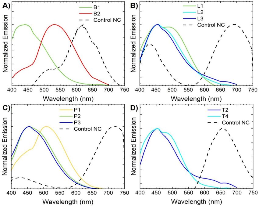
J. Phys. Mater. 3 (2020) 045002 https://doi.org/10.1088/2515-7639/ab8d90
Journal of Physics: Materials
PAPER
Amino-acid conjugated protein–Au nanoclusters with tuneable
OPEN ACCESS
fluorescence properties
RECEIVED
14 February 2020 Mark H Griep1 and Nicholas M Bedford2
REVISED 1
6 April 2020
Weapons and Materials Research Directorate, U.S. CCDC Army Research Laboratory, Aberdeen Proving Ground, MD, United States
of America
ACCEPTED FOR PUBLICATION 2
School of Chemical Engineering, University of New South Wales, Sydney, NSW, Australia
27 April 2020
PUBLISHED
E-mail: n.bedford@unsw.edu.au
3 September 2020
Keywords: protein Au nanocluster, supramolecular conjugate, amino acid, fluorescence
Original content from
this work may be used
under the terms of the Abstract
Creative Commons
Attribution 4.0 licence. Au-based protein nanoclusters (PNCs) represent an emerging class of fluorescence probes that are
Any further distribution
of this work must inherently biocompatible and combine the functionality of proteins and optical properties of Au
maintain attribution to nanoclusters. Here we report on a methodology to create conjugated Au PNCs using amino acid
the author(s) and the title
of the work, journal coupling strategies from a series of common laboratory proteins. We discover that the host protein
citation and DOI.
and the specific conjugation chemistry has a profound influence on the resulting fluorescence
properties. Synchrotron analyses showcase local Au NC aggeration upon PNC conjugation, which
causes local environment changes to invoke differences in fluorscence properties. The observed
aggeration does not give rise to plasmonic properties nor signifigant fluorescence quenching,
strongly indicating the PNCs are still in a near-native cluster state. Our methodology and findings
here could open new pathways for tuning PNC fluorescence properties in a rational fashion, having
a potential impact in host of biomedical and sensing applications.
1. Introduction
Metal nanoclusters (NCs) are engineered at dimensions approaching the Fermi wavelength of electrons,
allowing for the formation of discrete energy states that result in interesting fluorescence properties that
differ to those observed in larger metal nanoparticles with plasmonic properties [1–4]. While avoiding the
toxic components of traditional fluorescent quantum dots (QDs), metal NC have demonstrated high
quantum yield, tuneable photonic emission, highly efficient two-photon absorption, and tailorable
hybridization pathways. For biomedical applications in particular, the tailoring of NC properties and
biocompatibility through biomediated synthesis approaches has proven instrumental [5–9]. Not
surprisingly, protein-stabilized nanoclusters (PNC) have emerged as versatile hybrid platforms that can
leverage functional properties of both the NC and protein components [10–14].
In traditional metal NCs, ligand parameterization and corresponding electronic structure is very well
established [15–17]. In PNCs, however, identification of the stabilization region and specific ligand
interactions of NCs within a protein scaffold remains elusive and represents a major deficiency towards
controllable PNC synthesis and optimized functionality. If cluster location and protein interaction could be
better understood, one can learn how NCs are stabilized and what reaction mechanisms allow for growth of
the NC to their targeted size, electromagnetic properties, and responsive functionality. With this knowledge,
the rational identification and engineering of new proteins could be accomplished to grow NCs with
pre-determined properties. As Au–thiol interactions available through metal-binding residues within the
protein present likely target regions for NC stabilization, recent studies have targeted these through both
simulation and designer protein approaches [18]. While such studies provide secondary support for likely
stabilization regions, more direct characterization is required to definitively identify NC–ligand interactions
in a protein host.
Direct examination of the cluster region with a protein structure requires the utilization of x-ray and
crystallographic techniques. For the latter, highly crystalline materials are required which have yet to be
© 2020 The Author(s). Published by IOP Publishing Ltd
J. Phys. Mater. 3 (2020) 045002                                                        M H Griep and N M Bedford

achieved with PNC hybrids. One avenue to create highly-ordered PNC structures is through their
incorporation into metal–organic framework (MOF) scaffolds, however, solvent incompatibility has proven
a challenging issue for the integration of NCs into ordered MOFs [19, 20]. One method to overcome this
barrier involves the precipitation of nanoclusters with a metal ion solution, such as Zn2+ , to form aggregates
that are amenable to methanol dissolution. With proper precipitation, the NCs have been demonstrated
compatible for processing into zeolitic imidazole framework (ZIF-8) MOFs [20, 21]. While PNCs have, to
date, not been reported in composite form with MOF structures, proteins incorporation into ZIF-8 has been
shown [22]. In addition to ordering through MOFs, researchers have recently begun to examine large-pore
protein crystals (LPC) as scaffolds for NC incorporation. With LPC pore diameters up to 8 nm and aqueous
processing conditions, glutathione capped Au25 nanoclusters can be readily adsorbed into the patterned
crystal pores [23]. The ability to utilize purely organic frameworks reduces the processing requirements in
addition to providing a platform to enable precise patterning of the embedded materials within the LPC.
    More recently, a new methodology to directly conjugate PNCs and form crystal-like structures has been
developed [24]. By synthesizing PNCs in the presence of select amino acids (AA), protein conjugation
enabled the formation of supramolecular structures. Interestingly, through simple variations in pH and AA
composition, Ding et al demonstrated the ability to controllably tune NC fluorescence emission wavelength
across the visible spectrum for BSA-stabilized gold nanoclusters (AuNCs) conjugates [24]. As this approach
enables formation crystal-like structures composed nearly entirely of the PNC of interest with limited
additional materials, subsequent analysis of the gold-ligand stoichiometry can be more readily studied
without extraneous contaminating material signals. Additionally, it is hypothesized that the crystal-like
structure could generate PNCs of consistent orientation in a close-packed cluster; enabling more precise
x-ray analysis to resolve protein–Au NC interacations.
    Motivated by the aforementioned study, we report on the conjugation of PNCs using a series of protein
hosts and amino acid conjugation conditions. We found that the conjugated PNCs exhibit flourescence
characteristics that depend on the nature of conjugation chemistry and host protein using BSA, trypsin,
pepsin, and lysozyme. To study the atomic-scale origins of these phenomena, we further study the conjugated
PNCs using a x-ray absorption spectroscopy (XAS) and small angle x-ray scattering (SAXS). With these
methods, we observe unexpected clustering effects in the conjugated PNCs that are dependent on the protein
host. These findings should help guide and refine our ability to make Au PNCs with optimized properties.

2. Methods

2.1. Chemicals
All chemicals were purchased from Sigma-Aldrich (St. Louis, MO) and solutions prepared were aqueous,
unless otherwise noted: gold (III) chloride hydrate (HAuCl4 ), BSA, trypsin, lysozyme, pepsin, L-histidine,
glycine, hydrochloric acid (HCl), and sodium hydroxide (NaOH).

2.2. Synthesis of PNC’s and AA-conjugated PNCs
The synthesis of AA-conjugated PNC (AA-PNC) was performed with slight modifications of previously
reported methods [24]. In brief, hybrids were prepared with varied composition of AAs, including
L-histidine (L-His) and glycine (Gly), and protein hosts to include BSA, trypsin, lysozyme, and pepsin. Stock
solutions were prepared of the selected proteins (50 mg ml−1 ), L-His (31 mg ml−1 ), Gly (15 mg ml−1 ), and
HAuCl4 (16 mg ml−1 ). Samples were prepared according to table 1 by sequentially adding the protein stock
(1 ml), HAuCl4 (1 ml), AA (5 ml), and DI water (3 ml) to a glass vial. The pH was adjusted as outlined in
table 1 with 1 M solutions of NaOH or HCl. This procedure results in a final reaction solution of 5 mg ml−1
protein, 1.6 mg ml−1 HAuCl4 , and 7.5 mg ml−1 Gly (or 15.5 mg ml−1 L-His depending on amino acid
selection). Samples were stirred at 37 ◦ C for 12 h. The final solution is purified through centrifugation at
10 000 g for 30 min, with collection of the pellet to target formed conjugates. The pellet was resusped in DI
water and centrifugal purification repeated three times. The final pellet resuspended in 5 ml of DI water.
Control protein nanoclusters were made for each protein host utilizing the same procedure outlined above
without the addition of amino acids. Controls were synthesized at pH 12 and stirred for 12 h at 37 ◦ C.
Samples were purified through a 10 kDa molecular weight cut off (MWCO) centrifugal filter to remove
unreacted metal ions.

2.3. Characterization
Fluorescent emission of the protein-nanocluster hybrids was measured on a Horiba Fluorolog-3 with
excitation at 350 nm and emission capture range of 400–700 nm. All measurements were made in aqueous
solutions in a quartz cuvette with a path length of 1 cm. XAS measurements were performed at the 12-BM
beamline of the Advanced Photon Source (APS), Argonne National Laboratory (ANL). A sample from each

                                             2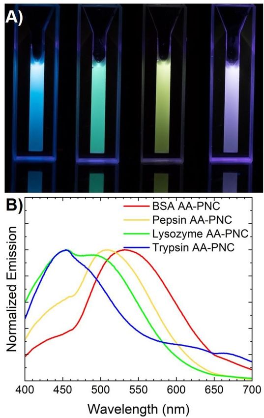
J. Phys. Mater. 3 (2020) 045002                                                                          M H Griep and N M Bedford

                                           Table 1. Synthesis parameters for each sample.

Protein                BSA                         Trypsin                        Lysozyme                        Pepsin
Sample      B1        B2     B3    B4     T1        T2     T3     T4      L1       L2       L3    L4      P1      P2    P3    P4

AA         L-His     Gly     Gly   Gly    L-His    Gly    Gly    Gly     L-His    Gly       Gly   Gly    L-His   Gly   Gly   Gly
pH         5.5       1.5     3.5   12.5   5.5      1.5    3.5    12.5    5.5      1.5       3.5   12.5   5.5     1.5   3.5   12.5

protein-host with glycine-assisted AA nanocluster formation was selected from table 1 to include B3 (BSA
PNC), L2 (Lysozyme PNC), T2 (Trypsin PNC), and P1 (Pepsin PNC). Solutions of Au NCs were loaded into
thin-walled quartz x-ray capillaries (1 mm OD) as synthesized and studied under a fluorescence
measurement geometry using a 13 channel Ge detector. XAS scans were performed from 200 eV below the
Au L3 -edge (11.919 keV) to 900 eV past the edge. Data reduction, analyzed, and modeling were performed
using the Demeter XAS software package [25]. EXAFS modeling was performed using scattering
contributions from the well-known crystal structures of bulk Au, Au2 O3 and/or experimental determined
structure of thiolated Au NCs [26]. A value of 0.798 for S0 2 was used in all fits as determined from the EXAFS
modeling of a reference foil. Small angle x-ray scattering (SAXS) measurements were performed at the
12-ID-C beamline of the APS. Samples were prepared in an identical fashion as to XAS experiments probed
in a transmission geometry using 12.0 keV x-rays. Data was processed onsite at 12-ID-C and further
modeled to obtain pair distance distribution functions, P(r), using the program ScÅtter [27].

3. Results and discussion

Motivated by the ability to better understand NC location within a protein host, AA conjugated PNCs were
synthesized using a series of proteins. While we were unable to get qaulity materials for single-crystal XRD,
we did observe interesting synthesis-dependent properties that warranted further invesitation. Utilizing the
methodology reported by Ding et al [24], AA conjugated PNC growth was successfully performed using four
different protein models: BSA, trypsin, lysozyme, and pepsin. To the authors’ knowledge, this is the first
reported AA-conjugated PNCs created with trypsin, lysozyme, and pepsin protein hosts. As shown in figure
1, this approach provides tailorable fluorescent emission wavelengths that can be obtained across the visible
spectrum. The control PNCs formed for each protein host yielded red emissions, which corresponds to
previous studies of pH 12 synthesis of BSA [5, 28], trypsin [29–31], pepsin [32], and lysozyme [33, 34].
While pepsin has been shown to readily create smaller clusters (i.e. blue-shifted emission) through
modulation of reaction pH [32], this tunability of BSA, lysozyme, and trypsin to the blue spectrum has been
limited. As can be seen in figure 2, the ability to tune each protein model to the blue spectrum has been
achieved through select AA integration into the synthesis process.
    Although AA conjugated PNCs were succesfully formed with each protein model, fluorescent PNCs were
not formed with every sample composition outlined in table 1. For non-fluorescent AA-conjugated PNCs,
plasmonic responses were observed, strongly suggesting the possibility of Au NC destabilization under
certain conjugation conditions. For the remaining PNCs, the fluorescent emission spectrum are shown in
figure 2. In all cases, the non-conjugated NCs exhibit fluoresence at lower energy (i.e. red-shifted)
wavelengths than the AA-conjugated PNCs. This suggests that the process promotes the formation of smaller
cluster sizes, as Au NC emission wavelengths directly correlate to the number of Au atoms in the cluster [32].
Previous studies with BSA have demonstrated the ability for Gly and L-His to migrate into the protein during
the unfolding/refolding process [24]. As the protein contains the residues required to reduce the metal salts,
the additional Gly or L-His can assist in the stabilization and capping of the cluster during formation. Both
amino acids, Gly and L-His, have previously shown the preferential capping of smaller metal cluster
structures, shifting the emission towards the blue spectrum [35, 36]. With respect to the formation of
conjugated PNC structures, the presence of additional AA capping residues within the protein structure
could inhibit proper protein refolding and induce aggregation. Although certain amino acids have shown the
ability to inhibit protein aggregration during the unfolding/folding process, this is not the case for glycine
and L-histidine [37].
    In the case of BSA, lysozyme, and pepsin; conjugated PNCs were formed with both L-His and Gly
approaches. Interestingly, the L-His conjugation of BSA stabilized PNCs yielded a spectral blue-shift in the
cluster emission, which is supported by reported literature (figure 2(A)); however yield a green cluster
emission in the lysozyme and pepsin models along with a blue emission of the trypsin AA-PNCs (figures
2(B)–(D)). As the incorporation of L-His into the synthesis mechanism shifts the traditional spectral
response of each protein model, it is likely that L-His plays a direct role in NC formation and stabilization

                                                     3
J. Phys. Mater. 3 (2020) 045002 M H Griep and N M Bedford
Figure 1. Fluorescent output of conjugated Au PNC as presented from optical images (left to right: trypsin, lysozyme, pepsin and
BSA PNC) (A) and fluorescence spectroscopy (B).
beyond simple protein conjugation and supports direct capping/stabilization of the cluster as it is
reduced/stabilized within the protein scaffold.
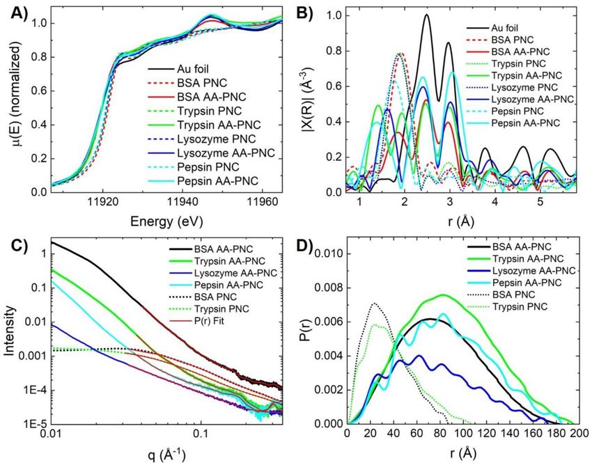
To better understand the effect of host protein and conjugation chemistry on the local structure of the Au
clusters, XAS was used to as an element specfic probe. The x-ray absoprtion near edge spectra (XANES) for
both as-synthesized Au PNC and the AA-conjugated PNCs are shown in figure 3(A). Overall, all probed
PNCs exhibit a ~+1–2 eV shift in E0 from the Au reference, indicating a partial increase in oxidation state.
The biggest shift in E0 is observed for PNCs stabilized within BSA, followed by trypsin, lyzosyme, and pepsin
stabilized PNCs. This shift in E0 is expected given the small cluster size and corresponding surface atoms
interactions with host protein moeties. Moreover, the XANES features observed in the as-synthesized PNCs
are signifigantly broad in comparison to the bulk Au reference as expected [38, 39], given the small cluster
size not replicating the bulk fcc lattice of Au. Upon conjugation, all PNCs exhibit a reduced E0 compared to
their as-synthesized counterparts that is more akin to the Au reference. Additionally, XANES features better
resemble that of the Au reference, albeit with an increase in white line intesity. These findings were
unexpected and incidate that the protein conjugation must force the Au atoms more into a larger aggerated
state, yielding XANES feature more resemblent of fcc Au. The increase in white line intensity as compared to
the reference Au additionally suggest sufficient Au–ligand interactions, incidating electron transfer from the
Au to the protein is still present.
After edge-step normalization, k2 -weighting, and Fourier transform, the extended x-ray absorption fine
structure spectra (EXAFS) reveal interesting local structural changes in the Au NCs (figure 3(B)). Note the
EXAFS is not corrected for phase shift, wherein peaks in the EXAFS are shifts ~0.3–0.4 Å lower from actual
distances. The as-synthesized PNCs all exhibit a prominate feature centered below 2.0 Å and smaller features
that somewhat align with those observed in from the bulk Au. This first distance is likely Au–thiol bonding
through interactions with the protein that are commonly observed in thiolated AuNC complexes with
organic ligands [38]. It is important to note that the pepsin PNC has a signifigantly shifted distance for the
Au–biotic interaction, perhaps indicating that diffent cluster binding motif may be present.
Upon conjugate formation, notable structural differences can be observed between the AA-conjugated
PNCs vs their non-conjugated counterparts. Features more resemblant of fcc Au become more evident, albeit
at slightly shifted distances. Qualitatvely, this indicates that protein conjugation condenses the clusters
4J. Phys. Mater. 3 (2020) 045002 M H Griep and N M Bedford Figure 2. Emission spectra of methodologies that yield nanocluster fluorescence for (A) BSA, (B) lysozyme, (C) pepsin, and (D) trypsin protein hosts. Non conjugated Au PNCs are provide for reference. together in an aggregated cluster arrangement that is likely perturbed signifigantly from the bulk lattice due to the likely imperfectious nature of cluster assembly during protein conjugation. The majority of the PNCs also exibit a feature
J. Phys. Mater. 3 (2020) 045002

                                                                             Table 2. EXAFS fitting results for Au PNCs.

                                                               Au-Au        Au-Au              Au-S                         Au-Au
                              Au-S                            (cluster)    (cluster)         (cluster)         Au-Au         (fcc)          u-Au                      Au-Z
    Sample     Au-S CN       NND (Å)          Au-S σ2            CN        NND (Å)              σ2            (fcc) CN     NND (Å)        (fcc) σ2       Au-Z CN     NND (Å)        Au-Z σ2

    BSA        2.00 ± 0.10   2.324 ± 0.003 Å 0.002 ± 0.001   0.68 ± 0.24   2.94 ± 0.02 Å 0.002 ± 0.0003           –            –             –             –             –              –
    PNCs

6
    Pepsin     2.47 ± 0.21   2.25 ± 0.02 Å   0.007 ± 0.003   1.37 ± 0.42   2.88 ± 0.04 Å 0.010 ± 0.001            –            –             –             –             –              –
    PNCs
    Trypsin    1.90 ± 0.11   2.30 ± 0.01 Å   0.002 ± 0.002   0.68 ± 0.27   2.84 ± 0.03 Å 0.002 ± 0.001            –            –             –             –             –              –
    PNCs
    Lyso       1.91 ± 0.08   2.30 ± 0.01 Å   0.002 ± 0.001   0.46 ± 0.11   2.84 ± 0.03 Å 0.002 ± 0.002            –            –             –             –             –              –
    PNCs
    BSA AA-    0.75 ± 0.04   2.288 ± 0.004 Å 0.004 ± 0.002       –             –                 –           5.18 ± 0.13   2.86 ± 0.02 Å 0.007 ± 0.001     –             –              –
    PNC
    Pepsin     0.15 ± 0.23   2.36 ± 0.03 Å   0.002 ± 0.001       –             –                 –           5.00 ± 0.51   2.85 ± 0.03 Å 0.003 ± 0.001 1.04 ± 0.71   1.91 ± 0.04 Å 0.002 ± 0.001
    AA-PNC
    Trypsin-   0.76 ± 0.21   2.31 ± 0.03 Å   0.002 ± 0.001       –             –                 –           3.45 ± 0.61   2.86 ± 0.02 Å 0.004 ± 0.002 1.02 ± 0.28   1.88 ± 0.03 Å 0.002 ± 0.001
    AA-PNC
    Lyso-      0.81 ± 0.16   2.18 ± 0.02 Å   0.002 ± 0.001       –             –                 –           8.84 ± 1.02   2.86 ± 0.01 Å 0.010 ± 0.002     –             –              –
    AA-PNC
                                                                                                                                                                                                   M H Griep and N M Bedford
J. Phys. Mater. 3 (2020) 045002 M H Griep and N M Bedford
Figure 3. Synchrotron characterization for selected PNCs; Au L3 -edge (A) XANES and (B) EXAFS along with (C) SAXS and (D)
corresponding P(r) fitting.
exhibit markedly different SAXS profiles. The form factor scattering is largely absent, which is coupled to a
highly linear region at lower q-range. This strongly suggests that protein-protein aggregation within the
sample is not prevalent and that PNCs are beginning to order in solution through the conjugation process.
Note that the ordering is not in a highly ordered periodic fashion to result in Bragg features, such as those
observed in DNA-programmed superlattices of Au nanoparticles [42, 43]. To further probe the effect of
conjugation on morphology, SAXS profiles we fitted to pair distance distribution functions, P(r) (red lines,
figure 3(C)), using known methods. The resulting P(r) functions are shown in figure 3(D) and showcase a
large difference in size and morphology for between the as-synthesized and crystallized materials. PNCs
synthesized with BSA and trypsin exhibit a main feature ~20 Å, and dmax values for 84 Å and 108 Å
respectively. The AA-conjugated PNCs, on the other hand, yield P(r) maxima at values >60 Å for all
materials and dmax values >170 Å over similar q-range values. This strongly indicates and organization of
PNCs into supramolecular structures caused by conjugation processes within the proteins. Taken together,
the synchrotron characterization demonstrate that AA-conjugation creates an aggregation of Au NCs
resembling some more similar to Au nanoparticle. However, the visibile light flourescence properties are
stongly suggest non-plasmonic behavior. This finding illistrates that cluster aggeration rates and the
appropriate protein host could be used to control flourescence propeties without inducing a plasmonic
response, and may provide insights in the generation of new PNCs for various applications.
4. Conclusions
In summary, AA-conjugated PNCs were created in multiple protein models, including BSA, trypsin, pepsin,
and lysozyme PNCs. Fluorescence properties were shown to be dependent on Au NC–protein interaction
and the aggreation of Au NCs that ocucrs during conjugation as revealed using synchortron characterization
methods. Our finding suggest that choice of host protein and the degree of NC aggeration allows for the
possible tunablity of fluorescence properties, priving new pathways to create new NC fluorescence materials
with user defined properties.
7J. Phys. Mater. 3 (2020) 045002 M H Griep and N M Bedford
Acknowledgments
We acknowledge research funding from the Army Research Laboratory for portions of this work. This
research used the 12-B and 12-ID-C beamlines of the Advanced Photon Source, a U.S. Department of Energy
(DOE) Office of Science User Facility operated for the DOE Office of Science by Argonne National
Laboratory under Contract No. DE-AC02-06CH11357. We would like to thank Benjamin Reinhart for
assistance with experiments at 12-BM and Soenke Seifert for assistance with experiments at 12-ID-C.
ORCID iDs
Mark H Griep https://orcid.org/0000-0001-6460-8304
Nicholas M Bedford https://orcid.org/0000-0002-4424-7094
References
[1] Zheng J, Nicovich P R and Dickson R M 2007 Highly fluorescent noble-metal quantum dots Annu. Rev. Phys. Chem. 58 409–31
[2] Lu Y and Chen W 2012 Sub-nanometre sized metal clusters: from synthetic challenges to the unique property discoveries Chem.
Soc. Rev. 41 3594–623
[3] Zhang L and Wang E 2014 Metal nanoclusters: new fluorescent probes for sensors and bioimaging Nano Today 9 132–57
[4] Mathew A and Pradeep T 2014 Noble metal clusters: applications in energy, environment, and biology Part. Part. Syst. Charact. 31
1017–53
[5] Xie J, Zheng Y and Ying J Y 2009 Protein-directed synthesis of highly fluorescent gold nanoclusters J. Am. Chem. Soc. 131 888–9 (in
Eng)
[6] Zuber G, Weiss E and Chiper M 2019 Biocompatible gold nanoclusters: synthetic strategies and biomedical prospects
Nanotechnology 30 352001
[7] Zhang Y, Zhang C, Xu C, Wang X, Liu C, Waterhouse G I N, Wang Y and Yin H 2019 Ultrasmall Au nanoclusters for biomedical
and biosensing applications: a mini-review Talanta 200 432–42
[8] Knoblauch C, Griep M and Friedrich C 2014 Recent advances in the field of bionanotechnology: an insight into optoelectric
bacteriorhodopsin, quantum dots, and noble metal nanoclusters Sensors 14 19731–66
[9] Griep M H, West A L, Sellers M S P, Karna M, Zhan E and Hoque N 2015 Biomediated atomic metal nanoclusters: synthesis and
theory Handbook of Nanoparticles, ed M Aliofkhazraei (Cham: Springer International Publishing) pp 1–24
[10] West A L, Griep M H, Cole D P and Karna S P 2014 DNase 1 retains endodeoxyribonuclease activity following gold nanocluster
synthesis Anal. Chem. 86 7377–82
[11] Sahu D K, Sarkar P, Singha D and Sahu K 2019 Protein-activated transformation of silver nanoparticles into blue and red-emitting
nanoclusters RSC Adv. 9 39405–9
[12] Wang B, Zhao M, Mehdi M, Wang G, Gao P and Zhang K-Q 2019 Biomolecule-assisted synthesis and functionality of metal
nanoclusters for biological sensing: a review Mater. Chem. Front. 3 1722–35
[13] Meng X, Zare I, Yan X and Fan K 2020 Protein-protected metal nanoclusters: an emerging ultra-small nanozyme WIREs Nanomed.
Nanobiotechnol. 12 e1602
[14] West A L, Schaeublin N M, Griep M H, Maurer-Gardner E I, Cole D P, Fakner A M, Hussain S M and Karna S P 2016 In situ
synthesis of fluorescent gold nanoclusters by nontumorigenic microglial cells ACS Appl. Mater. Interfaces 8 21221–7
[15] Jin R 2015 Atomically precise metal nanoclusters: stable sizes and optical properties Nanoscale 7 1549–65
[16] Jin R, Zeng C, Zhou M and Chen Y 2016 Atomically precise colloidal metal nanoclusters and nanoparticles: fundamentals and
opportunities Chem. Rev. 116 10346–413
[17] Aikens C M 2018 Electronic and geometric structure, optical properties, and excited state behavior in atomically precise
thiolate-stabilized noble metal nanoclusters Acc. Chem. Res. 51 3065–73
[18] Aires A, Llarena I, Moller M, Castro-Smirnov J, Cabanillas-Gonzalez J and Cortajarena A L 2019 A simple approach to design
proteins for the sustainable synthesis of metal nanoclusters Angew. Chem. Int. Ed. 58 6214–9
[19] Gao Q, Xu S, Guo C, Chen Y and Wang L 2018 Embedding nanocluster in MOF via crystalline ion-triggered growth strategy for
improved emission and selective sensing ACS Appl. Mater. Interfaces 10 16059–65
[20] Li Y, Hu X, Zhang X, Cao H and Huang Y 2018 Unconventional application of gold nanoclusters/Zn-MOF composite for
fluorescence turn-on sensitive detection of zinc ion Anal. Chim. Acta 1024 145–52
[21] Fan C, Lv X, Liu F, Feng L, Liu M, Cai Y, Liu H, Wang J, Yang Y and Wang H 2018 Silver nanoclusters encapsulated into
metal–organic frameworks with enhanced fluorescence and specific ion accumulation toward the microdot array-based
fluorimetric analysis of copper in blood ACS Sensors 3 441–50
[22] Lyu F, Zhang Y, Zare R N, Ge J and Liu Z 2014 One-pot synthesis of protein-embedded metal–organic frameworks with enhanced
biological activities Nano Lett. 14 5761–5
[23] Hartje L F, Munsky B, Ni T W, Ackerson C J and Snow C D 2017 Adsorption-coupled diffusion of gold nanoclusters within a
large-pore protein crystal scaffold J. Phys. Chem. B 121 7652–9
[24] Ding H, Li H, Wang X, Zhou Y, Li Z, Hiltunen J K, Shen J and Chen Z 2017 Expanding toolbox of imageable protein-gold hybrid
materials Chem. Mater. 29 8440–8
[25] Ravel B and Newville M 2005 ATHENA, ARTEMIS, HEPHAESTUS: data analysis for x-ray absorption spectroscopy using IFEFFIT
J. Synchrotron Radiat. 12 537–41
[26] Yang H, Wang Y, Edwards A J, Yan J and Zheng N 2014 High-yield synthesis and crystal structure of a green Au30 cluster co-capped
by thiolate and sulfide Chem. Commun. 50 14325–7
[27] Hura G L et al 2009 Robust, high-throughput solution structural analyses by small angle x-ray scattering (SAXS) Nat. Methods 6 606
[28] Griep M H, Demaree J D, Cole D P, Henry T C and Karna S P 2020 Protein-mediated synthesis of Au nanocluster decorated
reduced graphene oxide: a multifunctional hybrid nano-bio platform Plasmonics 15 897–903
[29] Kawasaki H, Yoshimura K, Hamaguchi K and Arakawa R 2011 Trypsin-stabilized fluorescent gold nanocluster for sensitive and
selective Hg2+ detection Anal. Sci. 27 591
8J. Phys. Mater. 3 (2020) 045002 M H Griep and N M Bedford
[30] Fan J, Li R, Xu P, Di J, Tu Y and Yan J 2014 Sensitive sulfide sensor with a trypsin-stabilized gold nanocluster Anal. Sci. 30 457–62
[31] Liu J-M, Chen J-T and Yan X-P 2013 Near infrared fluorescent trypsin stabilized gold nanoclusters as surface plasmon enhanced
energy transfer biosensor and in vivo cancer imaging bioprobe Anal. Chem. 85 3238–45
[32] Kawasaki H, Hamaguchi K, Osaka I and Arakawa R 2011 pH-dependent synthesis of pepsin-mediated gold nanoclusters with blue
green and red fluorescent emission Adv. Funct. Mater. 21 3508–15
[33] Lin Y-H and Tseng W-L 2010 Ultrasensitive sensing of Hg2+ and CH3 Hg+ based on the fluorescence quenching of lysozyme type
VI-stabilized gold nanoclusters Anal. Chem. 82 9194–200
[34] Lu D, Liu L, Li F, Shuang S, Li Y, Choi M M F and Dong C 2014 Lysozyme-stabilized gold nanoclusters as a novel fluorescence
probe for cyanide recognition Spectrochim. Acta A 121 77–80
[35] Zhang X, Wu F-G, Liu P, Gu N and Chen Z 2014 Enhanced fluorescence of gold nanoclusters composed of HAuCl4 and histidine
by glutathione: glutathione detection and selective cancer cell imaging Small 10 5170–7
[36] Kravets V, Culhane K, Dmitruk I and Pinchuk A 2012 Glycine-Coated Photoluminescent Silver Nanoclusters SPIE Proceedings Vol.
8232, Colloidal Nanocrystals for Biomedical Applications VII 2012
[37] Shiraki K, Kudou M, Fujiwara S, Imanaka T and Takagi M 2002 Biophysical effect of amino acids on the prevention of protein
aggregation J. Biochem. 132 591–5
[38] MacDonald M A, Zhang P, Qian H and Jin R 2010 Site-specific and size-dependent bonding of compositionally precise
gold–thiolate nanoparticles from x-ray spectroscopy J. Phys. Chem. Lett. 1 1821–5
[39] Simms G A, Padmos J D and Zhang P 2009 Structural and electronic properties of protein/thiolate-protected gold nanocluster with
“staple” motif: a XAS, L-DOS, and XPS study J. Chem. Phys. 131 214703
[40] MacDonald M A, Chevrier D M, Zhang P, Qian H and Jin R 2011 The structure and bonding of Au25 (SR)18 nanoclusters from
EXAFS: the interplay of metallic and molecular behavior J. Phys. Chem. C 115 15282–7
[41] Frenkel A I, Yevick A, Cooper C and Vasic R 2011 Modeling the structure and composition of nanoparticles by extended x-ray
absorption fine-structure spectroscopy Annu. Rev. Anal. Chem. 4 23–39
[42] Macfarlane R J, Lee B, Jones M R, Harris N, Schatz G C and Mirkin C A 2011 Nanoparticle superlattice engineering with DNA
Science 334 204
[43] Jones M R, Macfarlane R J, Lee B, Zhang J, Young K L, Senesi A J and Mirkin C A 2010 DNA-nanoparticle superlattices formed
from anisotropic building blocks Nat. Mater. 9 913–7
9You can also read