Construction of a sensitive and specic lead biosensor using a genetically engineered bacterial system with a luciferase gene reporter controlled ...
←
→
Page content transcription
If your browser does not render page correctly, please read the page content below

Construction of a sensitive and speci c lead
biosensor using a genetically engineered bacterial
system with a luciferase gene reporter controlled by
pbr and cadA promoters
Esmail Nourmohammadi
Mashhad University of Medical Sciences
Saman Hosseinkhani
Tarbiat Modares University Faculty of Biological Sciences
Reza Nedaeinia ( molecular_biology@mail.mui.ac.ir )
Isfahan University of Medical Sciences https://orcid.org/0000-0001-9922-7181
Hoda Khoshdel-Sarkarizi
Mashhad University of Medical Sciences
Mozhdeh Nedaeinia
Isfahan University of Medical Sciences
Maryam Ranjbar
Islamic Azad University Najafabad Branch
Neshat Ebrahimi
Cedars-Sinai Medical Center
Zahra Farjami
Mashhad University of Medical Sciences
Mohammad Nourmohammadi
Mashhad University of Medical Sciences
Ali Mahmoudi
Mashhad University of Medical Sciences
Mohammad Goli
Islamic Azad University Khorasgan Branch
Gordon A.Ferns
Brighton and Sussex Medical School
Majid Sadeghizadeh
Tarbiat Modares University Faculty of Biological Sciences
Research
Page 1/21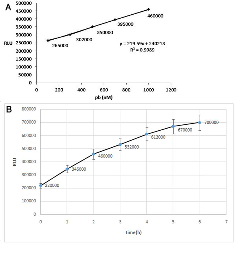
Keywords: Lead detection, Bacterial biosensor, Pollution control system
DOI: https://doi.org/10.21203/rs.3.rs-18087/v3
License: This work is licensed under a Creative Commons Attribution 4.0 International License.
Read Full License
Page 2/21
Abstract
Background: A bacterial biosensor refers to genetically engineered bacteria that produce an assessable
signal in the presence of a physical or chemical agent in the environment.

Methods: We have designed and evaluated a bacterial biosensor expressing a luciferase-reporter gene
controlled by pbr and cadA promoters in Cupriavidus metallidurans (previously termed Ralstonia
metallidurans) containing the CH34 and pI258 plasmids of Staphylococcus aureus, respectively, and that
can be used for the detection of heavy metals. In the present study, we have produced and evaluated
biosensor plasmids designated pGL3-luc/pbr-biosensor and pGL3-luc/cad-biosensor, that were based on
the expression of luc+ under the control of the cad promoter and the cadC gene of S. aureus plasmid
pI258 and pbr promoter and pbrR gene from plasmid pMOL30 of Cupriavidus metallidurans.

Results: We found that the biodegradable pGL3-luc/pbr-biosensor could be used to measure lead
concentrations between 1-100 μM in the presence of other metals, including: zinc, cadmium, tin and
nickel. The latter metals did not result in any signi cant gene expression of the reporter. The pGL3-
luc/cad-biosensor was able to detect lead concentrations between 10 nM to 10 μM.

Conclusions: This biosensor was found to be a speci c for measuring lead ions in both environmental
and biological samples.

Background
Ecological heavy metal pollution is a common problem that can exert damage to human health as well
as the environment [1]. Because heavy metal pollutants may lead to harmful ecological outcomes [2],
developing sensitive, e cient, rapid and cost-effective methods are necessary to e ciently screen for the
presence of harmful metals in the environment. Lead (Pb) is a toxic heavy metal that is extensively
utilized around the world [3, 4]. It has been estimated that the world production of lead is more than
3 million tons per year. It causes widespread environmental contamination in the air, water, soil, and food
[5]. This element can nd its way through human bodies as well as animals, entering the food chain; in
  sh and shrimp it can accumulate in the bone, liver, gills, kidney, ovary, and muscle [6].

Environmental lead may result in an increase in vascular endothelial growth factor (VEGF) and blood
concentrations [7, 8], and can lead to neurological and cardiovascular complications [9]. The reproductive
system may also be affected by developmental disorders that are highly likely to occur in children [10-13].
Lead can cross the placenta and cause damage to the developing fetal nervous system [14].

The assessment and monitoring for environment heavy metal contamination is very important to prevent
harm to human health. Currently, classical analytical methods, such as spectrometry, FIAAS ( ow
injection atomic absorption spectrometry), ion chromatography, and electrochemical techniques, are the
main methods used for measuring environmental heavy metals pollution. The main disadvantage of
these methods is the necessity for sample digestion under high temperature and pressure, or acidic

                                                 Page 3/21
conditions in which metal ions in solution are released [15]. Therefore, simpler methods for evaluating
heavy metals are required. More importantly, heavy metals are found to be present in biological systems
either in bioavailable/toxic or non-available/ non-toxic forms, and current measuring methods are unable
to distinguish between toxic and non-toxic fractions of these elements [16]. Furthermore, these methods
are both time-consuming and costly [17]. Biosensors have been developed that are an effective
alternative to conventional detecting systems. These may be highly sensitivity and simple to use [18].
Cell-based biosensors are a type of biologic sensor that contain a reporter gene under the control of a
promoter that is sensitive to the presence of an agent, such as environmental contaminants that include
heavy metals. Biosensors are used in various designs with different reporters and promoters. At low
concentration of heavy bioavailable metals, bioluminescence signals are likely to be suitable[19, 20].
They can also be applied to monitoring bioavailable concentrations of heavy metals [21-27] and
piezoelectric biosensors [28-30] as enzyme-based electrochemical biosensors. One of the most obvious
advantages of this method is the ability to measure the bioavailable heavy metal at very low
concentrations. It is also a cost-effective and time saving method [18]. In these biosensors, the expression
of a reporter gene is controlled by a promoter, such as the pbrR promoter in the
pMOL30plasmid of Cupriavidus metallidurans CH34 and cadC promoter in pI258 plasmid
of Staphylococcus aureus that is sensitive to heavy metals. Most of these promoters originate from
bacteria that have resistance systems against heavy metals [31, 32]. In this study, we have designed and
evaluated the luciferase reporter gene expression of bacterial biosensor under the control of pbr and cadC
promoters in Cupriavidus metalliduransCH34 and pI258 plasmids of Staphylococcus aureus, respectively,
for the measurement of lead.

Results
Sequencing

In order to ensure the integrity of the sequencing, the promoter region was sequenced in the modi ed
plasmid (Fig.1C and 1D).

Colony con rmation with PCR reaction

PCR was performed using primers designed for the pbr and cadA promoters, and the promoter sequence
and regulatory gene were ampli ed with 634 bp for pbr and 601 bp for cadA (Fig.2).

Biosensor activity of pGL3-luc/pbr

The expression of the luciferase gene, in the presence of different concentrations of lead, showed that 1
μM of lead was the lowest concentration that could stimulate the promoter and could be distinguished
from the basal expression of luciferase, and the highest measureable expression was seen at 100
μmol/L. A good biosensor should have two characteristics: speci city and sensitivity. According to the
data obtained from our experiments, this biosensor had a high speci city, and luciferase gene was only
expressed in the presence of lead.
                                                 Page 4/21
Biosensor speci city for lead in the presence of different concentrations of zinc (ZnCl2), tin (SnCl2) and
cadmium (CdCl2)
The biosensor was cultured in the presence of different concentrations of zinc, tin and cadmium, and did
not stimulate the pbrpromoter and expression of the reporter gene (Fig.3). Data obtained from the
expression of the luciferase gene in the presence of various concentrations of tin, zinc and cadmium,
indicated that these heavy metals did not stimulate the pbr promoter.
Biosensor activity in the presence of different concentrations of Lead (PbCl3)
Lead was the only metal that stimulated the pbr promoter. In the absence of lead, the regulator gene
prevents the promoter from activation. Lead ions bind to the regulator gene and inhibits its binding to the
operator. As a result, the promoter is activated and the luciferase is expressed. The minimum detectable
concentration of this biological sensor was approximately 1 µM and a maximum is 100 μmol/L. The
expression of luciferase was no longer linear for value of lead from 100 to 200μmol/L (Fig.4A).
The expression of pGL3-luc/ pbr-biosensor reporter gene at different times
In order to identify the appropriate time for biosensor growth, a biosensor was cultured at different
concentrations of lead for different durations (Fig.4B). The maximum expression of the luciferase gene
was at 12 h (Fig.5A).
The difference in the growth rate of pGL3-luc/ pbr-biosensor compared to E. coli strain DH5α
The sensor bacteria had a recombinant plasmid containing the pbr promoter region and the pbrR
regulatory gene. These bacteria have a greater resistance to lead than E. coliDH5α without plasmid. This
resistance may be related to the pbrR regulatory gene (Fig.5B). The resistance genes of metals have
heavy metal binding motifs, they can result in the non-toxicity of these metals inside the cell, because of
these proteins, the relative resistance of the cell to heavy metals.
The activity of pGL3-luc/cad-biosensor at the different concentrations of lead
The lowest and highest concentrations of lead that could stimulate the expression of the reporter gene
were 10 nmol/L and 10 μmol/L respectively (Fig.6 and 7A).
Expression of the Luciferase gene in the presence of 1 micro Molar concentration of Lead at different
times
The sensor bacteria were incubated at 0.2OD (1 μmol/L concentration) for different times in the
incubator. The expression of luciferase was measured at different times (Fig.7B). As shown in Fig. 7B, the
concentration of 1 μM lead can induce luciferase expression. The degree of expression increased with
time, with measureable change in Luciferase levels by 2 h measure, and in biological sensors pollution is
usually measured at low rates, we chose 2 h for culture of the pGL3-luc/cad-biosensor.
Page 5/21Discussion
There are several advantages to the use of bacterial biosensors, including speed, simplicity and cost.
Biological sensors containing cadA and pbr promoter regions have been designed by other researchers,
the optimization of this cell biological sensor with ability to measure lead comparing the cadA and pbr
promoters in a bioassay system was evaluated in this study. The use of biosensors or biological cell
sensors containing a reporter gene controlled by promoters susceptible to the heavy metal ions can
provide an e cient method to trace particular pollutants in the environment and in a biological
solution[33]. The present study assessed a biosensor system for detecting lead ions through construction
of a luminescent bacterial sensor containing the luc+ regulated by the cad promoter and cadC gene in
plasmid pI258 of S. aureus and the pbr promoter and pbrR gene in pMOL30 plasmid of Cupriavidus
metallidurans. Pb speci c bacterial biosensors were formerly de ned using reporter genes including lacZ,
lux,and luc in the transcription fusion constructs [34-36]. In our study, the luciferase reporter gene was
used. Luciferases, as a set of heterogeneous enzymes, are able to produce light as a byproduct of
catalyzing reactions. They are reporter genes extensively used by prokaryotic and eukaryotic organisms
due to their high sensitivity and ease of detection.The quanti cation of the emitted light, i.e.
bioluminescence,is of great importance; it can also be measured using a liquid scintillation counter ,a
luminometer, or even a X-ray lm [35]. It was concluded that a pGL3-luc/ pbr-biosensor can detect Pb+2 in
the range of 1–100 μM using the expression of re y luciferase as a detector system, and is highly
speci c, with no expression of reporter in the presence of other metals such as Sn+2, Ni+2, Cd+2 are
present. Moreover, this biosensor was 50 times more sensitive when compared with the previous
biosensors reported by Chakraborty et al [32]. The R. mettalidurans CH34 strain has several resistance
systems that can reduce the concentration of toxic substances to their non-toxic levels. A highly speci c
system for resistance to lead is known in plasmid pMOL30[37]. It effectively reduced the concentration of
lead ions and is equipped with speci c mechanisms for the transfer and separation of lead. The pbr
operon includes pbrA, pbrB, pbrC and pbrD genes in which pbrD has a role as a chaperone to accumulate
lead in the cell and pbrA eliminates lead ions[37]. Our results show that the pGL3-luc/pbr-biosensor is not
expressed in the presence of cadmium, zinc, ortin, indicating high sensitivity and speci city of the
designed system for lead detection. One of the most important heavy metal transfer systems in
Staphylococcus aureus is located in the plasmid pI258. The plasmid has an operon cadA that encodes an
ATPas of type P, which causes resistance to metals such as cadmium, lead, zinc, copper, and tin. The
expression of the cadA operon is controlled by the cadC homodimeric protein. This protein is able, in a
binary manner, to bind to the promoter and metal ions, such as cadmium, lead, zinc, and tin. The cad
belongs toArsR / SmtB, a regulating protein family[38]. In our study, the luciferase gene was used as a
reporter and E. coli strain of DH5α as a host. Our results showed that the pGL3-luc/cad-biosensor can
detect at least 10 nM of lead and the lead toxicity was not observed until a concentration of 300 μM.
However, the maximal expression of the reporter gene was performed at 10 μM. Our results are supported
by the report of Liao et al that showed the regulating role of cad promoter and the cadC gene in plasmid
pI258of S. aureus, the uorescence emission was intensi ed with increasing Cd(II), Pb(II), and Sb(III) ions
concentrations[39]. For Pb (II), just like our result in pGL3-luc/cad-Biosensor, to induce GFP expression
Page 6/21signi cantly, 10 nM was the low,and 10 μMwas the maximum concentration of lead that induced
signi cantly GFP expression[39]. The metallo regulatory α3N thiolate-rich site in cadC displays a practical
selectivity for larger, softer heavy metal like Pb(II), Cd(II), although smaller boundary metal ions such as
Zn(II) accommodated[40].
Conclusion
Our results show that the maximum expression of reporter gene was found in the presence of 100 μM of
Lead in pGL3-luc/pbr-biosensor and 1 μM of lead in pGL3-luc/cad-biosensor. In this study, the speci city
and sensitivity of the two heavy metal susceptive probes, pbr and cadA, was investigated. Sensors
composed of these two promoter regions were able to detect the concentration of lead between 1-100 μM
and 10 nM to 10 μM of lead, respectively. For other heavy metals such as mercury, copper, nickel,
manganese, zinc and cadmium, different biological sensors can be made and their presence in the
environment can be measured with very high accuracy. To determine the accuracy of biosensors, a
standard curve of Luciferase gene expression was plotted at different lead concentrations. The standard
curve was constructed from triplicates values, we evaluated the accuracy of the biosensor with the
speci c concentrations that we had obtained from lead metals. By developing these sensors, the time
required to identify environmental pollution can be minimized.
Methods
Chemicals
Analytical reagents, media and buffer solutions like TBE-EDTA buffer (Tris-borate-
Ethylenediaminetetraacetic acid), NaOH (Sodium hydroxide), CaCl2 (calcium chloride), boric acid, Tris
base, and agarose were all purchased from Merck (Germany). Fermentas (Lithuania) supplied the
restriction endonucleases Nco1 and Hind3, T4 DNA ligase, and molecular ladder 10000-300bp. We also
supplied the DNA polymerase (TaKaRa LA Taq®. DNA Polymerase), dNTP and MgCl2 from Takara
(Beijing, China). In addition, the plasmid extraction kit and primers were brought from Bioneer (Seoul,
South Korea).
Construction of biosensor plasmid
pMOL30 (X71400 AJ278984) and PI258 (GQ900378.1) containing the pbrR gene (634 bp) and CadC
gene (601bp) (Accession number: pbrR: WP_003103716.1and CadC: WP_000726009, respectively, were
synthesized and supplied by Millegen company. To ensure the accuracy of synthesized plasmid, the
promoter region was sequenced. PGl3-control as a vector containing the Luciferase gene and E.coli strain
DH5α as the host were used in our study. To obtain a large amount of pMA-T plasmid (a synthetic
plasmid) which contains p-promoter sequences and the regulatory gene was sent to MilliGen, after
evaluation at the NCBI site, for the synthesis of sequences. Synthesized sequences consisted of both
pbr_pMA-T plasmids containing the promoter sequence of the pRR operon and the pbrR regulator gene
Page 7/21including; cadA pMS-RQ-Bs plasmid containing the promoter region of the cadAp and the cadA gene regulating gene), it was cloned to E. coli host. Afterwards, pMA-T was extracted using plasmid extraction kit, and its quantity and quality were both examined by spectrophotometry and agarose gel, respectively, before they got digested by HindIII and NcoI. The promoter regions with the regulator genes were also puri ed from the gel electrophoresis. The received sequence and pGL3-control vector were cut using the same restriction enzyme (Nco1 and Hind3) and ligation reaction at 37°C for 3–4 h with ligase enzyme. The re y luciferase gene was placed under the control of the received promoter sequences and recombinant plasmids of cad and pbr promoters were named pGL3-luc/pbr-biosensor and pGL3-luc/Cad- biosensor, respectively. Recombinant plasmids pGL3-luc/pbr-biosensor (Fig. 1A) and pGL3-luc/Cad- biosensor (Fig. 1B) were transferred to the DH5α bacteria using the chemical method of CaCl2 and then were screened using selective plates containing antibiotic Ampicillin. After plasmid extraction, PCR was performed to detect colonies containing the promoter region of pbr and cadA using primers designed for the cloned fragments. After these processes, recombinant plasmids were used to evaluating and measuring different concentrations of heavy metals. Culture of bacteria and measuring biosensor activity of Luciferase enzyme To study the e ciency of promoters the detection of heavy metals, a luciferase enzyme measurement performedin the presence of lead and other heavy metals such as tin, zinc and cadmium. In this process, E.coli stains carrying pGL3-luc/Cad-Biosensor and pGL3-luc/pbr-Biosensor were cultured in Luriae Bertani (LB) broth that contained 100 µg/mL ampicillin at 37˚C, overnight. Then 50 µl from overnight grown culture of pGL3-luc/pbr-Biosensor for 12 h and pGL3-luc/Cad-Biosensor with optical density (OD 600) 0.8 for 2h were cultured in the presence of heavy metals at different concentrations [41]. Next, the culture was centrifuged at 5000 rpm for 10 min at 4°C metals for bacterial sedimentation. Then the medium was removed, and lysis buffer was added to the plate and sonicated at low temperature. Then, the amount of luciferase expression was measured by a luminometer (Berthold Company). Statistical analysis All the experiments were repeated at triplicate to minimize error. The Student t- test and one-way analysis of variance (ANOVA) were used to compare the statistically signi cant between the two groups and each group was compared with the baseline through the same method. . Statistical signi cance was set at *p
This article does not contain any studies with human participants or animals performed by any of the
authors.

Informed consent

For this type of study, formal consent is not required.

Competing interests

The authors have declared no con ict of interest.

Funding information

This study was supported by Department of Molecular Genetics, Faculty of Biological Sciences, Tarbiat
Modares University.

Consent for publication

All authors have given consent for publication

Availability of data and materials

All data generated or analyzed during this study are included in this published article

Author Contributions

MS and SH designed research; EN and RN performed research; HKS and NE analyzed data; EN and MN
wrote the manuscript; AM, MN and ZF performed statistical analysis; MG and MR contributed new
reagents or analytical tools. GAF revising the manuscript critically for important intellectual content. All
authors read and approved the manuscript.

Acknowledgements:

None

Abbreviations
Pb: Lead; VEGF: Vascular endothelial growth factor; Flow injection atomic absorption spectrometry:
FIAAS; LB: Luriae Bertani; RLU: Relative luminescence units.

References

                                                   Page 9/21
1. Adimalla N. Heavy metals pollution assessment and its associated human health risk evaluation of
urban soils from Indian cities: a review. Environmental Geochemistry and Health. 2020;42(1):173-90.
doi:10.1007/s10653-019-00324-4.
2. Hembrom S, Singh B, Gupta SK, Nema AK. A Comprehensive Evaluation of Heavy Metal
Contamination in Foodstuff and Associated Human Health Risk: A Global Perspective. In: Singh P,
Singh RP, Srivastava V, editors. Contemporary Environmental Issues and Challenges in Era of Climate
Change. Singapore: Springer Singapore; 2020. p. 33-63.
3. Ali H, Khan E, Ilahi I. Environmental chemistry and ecotoxicology of hazardous heavy metals:
environmental persistence, toxicity, and bioaccumulation. Journal of chemistry. 2019;2019.
4. Pourret O, Hursthouse A. It’s time to replace the term “heavy metals” with “potentially toxic elements”
when reporting environmental research. International journal of environmental research and public
health. 2019;16(22):4446.
5. Järup L. Hazards of heavy metal contamination. British Medical Bulletin. 2003;68(1):167-82.
doi:10.1093/bmb/ldg032.
6. Winder C, Stacey NH. Occupational toxicology. CRC press; 2004.
7. Yu B, Zhang Y, Shukla A, Shukla SS, Dorris KL. The removal of heavy metals from aqueous solutions
by sawdust adsorption—removal of lead and comparison of its adsorption with copper. Journal of
hazardous materials. 2001;84(1):83-94.
8. Machoń-Grecka A, Dobrakowski M, Kasperczyk A, Birkner E, Kasperczyk S. Angiogenesis and lead
(Pb): is there a connection? Drug and Chemical Toxicology. 2020:1-5.
9. Malik A, Ashraf MAB, Khan MW, Zahid A, Sha que H, Waquar S et al. Implication of Physiological
and Biochemical Variables of Prognostic Importance in Lead Exposed Subjects. Archives of
Environmental Contamination and Toxicology. 2019. doi:10.1007/s00244-019-00673-2.
10. Woolf AD, Goldman R, Bellinger DC. Update on the clinical management of childhood lead poisoning.
Pediatric Clinics of North America. 2007;54(2):271-94.
11. Zhou Q, Lin Y, Lin Y, Wei Q, Chen G, Tang D. Highly sensitive electrochemical sensing platform for
lead ion based on synergetic catalysis of DNAzyme and Au–Pd porous bimetallic nanostructures.
Biosensors and Bioelectronics. 2016;78:236-43.
12. Dolati S, Ramezani M, Abnous K, Taghdisi SM. Recent nucleic acid based biosensors for Pb2+
detection. Sensors and Actuators B: Chemical. 2017;246:864-78.
13. Mason LH, Harp JP, Han DY. Pb Neurotoxicity: Neuropsychological Effects of Lead Toxicity. BioMed
Research International. 2014;2014:8. doi:10.1155/2014/840547.
14. Meng Y, Tang C, Yu J, Meng S, Zhang W. Exposure to lead increases the risk of meningioma and
brain cancer: A meta-analysis. Journal of Trace Elements in Medicine and Biology. 2020;60:126474.
doi:https://doi.org/10.1016/j.jtemb.2020.126474.
15. Lemoine S, Bigot Y, Sellos D, Cosson R, Laulier M. Metallothionein isoforms in Mytilus edulis
(Mollusca, Bivalvia): complementary DNA characterization and quanti cation of expression in
Page 10/21different organs after exposure to cadmium, zinc, and copper. Marine Biotechnology. 2000;2(2):195-
203.
16. Vreeke M. Electrochemical biosensors for a nity assays. Part. 1997;1:39.
17. Gumpu MB, Sethuraman S, Krishnan UM, Rayappan JBB. A review on detection of heavy metal ions
in water–An electrochemical approach. Sensors and actuators B: chemical. 2015;213:515-33.
18. Ejeian F, Etedali P, Mansouri-Tehrani HA, Soozanipour A, Low ZX, Asadnia M et al. Biosensors for
wastewater monitoring: A review. Biosensors & bioelectronics. 2018;118:66-79.
doi:10.1016/j.bios.2018.07.019.
19. Jouanneau S, Durand MJ, Thouand Gr. Online detection of metals in environmental samples:
comparing two concepts of bioluminescent bacterial biosensors. Environmental science &
technology. 2012;46(21):11979-87.
20. Martín-Betancor K, Rodea-Palomares I, Munoz-Martin M, Leganés F, Fernández-Piñas F. Construction
of a self-luminescent cyanobacterial bioreporter that detects a broad range of bioavailable heavy
metals in aquatic environments. Frontiers in microbiology. 2015;6:186.
21. Dong S, Yang Q, Fu Y, Zhang D, Huang T. Carbon cloth-supported cobalt phosphide as an active
matrix for constructing enzyme-based biosensor. Journal of Solid State Electrochemistry.
2018;22(6):1689-96.
22. Hayat A, Marty JL. Aptamer based electrochemical sensors for emerging environmental pollutants.
Frontiers in chemistry. 2014;2:41.
23. Maleki N, Kashanian S, Maleki E, Nazari M. A novel enzyme based biosensor for catechol detection in
water samples using arti cial neural network. Biochemical Engineering Journal. 2017;128:1-11.
24. Moyo M, Okonkwo JO. Horseradish peroxidase biosensor based on maize tassel–MWCNTs
composite for cadmium detection. Sensors and Actuators B: Chemical. 2014;193:515-21.
25. Rao M, Scelza R, Acevedo F, Diez M, Gianfreda L. Enzymes as useful tools for environmental
purposes. Chemosphere. 2014;107:145-62.
26. Wei W, Dong S, Huang G, Xie Q, Huang T. MOF-derived Fe2O3 nanoparticle embedded in porous
carbon as electrode materials for two enzyme-based biosensors. Sensors and Actuators B: Chemical.
2018;260:189-97.
27. Zhang W, Liu Q, Guo Z, Lin J. Practical application of aptamer-based biosensors in detection of low
molecular weight pollutants in water sources. Molecules. 2018;23(2):344.
28. Asadnia M, Myers M, Akhavan N, O'Donnell K, Umana-Membreno GA, Mishra U et al. Mercury (II)
selective sensors based on AlGaN/GaN transistors. Analytica chimica acta. 2016;943:1-7.
29. Asadnia M, Myers M, Umana-Membreno GA, Sanders TM, Mishra UK, Nener BD et al. Ca2+ detection
utilising AlGaN/GaN transistors with ion-selective polymer membranes. Analytica chimica acta.
2017;987:105-10.
30. Teh HB, Li H, Li SFY. Highly sensitive and selective detection of Pb 2+ ions using a novel and simple
DNAzyme-based quartz crystal microbalance with dissipation biosensor. Analyst.
Page 11/212014;139(20):5170-5.
31. Corbisier P, Ji G, Nuyts G, Mergeay M, Silver S. luxAB gene fusions with the arsenic and cadmium
resistance operons of Staphylococcus aureus plasmid pI258. FEMS microbiology letters.
1993;110(2):231-8.
32. Chakraborty T, Babu PG, Alam A, Chaudhari A. GFP expressing bacterial biosensor to measure lead
contamination in aquatic environment. Current Science. 2008:800-5.
33. Gui Q, Lawson T, Shan S, Yan L, Liu Y. The Application of Whole Cell-Based Biosensors for Use in
Environmental Analysis and in Medical Diagnostics. Sensors (Basel). 2017;17(7):1623.
doi:10.3390/s17071623.
34. Shetty RS, Deo SK, Shah P, Sun Y, Rosen BP, Daunert S. Luminescence-based whole-cell-sensing
systems for cadmium and lead using genetically engineered bacteria. Analytical and bioanalytical
chemistry. 2003;376(1):11-7. doi:10.1007/s00216-003-1862-9.
35. Tauriainen S, Karp M, Chang W, Virta M. Luminescent bacterial sensor for cadmium and lead.
Biosensors and Bioelectronics. 1998;13(9):931-8.
36. Xu T, Close DM, Sayler GS, Ripp S. Genetically modi ed whole-cell bioreporters for environmental
assessment. Ecol Indic. 2013;28:125-41. doi:10.1016/j.ecolind.2012.01.020.
37. Mergeay M, Monchy S, Vallaeys T, Auquier V, Benotmane A, Bertin P et al. Ralstonia metallidurans, a
bacterium speci cally adapted to toxic metals: towards a catalogue of metal-responsive genes.
FEMS microbiology reviews. 2003;27(2-3):385-410.
38. Busenlehner LS, Pennella MA, Giedroc DP. The SmtB/ArsR family of metalloregulatory transcriptional
repressors: structural insights into prokaryotic metal resistance. FEMS microbiology reviews.
2003;27(2-3):131-43.
39. Liao VH-C, Chien M-T, Tseng Y-Y, Ou K-L. Assessment of heavy metal bioavailability in contaminated
sediments and soils using green uorescent protein-based bacterial biosensors. Environmental
Pollution. 2006;142(1):17-23. doi:https://doi.org/10.1016/j.envpol.2005.09.021.
40. Busenlehner LS, Weng T-C, Penner-Hahn JE, Giedroc DP. Elucidation of Primary (α3N) and Vestigial
(α5) Heavy Metal-binding Sites in Staphylococcus aureus pI258 CadC: Evolutionary Implications for
Metal Ion Selectivity of ArsR/SmtB Metal Sensor Proteins. Journal of Molecular Biology.
2002;319(3):685-701. doi:https://doi.org/10.1016/S0022-2836(02)00299-1.
41. Kumar A, Mathur R. Bioaccumulation kinetics and organ distribution of lead in a fresh water teleost,
Colisa fasciatus. Environmental technology. 1991;12(8):731-5.
Figures
Page 12/21Figure 1
Simpli ed schematic representation of the E. coli strain DH5α transfection. A Recombinant plasmid
(pGL3-luc/pbr-Biosensor). B Recombinant plasmid (pGL3-luc/cad-Biosensor). pGL3-luc/pbr-Biosensor
and pGL3-luc/Cad- biosensor were transferred to the E. coli strain DH5α using the chemical method of
Cacl2 and then were screened using selective plates containing antibiotic ampicillin. C Sequencing and
integrity of synthesis sequence. D PGL3-luc/pbr-Biosensor pGL3-luc/cad-Biosensor. The promoter region
was sequenced in the received plasmid.
Page 13/21Figure 2
A The proliferation region of the pbr promoter with 634 bp. B cadA promoter with 601 bp. The promoter
sequence and regulatory gene were ampli ed with 634 bp for pbr and 601 bp for cadA. 1 kb DNA Ladder
(containing 14 linear double-stranded DNA fragments).
Page 14/21Figure 3
Expression of luciferase gene in different concentrations of zinc, Tin and Cadmium. Heavy metal had no
effect on the stimulation of the pbr promoter.
Page 15/21Figure 4
A Luciferase expression in different concentrations of lead. The expression of luciferase was decreased
with a slight gradient from 100 to 200 micro molar. Relative luminescence units (RLU). B The expression
of pGL3-luc/pbr-biosensor reporter gene at different times.
Page 16/21Figure 5
A Linear expression ranges of Luciferase in the presence of lead with regression coe cient R2 = 0.960.
The maximum expression of the luciferase gene was 12h. B Difference in the growth rate of pGL3-
luc/pbr-biosensor compared to E. coli strain DH5α. Resistance may be related to the pbrR regulatory gene.
Page 17/21Figure 6
Expression of luciferase gene in different concentration of lead.
Page 18/21Figure 7
A linear expression ranges of Luciferase expression between 100-1000 nM concentrations of lead. B The
expression of luciferase at different times at 1 μM Pb concentration. During 2h, the amount of expression
is high enough to measure Luciferase, in biological sensors; the pollution is measured at low rates.
Page 19/21Figure 8
Difference in the growth rate of pGL3-luc/pbr-biosensor compared to E. coli strain DH5α. Resistance may
be related to the pbrR regulatory gene.
Figure 9
Expression of luciferase gene in different concentration of lead. Relative luminescence units (RLU).
Page 20/21Figure 10
linear expression ranges of Luciferase expression between 100-1000 nM concentrations of lead.
Figure 11
The expression of luciferase at different times at 1 μM lead concentration. During 2h, the amount of
expression is high enough to measure Luciferase, in biological sensors; the pollution is measured at low
rates. Relative luminescence units (RLU).
Page 21/21You can also read