Antioxidant Activity, Polyphenolic Content, and FT-NIR Analysis of Different Aspilia africana Medicinal Plant Tissues
←
→
Page content transcription
If your browser does not render page correctly, please read the page content below
Hindawi
Evidence-Based Complementary and Alternative Medicine
Volume 2021, Article ID 9917810, 11 pages
https://doi.org/10.1155/2021/9917810

Research Article
Antioxidant Activity, Polyphenolic Content, and FT-NIR
Analysis of Different Aspilia africana Medicinal Plant Tissues

 Denis Okello ,1,2,3 Yuseong Chung,1 Hyoseon Kim,1 Jun Lee,1,2 Endang Rahmat,1
 Richard Komakech,1,2,4 Yong-Goo Kim,1,2 Francis Omujal,4 and Youngmin Kang 1,2

 1
 Herbal Medicine Resources Research Center, Korea Institute of Oriental Medicine (KIOM), 111 Geonjae-ro, Naju-si,
 Jeollanam-do 58245, Republic of Korea
 2
 Korean Convergence Medicine Major, University of Science and Technology (UST), Daejeon, Republic of Korea
 3
 Gombe Secondary School, P.O. Box 192, Butambala, Mpigi, Uganda
 4
 Natural Chemotherapeutics Research Institute (NCRI), Ministry of Health, P.O. Box 4864, Kampala, Uganda

 Correspondence should be addressed to Youngmin Kang; ymkang@kiom.re.kr

 Received 2 April 2021; Accepted 1 September 2021; Published 15 September 2021

 Academic Editor: Caio P. Fernandes

 Copyright © 2021 Denis Okello et al. This is an open access article distributed under the Creative Commons Attribution License,
 which permits unrestricted use, distribution, and reproduction in any medium, provided the original work is properly cited.
 Aspilia africana has been used for generations to treat many diseases in Africa. Its biological activities, including antioxidant and
 anti-inflammatory potential, are attributed to a number of secondary metabolites, including alkaloids and polyphenolics. The
 antioxidant activities of A. africana callus (CA), juvenile in vitro leaf (IL) and root (IR), ex vitro root (SR) and leaf (SL), and wild
 leaf (WL) dried samples were assessed based on their diphenylpicrylhydrazyl (DPPH) free radical scavenging abilities. The total
 phenolic and flavonoid content of different plant samples was compared. Further, high-pressure liquid chromatography (HPLC)
 was used to quantitatively determine chlorogenic acid content in the A. africana plant samples. Fourier transform near-infrared
 (FT-NIR) analysis was also carried out to compare the antioxidant phytochemical content in the A. africana plant tissues. Among
 the samples, IR, with the highest total phenolic content (167.84 ± 1.057 mg GAE/g), total flavonoid content (135.06 ± 0.786 mg
 RUE/g), and chlorogenic acid (5.23 ± 0.298 mg/g) content, had the most potent antioxidant activity (IC50 � 27.25 ± 5.028 μg/mL),
 followed by WL. The lowest polyphenolic content and antioxidant activity were observed in SR. The antioxidant activities of
 A. africana tissues were positively correlated with the total phenolic and flavonoid content in the samples. The differences in
 antioxidant activities of A. africana tissues could be attributed to the difference in their polyphenolic content. Our study reports,
 for the first time, the antioxidant activities of A. africana callus and roots (in vitro and ex vitro). The A. africana samples IR, CA,
 and WL could be valuable natural sources of antioxidants that could be further exploited for the development of useful
 pharmaceutical products.

1. Introduction defend the body against diseases such as cancer, athero-
 sclerosis, neurodegenerative diseases, cardio\vascular dis-
Redox processes that occur during metabolism in aerobic eases, arthritis, diabetes mellitus, and nephritis [1, 4]. Plants
cells generate reactive oxygen and nitrogen species at low or possess powerful antioxidants, and the use of these plants in
moderate concentrations. These species possess important diets or as medicinal herbs reduces the occurrence of such
physiological functions, including transduction of cellular diseases [3, 5].
signals and defense against pathogens [1, 2]. If these reactive The medicinal plant Aspilia africana (Pers.) C. D. Adams
species are not regulated, they attack vital biological mol- belongs to the family Asteraceae and has been used for
ecules such as proteins, RNA, DNA, lipids, and carbohy- generations to treat diseases in many African countries [6].
drates, leading to cell death, tissue damage, and eventually to The plant is used to treat wounds, osteoporosis, stomach-
the development of chronic diseases [1, 3]. Antioxidants ache, rheumatic pains, ear infections, tuberculosis, cough,
2 Evidence-Based Complementary and Alternative Medicine

febrile headaches, wounds, gonorrhea, measles, diabetes, HO COOH
diarrhea, gastric ulcers, sores, malaria, and inflammatory O
conditions [5–7]. The biological activities of A. africana,
including antioxidant, anti-inflammatory, wound healing, HO O
anticancer, antidiabetic, and antiulcer activities, are mainly OH
 OH
attributed to the polyphenolic group of compounds [6, 7].
 OH
For the traditional treatment of diseases, the commonly used
plant parts of A. africana are leaves and roots collected from Figure 1: Chemical structure of chlorogenic acid.
the wild [5].
 Polyphenols are a large category of secondary metabo-
lites that are abundantly present in medicinal plants and derived from medicinal plants that have been utilized for
display excellent antioxidant properties [8]. These groups of generations to treat similar conditions. A. africana has been
chemical compounds are differentiated based on their used to treat some CNS-related disorders such as epilepsy
synthetic pathways [8]. A wide and highly heterogeneous [24], and its ethanolic leaf extract was confirmed to possess
group of polyphenols, which includes phenylpropanoids, sedative and antiseizure properties [25]. The sedative, an-
lignins, condensed tannins, and flavonoids, is derived from tiseizure, and apparent anxiolytic potentials of the plant were
an L-phenylalanine precursor, and the second largest group attributed to its richness mainly in flavonoids [25]. Flavo-
consisting of hydrolyzable tannins is derived from a gallic noids are apparently the main active principles responsible
acid precursor [8]. Currently, pharmacologists and re- for the sedative/anxiolytic effects of the most medicinal plant
searchers are interested in the phenolic and flavonoid [23]. Antioxidants eliminate reactive nitrogen and oxygen
content of plants, their antioxidant capacities, and their roles species and suppress the oxidative stress pathway, therefore
in preventing deadly diseases such as cardiovascular dis- protecting against neuronal damage triggered by oxidative
eases, neurodegenerative diseases, and cancer [4]. Poly- stress resulting in remission and functional recovery from
phenols possess hydroxyl (OH) groups, which act as positive symptoms of anxiety [26].
moieties for their antioxidant activities [9]. A. africana has been confirmed to contain polyphenolic
 Chlorogenic acid (Figure 1) is a phenylpropanoid compounds and vitamins, including ascorbic acid, ribofla-
compound produced during respiration (aerobic) in the vin, and thiamine, which possess antioxidant activities [5, 6].
shikimic acid pathway in plants [10]. It is a condensation The compound parahydroxybenzaldehyde, isolated from the
product of quinic and caffeic acids and is an active ingredient methanolic leaf extract of A. africana, competed favorably in
in several medicinal plants [10, 11]. Although chlorogenic comparison to standard antioxidant drugs [27]. There are a
acid is common in plants, only a few plants have it in high few studies on the antioxidant activity of A. africana leaves.
concentrations [9]. However, to the best of our knowledge, this is the first study
 Chlorogenic acid was previously detected in the most to determine the antioxidant activities of A. africana calli,
active leaf extracts of A. africana [12, 13]. It has been and juvenile in vitro regenerated A. africana leaves and roots
characterized as an important polyphenol in the Lamiaceae and to quantitatively compare total phenolic, flavonoid, and
and Asteraceae families [9]. It is a well-known antioxidant chlorogenic acid content in different plant tissues. In ad-
[9, 14, 15] and is used in the treatment of type 2 diabetes, dition, Fourier transform near-infrared spectroscopy (FT-
Alzheimer’s disease, stroke, eclampsia, and obesity [14]. NIR) was performed to compare the antioxidant chemical
Further, chlorogenic acid has been shown to improve lipid content of A. africana plant tissues. Therefore, this study
metabolism and glucose tolerance [16, 17], and its antimi- offers a basis for developing pharmaceutical products, in-
crobial [18, 19] and anxiolytic [19, 20] activities are well- cluding novel anxiolytic drugs, from this unique plant
documented. The antioxidant activity of plants such as coffee resource.
is dependent on phenolic compounds, especially its
chlorogenic acid content [21]. Chlorogenic acid significantly 2. Materials and Methods
inhibits the oxidative stress induced by interleukin-8 (IL-8)
secretion and mRNA expression [9]. Additionally, IL-8 2.1. Explant Sterilization, Callus Induction, and Culture.
production is suppressed by caffeic acid, a metabolite of Shoots of A. africana plants (100–120 mm from the tip) were
chlorogenic acid [9]. excised from three-month-old plants in a smart farm at
 Anxiety is a psychiatric and most prevalent Central Herbal Medicine Resources Research Centre, Korea Institute
Nervous System (CNS) disorder with high comorbidity and of Oriental Medicine (KIOM)-Naju. The shoots were washed
morbidity [22]. The increase in its prevalence worldwide is under running tap water for 5 min and moved to a laminar
due to the inhumane and highly competitive atmosphere in flow bench. The A. africana shoots were washed again with
man’s everyday life [23]. Thus, anxiolytic substances are autoclaved distilled water and sterilized using 100% (v/v)
among the most consumed drugs by humans [23]. The and 70% (v/v) ethanol for 20 and 30 s, respectively, and in 2%
current anxiolytic drugs are associated with many adverse (v/w) sodium hypochlorite for 3 min. Finally, the shoots
effects, including psychomotor function decay, dependence were rinsed four times with sterile water.
and abstinence syndromes, anterograde amnesia, and par- The leaves (except the terminal leaf pairs) were excised
adoxical reactions [22, 23]. Carlini [23] points out that al- from the shoots and cut into 7 mm2 segments, which were
ternative medication with fewer adverse effects could be used as explants for callus induction. Nine leaf segments
Evidence-Based Complementary and Alternative Medicine 3

were placed in each Petri dish containing 3 g/L gerlite-gelled concentrated A. africana sample (dried extract) was added to
MS medium with vitamins and supplemented with benzy- 5 mL of 80% aqueous ethanol to form a 10000 μg/mL stock
laminopurine (BAP, 0.5 mg/L) and 2,4-dichlorophenoxy- solution. The stock solution for each sample was diluted to
acetic acid (2,4-D, 1.0 mg/L). Sixty replicates were made, and varying concentrations (25, 100, 200, and 300 μg/mL) for the
the Petri dishes were placed in the dark at 25 ± 2°C and antioxidant assay.
relative humidity of 75% until the calli developed. The calli The antioxidant capacity of A. africana samples was
were subcultured twice at four-week intervals and main- determined using the diphenylpicrylhydrazyl (DPPH) rad-
tained under a 16-h photoperiod with light provided by cool ical scavenging method modified from Sarker and Oba [4].
white fluorescent tubes at 25°C and 70% relative humidity. To 0.1 mL solution of DPPH (Sigma-Aldrich, St. Louis, MO,
The calli were washed under running tap water to remove USA) in ethanol, 0.1 mL of the A. africana sample was added
traces of the medium and then dried for 24 h in an oven at at different concentrations in triplicate in a 96-well
60°C. The dried callus (CA) was homogenized into a fine microplate, wrapped in aluminum foil, and incubated at
powder and stored at 4°C until extraction for analysis. 37°C for 30 min. Spectrophotometric measurements were
 performed with Spectramax i3x (Molecular Devices,
 Wokingham, UK) at 517 nm. The radical scavenging activity
2.2. In Vitro Leaf and Root Induction, Culture, and Processing.
 was expressed in terms of antioxidant percentage calculated
The sterile A. africana shoot apices were cut into 30–45 mm
 from the following formula:
long pieces and end surfaces that were in contact with the
sterilizing agents were removed. The shoot apices were in- Acontrol − Asample
oculated in 3 g/L gerlite-gelled MS medium with vitamins antioxidant activity(%) � × 100, (1)
 Acontrol
fortified with 1-Naphthaleneacetic acid (NAA, 0.1 mg/L) for
in vitro root induction. Four shoot apices were placed in where Acontrol � absorbance of the control sample and
individual 125 × 100 mm culture vessels with 100 replica- Asample � absorbance of the test sample.
tions and then placed under a 16 h photoperiod with light Gallic acid was used as a positive control in this study.
provided by cool white fluorescent tubes at 25°C and 70% The IC50 values (sample concentration required to scavenge
relative humidity. After 6 wk, the in vitro leaves (IL) and 50% DPPH free radicals) were derived from a simple re-
roots (IR) were excised, washed, dried, homogenized, and gression analysis. The sample extraction and DPPH anti-
stored in the same way as the calli, ready for analysis. oxidant assays were carried out after storing the samples at
 4°C for one month.
2.3. Leaf and Root Samples from Three-Month-Old A. africana
Plants, Collecting, and Processing. Three-month-old 2.6. Determination of Total Polyphenolic Content
A. africana plants grown in a greenhouse at Herbal Medicine
Resources Research Centre, Korea Institute of Oriental 2.6.1. Total Phenolic Content. The total phenolic content of
Medicine (KIOM)-Naju were randomly chosen and care- the A. africana samples was estimated following the method
fully uprooted, and their roots were washed under running of Derakhshan et al. [28], with modifications. The previously
tap water. The leaves (SL) and roots (SR) were harvested and prepared stock extract of each sample was diluted to obtain
dried in an oven at 60°C for 24 h. The dried leaf and root 0.3 mg/mL sample, of which 0.5 mL aliquot was taken in a
samples were homogenized into a fine powder and stored at 1.5 mL microcentrifuge tube and mixed with the same
4°C until extraction for analysis. volume of Folin-Ciocalteu’s reagent. After 4 min, 0.5 mL of
 10% Na2CO3 was added to the mixture, mixed thoroughly
2.4. Wild A. africana Leaf Sample Collection and Processing. and incubated in the dark for 60 min at 25°C. Spectro-
Leaves of wild A. africana plants (WL) were randomly photometric measurements were performed with Spec-
collected from more than 50 plants in Pece, Gulu district, tramax i3x (Molecular Devices, Wokingham, UK) in
Uganda, East Africa. The leaves were sun-dried for 5 d, triplicate for each sample, and absorbance was read at
homogenized into a fine powder, packed in airtight bags, and 725 nm. A calibration curve of standard gallic acid was
posted to the Korea Institute of Oriental Medicine (KIOM), constructed to determine the total phenolic content of each
Herbal Medicine Resources Research Centre, Republic of sample. The total phenolic content in each sample was
South Korea. The samples were stored at 4°C until analysis. expressed as mg gallic acid equivalent (mg GAE/g).

2.5. Antioxidant Capacity Assay. A month after harvesting, 2.6.2. Total Flavonoid Content. The method used by Lee
about 2 g of each powdered sample (wild leaves, in vitro et al. [29] was modified to determine the total flavonoid
roots and leaves, green house roots and leaves, and callus of content of A. africana samples. In brief, 0.1 mL of each
A. africana) was added to 50 mL of 80% ethanol and son- sample extract (1 mg/mL) was taken in a 1.5 mL micro-
icated at 40°C for 1 h. The sample extracts were filtered centrifuge tube, and 0.8 mL diethyl glycol (90%), and 10 μL
(using a syringe filter with a 0.45 μm pore size membrane) of 1 N sodium hydroxide solution was added to the tube.
and the filtrate was concentrated in a rotary evaporator This mixture was then vortexed for about 3 sec and incu-
(EYELA N-1200B, Tokyo Rikakikai Co. Ltd., Japan) at 40°C bated for 60 min in a water bath at 37°C. Spectrophotometric
under reduced pressure. Fifty milligrams of each measurements were performed with Spectramax i3x
4 Evidence-Based Complementary and Alternative Medicine

(Molecular Devices, Wokingham, UK) in triplicate for each 2.9. Statistical Analysis. All experimental data were sub-
sample and absorbance was read at 420 nm. The total fla- jected to one-way analysis of variance (ANOVA) with
vonoid content in each sample was determined from the Tukey’s post hoc test using Prism (GraphPad software, v
calibration curve of the standard, rutin, and expressed as mg 5.03). All means were considered statistically significantly
rutin equivalent (mg RUE/g). different at p ≤ 0.05.

2.7. Sample Preparation for HPLC Analysis and Quantifica- 3. Results
tion of Chlorogenic Acid. Powdered A. africana WL, SL, SR, 3.1. Callus Induction. A high rate of callus induction (87%)
IL, IR, and CA samples (0.2 g each) were extracted using from the leaf segment explants in the callus induction
10 cm3 of 80% high-pressure liquid chromatography medium was attained within 4-5 wk. The calli generated were
(HPLC) grade methanol for 60 min using sonication. The cream to brown, friable, and structurally compact (Figure 2).
extract was filtered through Whatman grade 1 filter paper
and the filtrate was refiltered using a syringe filter (13 mm,
0.45 μm pore size PTFE membrane) for HPLC analysis. 3.2. Antioxidant Capacity of A. africana Tissues. The DPPH
Standard chlorogenic acid (1.2 mg) was dissolved in 1 mL of antioxidant activity of A. africana samples was assessed at
80% HPLC grade methanol to make a stock solution varying concentrations. The antioxidant activity of the
(1200 μg/mL) and diluted to varying concentrations (120, 60, A. africana samples was generally good, with over 50% DPPH
30, 15, and 7.5 μg/mL). inhibition for all samples at concentrations of 200 and
 An HPLC system (1200 series, Agilent Technologies, Palo 300 μg/mL (Figure 3). Overall, the highest antioxidant activity
Alto, CA, USA) equipped with a photodiode array detector was observed for IR (Figures 3 and 4). The DPPH antioxidant
(PDA) was used for the analysis. Reversed-phase chroma- activity of all the tested samples increased with increasing
tography was performed in a binary mode of gradient and concentrations (Figure 3). The highest DPPH free radical
isocratic mobile phase with a reversed-phase C-18 column scavenging activity was observed for IR with values of
(Luna 5 μm C-18(2) 100 Å, New Column 250 × 4.6 mm) at 86.32 ± 0.592% and 88.43 ± 0.796% at a concentration of 200
25°C. The following running conditions were maintained: and 300 mg/mL, respectively. These values did not differ
10 μL injection volume; mobile phase consisting of acetoni- significantly from those of the positive control, gallic acid,
trile (A) and 0.1% acetic acid in water (B) was used as follows: even at the highest concentration used (Figure 3). CA had the
5% A (0 min), 5–20% A (0–15 min) and 20% A (15–50 min); highest overall antioxidant activity after IR, with values of
flow rate: 0.7 mL/min; run time: 50 min; ultraviolet (UV) 82.85 ± 1.098% and 85.69 ± 0.906% DPPH inhibition at a
detection wavelength of 200–400 nm. Chlorogenic acid was concentration of 200 and 300 mg/mL, respectively (Figure 3).
identified by comparing the sample chromatographic peaks The antioxidant activity of CA did not differ significantly
with the standard retention time. After the analysis, the peak from that of WL, nor did the antioxidant activity of IL differ
areas were calculated using a Winchrom integrator. The five significantly from that of SL (Figures 3 and 4). IR had the
concentrations of chlorogenic acid (7.5, 15, 30, 60, and lowest IC50 value (27.25 ± 5.028 μg/mL), confirming its good
120 μg/mL) were subjected to regression analysis to calculate DPPH antioxidant activity. It was followed by CA and WL,
the calibration equation and correlation. The amount of whose IC50 values did not differ significantly (p < 0.05)
chlorogenic acid in each sample was expressed as mg/g of the (Figure 4). In contrast, the lowest antioxidant activity among
extract. the samples was of SR, with a significantly higher IC50 value
 (178.45 ± 1.609 μg/mL) compared to the rest of the samples
 (Figure 4). For the positive standard, the IC50 value was as
2.8. Fourier Transform Near-Infrared Spectroscopy (FT-NIR)
 low as 2.62 ± 0.307 μg/mL (Figure 4).
Analysis. The analysis was performed using a TANGO FT-
NIR spectrometer (Bruker Optics, Billerica, MA, USA) on
the powdered WL, SL, SR, IL, IR, and CA samples. Cali- 3.3. Total Phenolic and Flavonoid Content. The total phenolic
bration of the spectrometer was performed with a Light and flavonoid content varied significantly in the A. africana
Trap: (Type 1002961, ECL 00) and Gold standard (Type samples (Figure 5). The total phenolic content of the
1024957, ECL: 01). Further, 2 g of each sample was analyzed A. africana tissues ranged from 16.77 ± 0.282 to
in glass vials (22 mm). The absorbance spectra were obtained 167.84 ± 1.057 mg GAE/g, whereas the total flavonoid
at 12487–3948 cm−1 wave numbers, which determined the content ranged from 55.92 ± 0.939 to 135.06 ± 0.786 mg
different classes of compounds, including antioxidant RUE/g (Figure 5). The highest total phenolic and flavonoid
constituents, in the samples based on their functional content was observed in IR at 167.84 ± 1.057 mg GAE/g and
groups. Dendrograms were constructed for the samples 135.06 ± 0.786 mg RUE/g, respectively. These values were
based on Ward’s clustering algorithm upon characteristic significantly higher than the values of WL, which possessed
preprocessing of data (first derivative) and vector normal- the second-highest total phenolic (149.98 ± 1.032 mg GAE/g)
ization and standardization of the Euclidean distance in the and flavonoid (97.98 ± 0.749 mg RUE/g) content (Figure 5)
9981–4014 cm−1 frequency range. The software OPUS among the samples. Among the samples, the lowest total
TANGO-R was used for the Ward algorithm. Homogeneous phenolic and flavonoid content was observed in SR at
categories were sorted maximally using the minimum 55.92 ± 0.939 mg GAE/g and 33.41 ± 2.351 mg RUE/g, re-
variance method analysis of clusters. spectively (Figure 5).
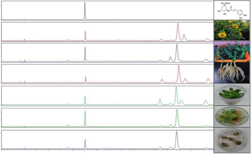
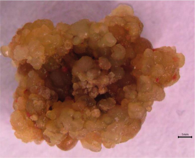
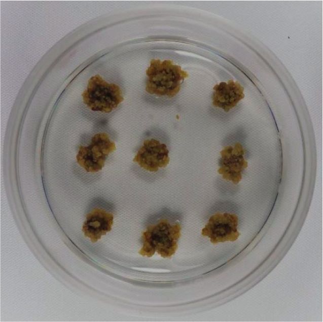
Evidence-Based Complementary and Alternative Medicine 5

 (a) (b)

 Figure 2: Aspilia africana callus generated from leaf explants. (a) Callus in Petri dish. (b) Callus nature and structure.

 100

 a a a a
 a a
 ab b ab ab
 b b
 80 b
 b b b
 DPPH inhibition (%)

 c c c cde
 60 cd c
 de
 e e

 f f
 40 f f
 g
 g g g
 g g
 20 h

 i
 0
 25
 50
 100
 200
 300
 25
 50
 100
 200
 300

 25
 50
 100
 200
 300
 25
 50
 100
 200
 300
 0
 5
 10
 50
 100
 250
 500
 25
 50
 100
 200
 300
 25
 50
 100
 200
 300

 WL SL SR IL IR CA GA (µM)
 A. africana samples and concentrations (µg/ml)
Figure 3: DPPH antioxidant activities of A. africana samples and gallic acid. Values are presented as means ± standard deviation. Same
letters are not significantly different by Tukey’s test and p � 0.05.

 200
 e 3.4. Chlorogenic Acid Content of A. africana Tissues.
 Chlorogenic acid in the A. africana samples was identified by
 150
 IC50 Value (µg/ml)

 comparing the HPLC retention time, UV absorption, and
 d d mass spectra with those of the chlorogenic acid standard
 100 c c (Figure 6). Chlorogenic acid was detected in all the samples
 analyzed, as shown in the HPLC chromatograms in Figure 6.
 A quantitative assessment of chlorogenic acid was
 50
 b performed at 280 nm. The highest quantity of chlorogenic
 a acid was observed in IR at 5.23 ± 0.298 mg/g. This value was
 0 significantly higher (p < 0.05) than that in the rest of the
 WL SR SL IL IR CA GA A. africana samples analyzed, except in SL
 A. africana samples and GA standard (4.308 ± 0.394 mg/g) (Figure 7). The chlorogenic acid con-
Figure 4: DPPH antioxidant activities of A. africana samples and tent in WL, IL, SA, and CA did not differ significantly
gallic acid. Values are presented as means ± standard deviation. Same (Figure 7). The lowest chlorogenic acid content was observed
letters are not significantly different by Tukey’s test and p � 0.05. in SR at 1.511 ± 0.055 mg/g (Figure 7).
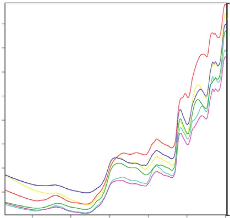
6 Evidence-Based Complementary and Alternative Medicine

 200 200
 a

 Total flavonoids (mgGAE/g)
 Total phenolics (mgGAE/g) b
 150 150
 a
 c

 d d b
 100 100
 c c
 c
 e
 50 50 d

 0 0
 WL SR SL IL IR CA WL SR SL IL IR CA
 A. africana samples A. africana samples
 (a) (b)

Figure 5: Total phenolic and flavonoid contents in A. africana samples. (a) Total phenolic contents. (b) Total flavonoid contents. Values are
presented as means ± standard deviation. Same letters are not significantly different by Tukey’ s test and p � 0.05.

 0.40
 ChA
 0.20
 AU

 0.00
 0.40
 WL
 0.20
 AU

 0.00

 SL 0.40
 0.20
 AU

 0.00
 0.40
 SR
 0.20
 AU

 0.00
 0.40
 IL
 0.20
 AU

 0.00
 0.40
 IR
 0.20
 AU

 0.00
 0.40
 CA
 0.20
 AU

 0.00
 0.00 2.00 4.00 6.00 8.00 10.00 12.00 14.00 16.00 18.00 20.00 22.00 24.00 26.00 28.00 30.00 32.00 34.00
 Minutes
Figure 6: Comparative chromatograms of the different samples of A. africana at ultraviolet (UV) detection wavelength of 200–400 nm.

3.5. Fourier Transform Near-Infrared Spectrometry (FT-NIR) and SR also had a close similarity, with a small heterogeneity
Analysis. The FT-NIR spectra showed some degree of value of 0.57 (Figure 8). The highest dissimilarity was
similarity in the chemical composition of the different recorded between CA and the root samples (IR and SR), with
A. africana samples (Figure 8). The highest degree of sim- a heterogeneity value of 1.45 (Figure 8).
ilarity in the chemical composition was observed among the
leaf samples (WL, IL, and SL), which formed up to eight 4. Discussion
peaks between 9000 and 4000 cm−1 (Figure 8).
 The root samples (SR and MR) had peaks similar to those Although A. africana has been studied extensively, there
of the leaf samples, except between 5000 and 4000 cm−1 have been few studies on the antioxidant properties of the
wavenumbers (Figure 8). Unlike the rest of the samples, the plant. Most of the previous studies are centered on the
spectrum of CA exhibited a peak at 5100 cm−1 (Figure 7). antioxidant activities of the plant leaves without exploring
Ward’s algorithm-based sample clustering showed the other plant parts/tissues [12, 27, 30, 31]. The antioxidant
closest homogeneity (0.35) between IL and SL (Figure 8). IR properties of natural products have been widely investigated
Evidence-Based Complementary and Alternative Medicine 7

 6 standard antioxidant, gallic acid, and the lowest IC50 value
 a
 (27.25 ± 5.028 μg/mL) among all A. africana tissues. The
 Chlorogenic acid contents (mg/g)

 a antioxidant properties of the samples were positively cor-
 related with chlorogenic acid content in the sample tissues,
 4
 except for SL. This indicates that although chlorogenic acid
 may contribute to the antioxidant capacity of A. africana
 b b tissues, it is not the main contributor. Rather, it is a part of a
 b
 2 large group of polyphenolic compounds present in
 b
 A. africana tissues. In contrast to our observations, there are
 plant species of medicinal value, for which the antioxidant
 capacity of the tissues is dependent mainly on chlorogenic
 0
 acid content [35, 36]. Wu [36] showed that the antioxidant
 WL SR SL IL IR CA
 capacity of Flos Lonicerae is dependent on its chlorogenic
 A. africana samples and GA standard
 acid content. In his study, Wu [36] demonstrated that
Figure 7: Chlorogenic acid contents in different samples of F. Lonicerae samples with higher chlorogenic acid content
A. africana samples. Values are presented as means ± standard exhibited higher efficiency in scavenging DPPH radicals and
deviation. Same letters are not significantly different by Tukey’s test reducing Fe3+ to Fe2+. Tomac et al. [35] demonstrated that
and p � 0.05.
 the antioxidant activity of coffee beans is positively corre-
 lated with their chlorogenic acid content. In their study,
using DPPH assays [32]. The DPPH antioxidant test dem- Tomac et al. [35] compared the antioxidant activity of coffee
onstrated the ability of a test sample to scavenge free radicals brands and their chlorogenic acid content. In the same
[32]. The assay is based on the reduction of DPPH (colored study, Tomac et al. [35] further demonstrated that the
free radical) methanolic solution by the free radical scav- mechanism of action of chlorogenic acid as an antioxidant
enging action of bioactive compounds or test samples [32]. was the direct scavenging of hydroxyl radicals (OH−) and
The DPPH free radical scavenging test on A. africana in- direct chemical reactions between chlorogenic acid and the
dicated that all the investigated tissues possessed antioxidant radicals formed from DNA during nucleic acid damage. The
properties. Previous studies on A. africana leaves have also antioxidant property of A. africana tissues is possibly due to
reported their antioxidant potential [12, 27, 30]. the total phenolic and total flavonoid content in the plant
 The antioxidant activity of the plant tissues is largely at- tissues.
tributed to the presence of bioactive polyphenols, which also The difference in antioxidant activities of the different
possess antimicrobial and anti-inflammatory properties parts of A. africana could be attributed to the difference in
[32, 33]. A number of studies have demonstrated a positive the concentration of antioxidant content, mainly total
correlation between the polyphenol content of plant samples phenolic and flavonoid content in these plant parts. Simi-
and their antioxidant capacities [32, 34]. Similarly, our study larly, Feduraev et al. [37] demonstrated in their study that
clearly shows that the higher the total phenolic and flavonoid the antioxidant activities of different parts of Rumex crispus
content in the tissues, the higher the antioxidant activity. Thus, L. and R. obtusifolius differed from each other. They at-
IR, with the highest total phenolic, flavonoid, and chlorogenic tributed these differences to the varying phenolic content in
acid content, displayed the highest DPPH free radical scav- the different plant parts. Secondary metabolite content in
enging capacity and the lowest IC50 value. In contrast, SR had plant organs of different species varies depending on the
the lowest polyphenolic content and exhibited the lowest developmental stage of the plant and environmental con-
antioxidant activity. Polyphenols are abundant in A. africana ditions [38, 39]. In our study, IR had the highest concen-
[5, 12] as confirmed by our study and may be the main tration of polyphenolic compounds (total phenolic and total
contributor to the antioxidant potential exhibited by its tissues. flavonoid content), giving it the highest antioxidant capacity,
In a previous study by Niyonizigiye et al. [12], the highest total while SR had the lowest polyphenolic content and exhibited
phenolic and flavonoid content in A. africana leaves was the lowest antioxidant activity.
76.61 ± 3.90 mg GAE/g and 62.71 ± 2.10 mg quercetin equiva- In comparison to a previous antioxidant study on
lent (QE)/g, respectively. These values were obtained through A. africana, Johnson et al. [27] revealed that the plant leaf
heating and agitation extraction techniques. The total phenolic extract had an IC50 value of 95.50 μg/mL. This value did not
and flavonoid content in all the leaf samples in our study was differ much from the IC50 values of A. africana leaf extracts
higher than that seen in the samples of Niyonizigiye et al. [12]. in our study, which were 99.08 ± 4.483 μg/mL (WL),
This disparity could be attributed to the differences in ex- 124 ± 14.994 μg/mL (SL), and 128 ± 4.606 μg/mL (IL). The
traction technique and the environment since the A. africana minor difference between the IC50 value of WL and the IC50
samples in the two studies were obtained from different value of the leaf extract from the previous study could be
geographical regions. attributed to the difference in extracting solvents ([27] used
 Chlorogenic acid, a phenolic compound and well-known methanol), while the relatively large differences between the
antioxidant [9, 35], was shown in this study to be present in IC50 value of the leaf extract from the previous study and the
all A. africana tissues, with the highest concentration being IC50 values of SL and IL could be due to differences in plant
observed in IR. IR also had the highest antioxidant potential, developmental stages, in addition to the extraction solvents
as exhibited by high DPPH inhibition comparable to the used. Unlike in our study, Niyonizigiye et al. [12] obtained a
8 Evidence-Based Complementary and Alternative Medicine

 0.45 IL SL WL CA IR SR

 0.40

 0.35
 Absorbance Units

 0.30 0.0 IL IL IL SL SL SL WLWL WLCACACA IR IR IR SR SR SR 0.0

 0.25
 0.2 0.2

 0.20
 0.4 0.4
 0.15

 Heterogeneity
 0.6 0.6
 0.10
 0.8 0.8

 9000 8000 7000 6000 5000 4000
 Wavenumber /cm 1.0 1.0

 A. africana wild leaf (WL) A. africana roots from
 soil grown plants (SR) 1.2 1.2
 In vitro A. africana root (IR)
 Callus (CA) In vitro A. africana leaf (IL)
 A. africana leaf from 1.4 1.4
 soil grown plant (SL)
 (a) (b)

 Figure 8: (a) FT-NIR spectra for A. africana samples. (b) Dendogram of A. africana samples analyzed from FT-NIR.

very large DPPH antioxidant activity for A. africana etha- contributes to the NIR spectra [47, 48]. FT-NIR spectra have
nolic leaf extract (IC50 value: 3 ± 0.03 mg/mL). This differ- been used in many studies for the assessment of antioxidant
ence could be attributed chiefly to the difference in chemical constituents in several plants [47, 48, 50].
extraction techniques since the green extraction technique The FT-NIR spectral peaks from 4200 to 4900 cm−1 are
was used in the previous study. This difference could also be assigned to combination (stretching and deformation)
attributed to differences in the geographical regions from modes due to C-H and O-H groups that belong to phenolic
which the plant leaf samples were collected. Guimarães et al. rings [48, 50]. The FT-NIR spectra in our study showed three
[38] emphasize that the environment influences the sec- prominent peaks for all the A. africana plant samples within
ondary metabolite content in plants and thus their activity. these wavenumbers except for SR, indicating the absence of
 Through scientific studies, calli obtained from various an antioxidant phenolic phytochemical compound in SR at
plant tissues have been demonstrated to possess various 4336 cm−1, which possibly contributed to its low antioxid-
secondary metabolites and potent biological activity, in- izing property in comparison to other A. africana samples.
cluding antioxidant potential. These plants include Artemisia The peaks at intervals 5050–5200 cm−1 are associated with
amygdalina [40], Harpagophytum procumbens [41], Vigna the combination modes of the O-H group in phenols and the
unguiculata [42], Randia echinocarpa [43], and Oroxylum corresponding aromatic ring-related vibrations [48]. Spec-
indicum [44]. Based on this background, we generated tral features from 5400–6000 cm−1 are due to the first
calli from A. africana leaf explants and used them as part of overtones of C-H stretching modes from the corresponding
our study material. A. africana calli had good antioxidant aromatic rings [48, 51]. The spectral peaks from
activity, with an IC50 value of 96.08 ± 4.144 μg/mL. The an- 6050–7200 cm−1 are due to second overtones due to C�O
tioxidant potency of calli from other plants species has stretching in flavonols [50] and O-H combinations in
also been reported previously, including H. procumbens phenols [51]. As shown in the FT-NIR spectra of all the
(IC50 � 46.74–163.66 μg/mL) [41], Inula crithmoides samples, there is a close similarity in the groups of anti-
(IC50 � 0.09–10.2 mg/mL) [45], and Justicia gendarussa oxidant chemical compounds present in the A. africana
(IC50 � 15.81–40.75 mg/mL) [46]. The differences observed in samples. The difference in antioxidant abilities could be
the antioxidant activities of the calli are due to varying media attributed to the differences in the relative quantities of the
and hormonal combinations, explant parts, calli culture antioxidant chemical constituents in the samples, as indi-
duration, and plant species [41, 45, 46]. cated by the differences in their absorbance.
 The FT-NIR technique, known to be a fingerprint tech-
nique, captures chemical data in relation to C-H, S-H, N-H, 5. Conclusion
and O-H bonds in the sample [47]. Infrared spectrometry
provides valuable information for medicinal plant analysis and The antioxidant activity of A. africana tissues was positively
quantification of chemical content [48, 49]. The phytochemical correlated with the total phenolic and flavonoid content in
content of plant tissues that exhibit antioxidant properties the samples. A. africana in vitro regenerated roots (IR) had
Evidence-Based Complementary and Alternative Medicine 9

the highest total phenolic content (167.84 ± 1.057 mg GAE/g), Institute of Oriental Medicine through the Ministry of
total flavonoid content (135.06 ± 0.786 mg RUE/g), and Science and ICT, Republic of Korea. The authors greatly
chlorogenic acid content (5.23 ± 0.298 mg/g) and the highest thank Mr. Gang Roggers (National Agricultural Research
antioxidant capacity, with the lowest IC50 value of Organization, Uganda) for collecting the seeds of the plant.
27.25 ± 5.028 μg/mL. In contrast, SR had the lowest poly-
phenolic content and the lowest antioxidant potential. The References
antioxidant activities of all analyzed A. africana tissues varied
significantly. Although chlorogenic acid, an important anti- [1] J.-G. Xu, Q.-P. Hu, and Y. Liu, “Antioxidant and DNA-
oxidant phytochemical compound, is present in a number of protective activities of chlorogenic acid isomers,” Journal of
plants, to the best of our knowledge, this is the first report of Agricultural and Food Chemistry, vol. 60, no. 46, pp. 11625–
this compound in A. africana callus (CA) and root tissues. 11630, 2012.
 [2] M. Valko, D. Leibfritz, J. Moncol, M. T. D. Cronin, M. Mazur,
Our study reports, for the first time, the antioxidant activities
 and J. Telser, “Free radicals and antioxidants in normal
of A. africana callus and roots (in vitro and ex vitro). IR, CA, physiological functions and human disease,” The Interna-
and WL tissues of A. africana could be valuable natural tional Journal of Biochemistry & Cell Biology, vol. 39, no. 1,
sources of antioxidants that could be further exploited for the pp. 44–84, 2007.
development of useful pharmaceutical products including [3] R. Niggeweg, A. J. Michael, and C. Martin, “Engineering
anxiolytic drugs, given the fact that these tissues are rich in plants with increased levels of the antioxidant chlorogenic
flavonoids and chlorogenic acid that are known to possess acid,” Nature Biotechnology, vol. 22, no. 6, pp. 746–754, 2004.
anxiolytic effects. We recommend further screening and [4] U. Sarker and S. Oba, “Phenolic profiles and antioxidant
isolation of the phytochemical constituents responsible for the activities in selected drought-tolerant leafy vegetable ama-
high antioxidant activities of A. africana plant tissues, es- ranth,” Scientific Reports, vol. 10, no. 1, pp. 18287–18311,
 2020.
pecially in in vitro regenerated roots, wild leaves, and calli.
 [5] D. Okello, J. Lee, and Y. Kang, “Ethnopharmacological po-
 tential of Aspilia africana for the treatment of inflammatory
Data Availability diseases,” Evidence-Based Complementary and Alternative
 Medicine, vol. 2020, Article ID 8091047, 11 pages, 2020.
The data used to support the findings of this study are [6] G. U. I. Ogbuehi and J. B. O. Echeme, “Chemical constituents
available from the corresponding author upon request. of methanol leaf extract of Aspilia africana C.D. Adams by GC
 MS,” International Journal of Advanced Research in Chemical
Conflicts of Interest Science, vol. 5, no. 10, pp. 21–29, 2018.
 [7] D. Okello and Y. Kang, “Exploring antimalarial herbal plants
The authors declare no conflicts of interest. across communities in Uganda based on electronic data,”
 Evidence-Based Complementary and Alternative Medicine,
Authors’ Contributions vol. 2019, Article ID 3057180, 27 pages, 2019.
 [8] L. Bragazza and C. Freeman, “High nitrogen availability re-
DO conceived the research idea and, together with YK, duces polyphenol content in Sphagnum peat,” The Science of
designed the experimental plan. DO, ER, RK, and YC the Total Environment, vol. 377, no. 2-3, pp. 439–443, 2007.
cultured the A. africana tissues, prepared the samples, and [9] M. Naveed, V. Hejazi, M. Abbas et al., “Chlorogenic acid
performed the antioxidant assay. DO and YC performed (CGA): a pharmacological review and call for further re-
total phenolic and flavonoid content analysis of the samples. search,” Biomedicine & Pharmacotherapy, vol. 97, pp. 67–74,
HK, JL, and DO performed HPLC-based quantitative as- 2018.
 [10] Y. Yan, X. Zhou, K. Guo, F. Zhou, and H. Yang, “Use of
sessment of chlorogenic acid contents in the samples. YGK
 chlorogenic acid against diabetes mellitus and its complica-
performed FT-NIR analysis and, together with DO, statis- tions,” Journal of Immunology Research, vol. 2020, Article ID
tically analyzed all data. DO wrote the manuscript. FO read 9680508, 6 pages, 2020.
and improved the manuscript. YK provided technical [11] C. Chaowuttikul, C. Palanuvej, and N. Ruangrungsi, “Phar-
guidance, supervised the whole research work, and read and macognostic specification, chlorogenic acid content, and in
improved the manuscript. All authors read and approved the vitro antioxidant activities of Lonicera japonica flowering
final manuscript. bud,” Pharmacognosy Research, vol. 9, no. 2, pp. 128–132,
 2017.
Acknowledgments [12] I. Niyonizigiye, D. Nkurunziza, D. Ngabire, A. T. Gitachew,
 B. S. Chun, and G.-D. Kim, “Characterization and in vitro
This study was supported under the framework of the In- cytotoxicity of phytochemicals from Aspilia africana obtained
ternational Cooperation Program (Korea-South Africa using green extraction techniques,” South African Journal of
Cooperative Research Project for Excavation of Candidate Botany, vol. 128, pp. 231–238, 2020.
 [13] S. Oguntimehin, E. Ajaiyeoba, O. Ogbole, H. Dada-Adegbola,
Resources of Complementary and Alternative Medicine)
 B. Oluremi, and A. Adeniji, Evaluation of Selected Nigerian
managed by National Research Foundation of Korea (grant Medicinal Plants for Antioxidant, Antimicrobial and Cytotoxic
no. 2017093655 and KIOM: D17470). Additionally, this Activities, Research Square, Durham, NC, USA, 2021.
work was also supported by Development of Foundational [14] Y. Zhao, J. Wang, O. Ballevre, H. Luo, and W. Zhang,
Techniques for the Domestic Production of Herbal Medi- “Antihypertensive effects and mechanisms of chlorogenic
cines (K18405), Development of Sustainable Application for acids,” Hypertension Research, vol. 35, no. 4, pp. 370–374,
Standard Herbal Resources (KSN2013320), and Korea 2012.
10 Evidence-Based Complementary and Alternative Medicine

[15] Y. Sato, S. Itagaki, T. Kurokawa et al., “In vitro and in vivo (Pers) CD Adams,” International Research Journal of Phar-
 antioxidant properties of chlorogenic acid and caffeic acid,” macy, vol. 655, pp. 135–138, 2012.
 International journal of pharmaceutics, vol. 403, no. 1-2, [31] H. Maduka, C. Ugwu, A. Okpogba et al., “Phytochemical
 pp. 136–138, 2011. studies, antioxidant properties and development of dye in-
[16] A.-S. Cho, S.-M. Jeon, M.-J. Kim et al., “Chlorogenic acid dicator from Aspilia africana leaves,” Journal of Applied Life
 exhibits anti-obesity property and improves lipid metabolism Sciences International, vol. 18, no. 3, pp. 1–7, 2018.
 in high-fat diet-induced-obese mice,” Food and Chemical [32] O. Amaeze, G. Ayoola, M. Sofidiya, A. Adepoju-Bello,
 Toxicology, vol. 48, no. 3, pp. 937–943, 2010. A. Adegoke, and H. Coker, “Evaluation of antioxidant activity
[17] D. V. Rodriguez de Sotillo, M. Hadley, and J. E. Sotillo, of Tetracarpidium conophorum (Müll. Arg) Hutch & Dalziel
 “Insulin receptor exon 11+/− is expressed in Zucker (fa/fa) leaves,” Oxidative Medicine and Cellular Longevity, vol. 2011,
 rats, and chlorogenic acid modifies their plasma insulin and Article ID 976701, 7 pages, 2011.
 liver protein and DNA,” The Journal of Nutritional Bio- [33] D. X. Cuong, V. N. Boi, T. T. T. Van, and L. N. Hau, “Effect
 chemistry, vol. 17, no. 1, pp. 63–71, 2006. of storage time on phlorotannin content and antioxidant
[18] X. Zhu, H. Zhang, and R. Lo, “Phenolic compounds from the activity of six Sargassum species from Nhatrang bay, Viet-
 leaf extract of artichoke (Cynara scolymus L.) and their an- nam,” Journal of Applied Phycology, vol. 28, no. 1,
 timicrobial activities,” Journal of Agricultural and Food pp. 567–572, 2016.
 Chemistry, vol. 52, no. 24, pp. 7272–7278, 2004. [34] G.-F. Deng, X. Lin, X.-R. Xu, L.-L. Gao, J.-F. Xie, and H.-B. Li,
[19] P. Shao, J. Zhang, Z. Fang, and P. Sun, “Complexing of “Antioxidant capacities and total phenolic contents of 56
 chlorogenic acid with β-cyclodextrins: inclusion effects, vegetables,” Journal of Functional Foods, vol. 5, no. 1,
 antioxidative properties and potential application in grape pp. 260–266, 2013.
 juice,” Food Hydrocolloids, vol. 41, pp. 132–139, 2014. [35] I. Tomac, M. Šeruga, and J. Labuda, “Evaluation of antioxidant
[20] J. Bouayed, H. Rammal, A. Dicko, C. Younos, and activity of chlorogenic acids and coffee extracts by an elec-
 R. Soulimani, “Chlorogenic acid, a polyphenol from Prunus trochemical DNA-based biosensor,” Food Chemistry, vol. 325,
 domestica (Mirabelle), with coupled anxiolytic and antioxi- Article ID 126787, 2020.
 dant effects,” Journal of the Neurological Sciences, vol. 262, [36] L. Wu, “Effect of chlorogenic acid on antioxidant activity of
 no. 1-2, pp. 77–84, 2007. Flos Lonicerae extracts,” Journal of Zhejiang Uni-
[21] M. Jeszka-Skowron, A. Sentkowska, K. Pyrzyńska, and versity—Science B, vol. 8, no. 9, pp. 673–679, 2007.
 M. P. De Peña, “Chlorogenic acids, caffeine content and [37] P. Feduraev, G. Chupakhina, P. Maslennikov, N. Tacenko, and
 antioxidant properties of green coffee extracts: influence of L. Skrypnik, “Variation in phenolic compounds content and
 green coffee bean preparation,” European Food Research and antioxidant activity of different plant organs from Rumex
 Technology, vol. 242, no. 8, pp. 1403–1409, 2016. crispus L. and Rumex obtusifolius L. at different growth
[22] K. Kumar, S. Sharma, P. Kumar, and R. Deshmukh, “Thera- stages,” Antioxidants, vol. 8, no. 7, p. 237, 2019.
 peutic potential of GABAB receptor ligands in drug addiction, [38] S. F. Guimarães, I. M. Lima, and L. V. Modolo, “Phenolic
 anxiety, depression and other CNS disorders,” Pharmacology content and antioxidant activity of parts of Passiflora edulis as
 Biochemistry and Behavior, vol. 110, pp. 174–184, 2013. a function of plant developmental stage,” Acta Botanica
[23] E. A. Carlini, “Plants and the central nervous system,” Brasilica, vol. 34, no. 1, pp. 74–82, 2020.
 Pharmacology Biochemistry and Behavior, vol. 75, no. 3, [39] J. Y. Jang, J. H. Ahn, Y. H. Jo, B. Y. Hwang, and M. K. Lee,
 pp. 501–512, 2003. “Antioxidant activity and phenolic content of different parts
[24] E. Noumi and F. L. Fozi, “Ethnomedical botany of epilepsy of lotus and optimization of extraction condition using re-
 treatment in Fongo-Tongo village, Western province, sponse surface methodology,” Natural Product Sciences,
 Cameroon,” Pharmaceutical Biology, vol. 41, no. 5, vol. 25, no. 1, pp. 44–48, 2019.
 pp. 330–339, 2003. [40] R. Rasool, B. A. Ganai, A. N. Kamili, and S. Akbar, “Anti-
[25] O. Kemelayefa and H. Kagbo, “Anticonvulsant potential of oxidant potential in callus culture of Artemisia amygdalina
 ethanolic extract of Aspilia africana leaf in mice,” Journal of Decne,” Natural Product Research, vol. 26, no. 22,
 Applied Life Sciences International, vol. 16, no. 2, pp. 1–7, 2018. pp. 2103–2106, 2012.
[26] Y. Xu, C. Wang, J. Klabnik, and J. O’Donnell, “Novel ther- [41] R. Gra˛bkowska, A. Matkowski, I. Grzegorczyk-Karolak, and
 apeutic targets in depression and anxiety: antioxidants as a H. Wysokińska, “Callus cultures of Harpagophytum pro-
 candidate treatment,” Current Neuropharmacology, vol. 12, cumbens (Burch.) DC. ex Meisn.; production of secondary
 no. 2, pp. 108–119, 2014. metabolites and antioxidant activity,” South African Journal of
[27] E. Johnson, E. Etim, and E. Archibong, “Isolation and anti- Botany, vol. 103, pp. 41–48, 2016.
 oxidant potentials of parahydroxybenzaldehyde from the [42] S. Vats, “Antioxidant activity of callus culture of Vigna
 methanol leaf extract of Aspilia africana (Pers.) CD Adams unguiculata (L.) Walp,” Researcher, vol. 4, no. 6, pp. 22–24,
 (Asteraceae),” Nigerian Journal of Pharmaceutical and Ap- 2012.
 plied Science Research, vol. 6, no. 1, pp. 26–32, 2017. [43] D. A. Valenzuela-Atondo, F. Delgado-Vargas, G. López-
[28] Z. Derakhshan, M. Ferrante, M. Tadi et al., “Antioxidant Angulo, C. L. Calderón-Vázquez, M. L. Orozco-Cárdenas,
 activity and total phenolic content of ethanolic extract of and A. Cruz-Mendı́vil, “Antioxidant activity of in vitro
 pomegranate peels, juice and seeds,” Food and Chemical plantlets and callus cultures of Randia echinocarpa, a me-
 Toxicology, vol. 114, pp. 108–111, 2018. dicinal plant from northwestern Mexico,” In Vitro Cellular &
[29] O.-H. Lee, B.-Y. Lee, J. Lee et al., “Assessment of phenolics- Developmental Biology—Plant, vol. 56, no. 4, pp. 440–446,
 enriched extract and fractions of olive leaves and their an- 2020.
 tioxidant activities,” Bioresource Technology, vol. 100, no. 23, [44] R. Faraz, M. Gokhale, and R. Gothalwal, “Callus extracts of
 pp. 6107–6113, 2009. Oroxylum indicum (L.) vent containing baicalein have in
[30] F. Faleye and O. Ogundaini, “Evaluation of antioxidant and vitro antioxidant and antibacterial activities,” Biotecnologı́a
 antimicrobial activities of two isolates from Aspilia africana Vegetal, vol. 20, no. 1, pp. 51–62, 2020.
Evidence-Based Complementary and Alternative Medicine 11

[45] A. Bucchini, L. Giamperi, and D. Ricci, “Total polyphenol
 content, in vitro antifungal and antioxidant activities of callus
 cultures from Inula crithmoides,” Natural Product Commu-
 nications, vol. 8, no. 11, pp. 1587–90, 2013.
[46] A. Amid, N. N. Johan, P. Jamal, and W. N. W. M. Zain,
 “Observation of antioxidant activity of leaves, callus and
 suspension culture of Justicia gendarusa,” African Journal of
 Biotechnology, vol. 10, no. 81, pp. 18653–18656, 2011.
[47] R. N. Páscoa, M. J. Gomes, and C. Sousa, “Antioxidant activity
 of blueberry (Vaccinium spp.) cultivar leaves: differences
 across the vegetative stage and the application of near infrared
 spectroscopy,” Molecules, vol. 24, no. 21, p. 3900, 2019.
[48] B. Carbas, N. Machado, D. Oppolzer et al., “Prediction of
 phytochemical composition, in vitro antioxidant activity and
 individual phenolic compounds of common beans using MIR
 and NIR spectroscopy,” Food and Bioprocess Technology,
 vol. 13, no. 6, pp. 962–977, 2020.
[49] A. A. Bunaciu, H. Y. Aboul-Enein, and S. Fleschin, “FTIR
 spectrophotometric methods used for antioxidant activity
 assay in medicinal plants,” Applied Spectroscopy Reviews,
 vol. 47, no. 4, pp. 245–255, 2012.
[50] V. Wiedemair, R. Ramoner, and C. W. Huck, “Investigations
 into the total antioxidant capacities of cultivars of gluten-free
 grains using near-infrared spectroscopy,” Food Control,
 vol. 95, pp. 189–195, 2019.
[51] I. R. N. de Oliveira, J. V. Roque, M. P. Maia, P. C. Stringheta,
 and R. F. Teófilo, “New strategy for determination of an-
 thocyanins, polyphenols and antioxidant capacity of Brassica
 oleracea liquid extract using infrared spectroscopies and
 multivariate regression,” Spectrochimica Acta Part A: Mo-
 lecular and Biomolecular Spectroscopy, vol. 194, pp. 172–180,
 2018.
You can also read