Applications of Extended Reality in Ophthalmology: Systematic Review - XSL FO
←
→
Page content transcription
If your browser does not render page correctly, please read the page content below
JOURNAL OF MEDICAL INTERNET RESEARCH                                                                                                          Ong et al

     Review

     Applications of Extended Reality in Ophthalmology: Systematic
     Review

     Chee Wui Ong1, MBBS; Marcus Chun Jin Tan2, MBBS, MMed; Michael Lam3, MBBS, MMed; Victor Teck Chang
     Koh2,4, MBBS, MMed, MRCSEd
     1
      Yong Loo Lin School of Medicine, National University of Singapore, Singapore, Singapore
     2
      Department of Ophthalmology, National University Hospital, Singapore, Singapore
     3
      Department of Ophthalmology, Ng Teng Fong General Hospital, Singapore, Singapore
     4
      Department of Ophthalmology, Yong Loo Lin School of Medicine, National University of Singapore, Singapore, Singapore

     Corresponding Author:
     Victor Teck Chang Koh, MBBS, MMed, MRCSEd
     Department of Ophthalmology
     Yong Loo Lin School of Medicine
     National University of Singapore
     1E Kent Ridge Road NUHS Tower Block, Level 7
     Singapore, 119228
     Singapore
     Phone: 65 90230056
     Email: victor_koh@nuhs.edu.sg

     Abstract
     Background: Virtual reality, augmented reality, and mixed reality make use of a variety of different software and hardware,
     but they share three main characteristics: immersion, presence, and interaction. The umbrella term for technologies with these
     characteristics is extended reality. The ability of extended reality to create environments that are otherwise impossible in the real
     world has practical implications in the medical discipline. In ophthalmology, virtual reality simulators have become increasingly
     popular as tools for surgical education. Recent developments have also explored diagnostic and therapeutic uses in ophthalmology.
     Objective: This systematic review aims to identify and investigate the utility of extended reality in ophthalmic education,
     diagnostics, and therapeutics.
     Methods: A literature search was conducted using PubMed, Embase, and Cochrane Register of Controlled Trials. Publications
     from January 1, 1956 to April 15, 2020 were included. Inclusion criteria were studies evaluating the use of extended reality in
     ophthalmic education, diagnostics, and therapeutics. Eligible studies were evaluated using the Oxford Centre for Evidence-Based
     Medicine levels of evidence. Relevant studies were also evaluated using a validity framework. Findings and relevant data from
     the studies were extracted, evaluated, and compared to determine the utility of extended reality in ophthalmology.
     Results: We identified 12,490 unique records in our literature search; 87 met final eligibility criteria, comprising studies that
     evaluated the use of extended reality in education (n=54), diagnostics (n=5), and therapeutics (n=28). Of these, 79 studies (91%)
     achieved evidence levels in the range 2b to 4, indicating poor quality. Only 2 (9%) out of 22 relevant studies addressed all 5
     sources of validity evidence. In education, we found that ophthalmic surgical simulators demonstrated efficacy and validity in
     improving surgical performance and reducing complication rates. Ophthalmoscopy simulators demonstrated efficacy and validity
     evidence in improving ophthalmoscopy skills in the clinical setting. In diagnostics, studies demonstrated proof-of-concept in
     presenting ocular imaging data on extended reality platforms and validity in assessing the function of patients with ophthalmic
     diseases. In therapeutics, heads-up surgical systems had similar complication rates, procedural success rates, and outcomes in
     comparison with conventional ophthalmic surgery.
     Conclusions: Extended reality has promising areas of application in ophthalmology, but additional high-quality comparative
     studies are needed to assess their roles among incumbent methods of ophthalmic education, diagnostics, and therapeutics.

     (J Med Internet Res 2021;23(8):e24152) doi: 10.2196/24152

     KEYWORDS
     extended reality; virtual reality; augmented reality; mixed reality; ophthalmology; ophthalmic
     https://www.jmir.org/2021/8/e24152                                                                J Med Internet Res 2021 | vol. 23 | iss. 8 | e24152 | p. 1
                                                                                                                     (page number not for citation purposes)
XSL• FO
RenderX
JOURNAL OF MEDICAL INTERNET RESEARCH Ong et al
Introduction Methods
The rapid development of extended reality technologies has Eligibility Criteria
necessitated recent efforts to define and draw lines between new We included studies evaluating the use of extended reality for
concepts and subgroups of extended reality applications [1]. ophthalmic applications in education, diagnostics, and
Virtual reality has been defined as one in which our natural therapeutics for eye care professionals and ophthalmic patients.
surroundings are completely replaced with a 3D All study designs were included with the exception of systematic
computer-generated environment via wearable screens in the reviews, case reports, and case series with ≤3 patients.
form of head-mounted displays [2]. Augmented reality is a Non-English publications and publications on the technical
superimposition of computer-generated content with limited engineering of extended reality were excluded.
interactivity onto our visible surroundings. Mixed reality is
similar to augmented reality, except that the user is able to Search Methods
interact vividly with computer-generated content [1]. Mixed Three databases served as the source of our search—PubMed
reality can be considered an amalgamation of the features of MEDLINE, Embase, and Cochrane Register of Controlled
both virtual reality and augmented reality, as both highly Trials. Search terms included “Virtual Reality,” “Augmented
interactive computer-generated objects and the real physical Reality,” “Mixed Reality,” “Simulation,” “Simulated,” “3D,”
world are integrated to dynamically coexist within a single “Ophthalmology,” “Ophthalmic,” and “Eye.” The search was
display [3,4]. Whereas virtual reality, augmented reality, and performed on April 15, 2020. Publications from January 1, 1956
mixed reality make use of a variety of different software and to April 2020 were searched without language or
hardware, these extended reality technologies share 3 main publication-type restrictions. References in studies meeting the
characteristics: immersion, presence, and interaction [2,5]. eligibility criteria were searched to identify additional eligible
Immersion refers to a perception of physical existence within studies. EndNote X9 (2020; Clarivate Analytics) was used to
the extended reality environment, presence describes the manage all identified publications and remove duplicates
perception of connection to the environment, whereas (Multimedia Appendix 1). Search results were recorded
interaction is the ability to act and receive feedback within the according to PRISMA (Preferred Reporting Items for Systematic
environment [2]. Reviews and Meta-analyses) guidelines [22].
In medicine, the nascent influence of extended reality is Study Selection
prevalent. Virtual reality platforms have been designed to teach
Two authors (CWO and MCJT) read all titles returned by the
foundational subjects, such as human anatomy [6,7], and train
search. All abstracts of relevant titles and full texts of the
surgeons in complex surgical procedures [8-11]. Augmented
relevant abstracts were read by the same authors to evaluate
and mixed reality offer methods of visualizing intraoperative
eligibility. Any uncertainties was resolved by discussion among
procedures and diagnostic images with devices, such as Google
all authors.
Glass (Google Inc) or Microsoft HoloLens (Microsoft Inc), that
have the potential to improve procedure safety and success Data Collection and Analysis
[12-14]. The ability of virtual reality to distract patients from For each study that met eligibility criteria, the quality of study
the physical environment also offers therapeutic approaches for was evaluated using Oxford Centre for Evidence-Based
rehabilitation and for treating pain or psychiatric disorders Medicine (OCEBM) levels of evidence [23].
[15-17]. Likewise, ophthalmology has seen a growing influence
of extended reality. Ophthalmic graduate medical education in Information from each study was extracted, including aim,
the United States has seen an increase in the use of virtual eye design, population, sample size, extended reality technology
surgery simulators, from 23% in 2010 to 73% in 2018 [18,19]. type, application, outcomes, and findings.
Extended reality technologies have also been explored as a A number of eligible studies investigated the use of extended
method of therapy in ophthalmic diseases such as amblyopia reality educational training simulators as training and assessment
and visual field defects [20,21]. Although the versatility of tools. Evidence of validity should be used to support the
extended reality platforms can influence the practice of appropriateness of interpretation of results from assessments of
ophthalmology, health care providers should be well informed performance using these simulators [24,25]. Validation is critical
of the benefits and limitations of such technologies. This will to be able to trust the results of a given education tool, and
allow evidence-based decision making when adopting nascent educators need evidence of validity to identify the appropriate
methods of ophthalmic education, diagnosis, and treatment. The assessment tool to meet specific educational needs with finite
focus of this review was to systematically evaluate current resources [26]. We chose a contemporary model of validity
evidence of the efficacy, validity, and utility of the application [24], comprising 5 sources of validity evidence—Content,
of extended reality in ophthalmic education, diagnostics, and Response process, Internal structure, Relationship to other
therapeutics. variables, and Consequences [25,27] (Multimedia Appendix
2), to evaluate the extent to which the validity of these
simulator-based assessments had been established by evidence.
Due to a high degree of heterogeneity between studies,
quantitative statistical analysis was not conducted.
https://www.jmir.org/2021/8/e24152 J Med Internet Res 2021 | vol. 23 | iss. 8 | e24152 | p. 2
(page number not for citation purposes)
XSL• FO
RenderXJOURNAL OF MEDICAL INTERNET RESEARCH Ong et al
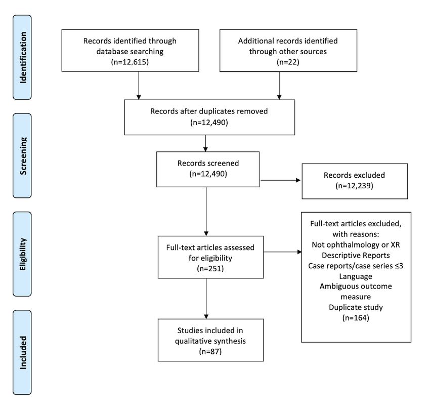
for assessment for final eligibility. Of these, 164 were excluded,
Results and 87 studies met the final eligibility criteria (Figure 1). Of
General these, 54 were relevant to the use of extended reality in
education, 5 were relevant to the use of extended reality in
A total of 12,490 unique records were identified. After screening diagnostics, and 28 were relevant to the use of extended reality
by title and abstract, 251 full-text publications were retrieved in therapeutics.
Figure 1. Flow Diagram showing inclusion process for identified records. XR: extended reality.
included MicroVisTouch (ImmersiveTouch) (n=1), PixEye
Education Ophthalmic Simulator (SimEdge SA) (n=1), and 6 self-designed
Overview simulators. The most common surgical procedure simulated in
these studies was cataract surgery (n=36), followed by
Applications of extended reality in education included surgical
vitreoretinal procedures (n=9), laser trabeculoplasty (n=1), and
simulators (46/54), ophthalmoscopy simulators (6/54), and
corneal laceration repair (n=1).
optometry training simulators (2/54), with medical students,
optometry students, trainee, or trained ophthalmologists as Of the 46 studies, 24 were studies that evaluated the efficacy
participants. of surgical simulators using evaluations of surgical performance
on real patients, objective assessments by subspecialty experts,
Surgical Simulators
or simulator-based metrics as outcome measures. There were
Of 46 studies evaluating surgical simulators, the EyeSi surgical 14 [28-47] studies that used surgical performance on real
simulator (VR Magic) was most commonly used (n=38). Others patients as outcome measures, of which 4 were randomized
https://www.jmir.org/2021/8/e24152 J Med Internet Res 2021 | vol. 23 | iss. 8 | e24152 | p. 3
(page number not for citation purposes)
XSL• FO
RenderXJOURNAL OF MEDICAL INTERNET RESEARCH Ong et al
trials [48-51] (Table 1); these randomized trials compared the efficiency, efficacy, or complication rates. In particular,
use of virtual reality ophthalmic surgical simulators with Deuchler et al [50] found that warm-up simulation training
conventional methods of surgical training. Objective assessment improved performance for surgeons who had not operated for
metrics of participants’ surgical performance on real patients a significant period of time. Daly et al [49] showed that residents
were evaluated by subspecialty experts. All studies [48,50,51], who underwent EyeSi training were significantly slower at
except one [49], showed that simulator-training resulted in performing their first continuous curvilinear capsulorhexis than
surgical performance significantly superior to that of participants who underwent wet-lab training but achieved similar
conventional training methods in terms of quality, time surgical performance scores.
Table 1. Randomized trials evaluating efficacy of surgical simulators in improving surgical performance on real patients.
Study Design Intervention group training Control group Simulated task Population Findings
(OCEBMa level) training
Peugnet Randomized trial Laser photocoagulation Real patients Retinal photo-coagu- Eye residents Simulator training was
(1998) [48] (2b) simulator (n=5) (n=4) lation significantly more time ef-
ficient (time efficiency in-
dex of 0.59 vs 0.28,
PJOURNAL OF MEDICAL INTERNET RESEARCH Ong et al
Table 2. Nonrandomized trials evaluating efficacy of surgical simulators in improving surgical performance on real patients.
Study Design Comparison or control Outcome measure Participants and Findings
(OCEBMa level) group cataract surgeries, (n)
Thomsen Cohort (2b) Before EyeSi use OSACSSb Cataract surgeons (19) Novices and less-experienced surgeons
(2017a) [28] showed significant improvements in the
operating room (32% and 38% improve-
ment, P=.008 and P=.018 respectively)
after EyeSi training
La Cour Cohort (2b) Before EyeSi use OSACSS Cataract surgeons (19) EyeSi training resulted in significantly
(2019) [29] improved surgical performance in less-
experienced surgeons. Skill-transfer be-
tween modules was not demonstrable
Roohipoor Cohort (2b) N/Ac GRASISd Ophthalmology resi- Significant correlations between residents’
(2017) [30] dents (30) EyeSi simulator-based scores and their
eventual surgery count and GRASIS
scores
Belyea (2011) Cohort (3b) No EyeSi use Phaco time, per- Surgeries by ophthal- EyeSi training resulted in significantly
[31] centage power, mology residents lower procedure duration (P=.002), per-
complications (592) centage power (P=.001), and nonsignifi-
cantly fewer intraoperative complications
Pokroy (2013) Cohort (2c) No EyeSi use Incidence of poste- Surgeries by ophthal- EyeSi training resulted in nonsignificantly
[32] rior capsule tears, mology residents fewer posterior capsule tears and shorter
operation duration (1000) learning curves
Ferris (2020) Cohort (2b) No EyeSi access Posterior capsule Surgeries by ophthal- Residents with EyeSi access had a signifi-
[33] rupture rates mology residents cant reduction in posterior capsule rupture
(17831) rates (4.2% vs 2.6%, Difference in Propor-
tions 1.5%, 95% CI 0.5-2.6%, P=.003).
Posterior capsule rupture rates significant-
ly lower after access to EyeSi (3.5% to
2.6%, Difference in proportions 0.9%,
95% CI 0.4-1.5%, P=.001)
Lucas (2019) Cohort (2b) No EyeSi use Complication rates Surgeries by ophthal- EyeSi training resulted in significantly
[34] mology residents fewer complications (12.86 vs 27.14%,
(140) P=.031)
Staropoli Cohort (2b) No EyeSi use Complication rates Surgeries by ophthal- EyeSi training resulted in significantly
(2018) [35] mology residents fewer complications (2.4 vs 5.1%, P=.037)
(955)
McCannel Case series (4) Reduced EyeSi use Errant continuous Surgeries by ophthal- EyeSi training resulted in significantly
(2013) [36] curvilinear capsu- mology residents lower errant continuous curvilinear capsu-
lorhexis rates (1037) lorhexis rates (5.0 vs 15.7%, PJOURNAL OF MEDICAL INTERNET RESEARCH Ong et al
significantly improve performance at the other complications than those who did not train on the simulator.
(OSACSS—training: 8, no training: 8, P=.64; OSATS These trials suggest that simulator training improves
score—training: 7, no training: 10, P=.52); however, repeated performance only for the specific procedure being trained. The
practice with each module significantly improved other 4 studies [41-44] were prospective cohort studies or case
simulator-based scoring for the respective modules. Bergqvist series (Table 4) that demonstrated that extended reality surgical
et al [38] found that medical students who trained with simulated training resulted in significant improvements on subsequent
cataract surgery had higher overall simulator scores and fewer simulator-based performance scores.
Table 3. Randomized trials evaluating efficacy of surgical simulators in improving surgical performance as measured by the same simulator.
Study Design Intervention group Control group Participants Simulated task
(OCEBMa level)
Bergqvist Randomized trial EyeSi training No EyeSi training (n=10) Medical students Cataract surgery
(2014) [38] (1b) (n=10)
Thomsen Randomized trial EyeSi training (n=6) No EyeSi use (n=6) Eye residents Cataract surgery, vitreoretinal surgery
(2017b) [39] (2b)
Selvander Randomized trial EyeSi cataract navi- EyeSi capsulorhexis Medical students Capsulorhexis, cataract navigation
(2012) [40] (2b) gation training first training first (n=18)
(n=17)
a
OCEBM: Oxford Centre for Evidence-Based Medicine.
Table 4. Nonrandomized trials evaluating efficacy of surgical simulators in improving surgical performance as measured by the same simulator.
Study Design Surgical simulator Participants Simulated task Findings
a
(OCEBM level)
Saleh (2013a) Case series (4) EyeSi Eye residents (n=17) Cataract surgery EyeSi training resulted in significantly
[41] improved scores (PJOURNAL OF MEDICAL INTERNET RESEARCH Ong et al
Table 5. Studies evaluating efficacy of surgical simulators in improving surgical performance in the wet lab.
Study Design (OCEBMa Intervention group Control group Participants Outcome measure tool
level)
Jonas (2003) [45] Randomized trial Simulator training No simulator training Medical students, eye Amount of vitreous removed, retinal
(2b) (n=7) (n=7) residents lacerations, residual retinal detach-
ment, duration
Feldman (2007) Randomized trial EyeSi training (n=8) No EyeSi training Medical students Corneal Laceration Repair Assess-
[46] (2b) (n=8) ment
Feudner (2009) Randomized trial EyeSi training No EyeSi training Eye residents Scoring based on capsulorhexis
[47] (1b) (n=31) (n=31) video
a
OCEBM: Oxford Centre for Evidence-Based Medicine.
Of 46 studies, 20 studies evaluated the validity of surgical Two randomized trials [74,75], with OCEBM evidence levels
simulator-based assessments (Multimedia Appendix 3). Most 2b, assessed the efficacy of the EyeSi Binocular Indirect
validity studies achieved an OCEBM level of evidence of 2b, Ophthalmoscopy simulator. Both studies showed that
corresponding to exploratory cohort studies with good reference participants who trained with the EyeSi Binocular Indirect
standards. The most common source of validity evidence was Ophthalmoscopy simulator performed significantly better than
Relationship with other variables, addressed in 19 of 20 studies participants who underwent conventional training.
(95%). Studies achieved this by statistically evaluating the
Three studies [75-77], with OCEBM evidence levels of 2b,
relationship between surgical performance on the simulator and
assessed the validity of the EyeSi Binocular Indirect
participants’ levels of expertise. Content validity was addressed
Ophthalmoscopy simulator (n=2) and the EyeSi Binocular Direct
in 18 studies (90%), Response process was addressed in 9
Ophthalmoscopy simulator (n=1) for training and assessment.
studies (45%), Internal structure was addressed in 5 studies
All studies demonstrated Relationships with other variables as
(25%), and Consequences was addressed in 2 studies (10%).
a source of validity evidence and found that participants with
Only 2 of 20 (10%) studies addressed all 5 sources.
more experience had significantly higher ophthalmoscopy
Of these 20 studies, 12 assessed surgical performance using evaluation scores. Content validity was addressed in all studies.
simulator-based scoring only [39,43,52-61], 6 studies compared Only 1 study [77] addressed Internal structure by evaluating
simulator-based scores with video-based scoring (OSACSS, internal consistency between simulator modules and evaluated
OSATS, or motion-tracking software) [62-67], and 2 studies Consequences by calculating a pass or fail score.
used video-based scoring only [40,68].
Two user perception studies [78,79] found that medical students
For the EyeSi surgical simulator, most studies found that the felt that self-assembled virtual reality direct ophthalmoscopy
surgical performance of experienced participants was simulators were usable and useful in improving ophthalmoscopy
significantly better than that of less-experienced participants. skills.
Sikder et al [52] found that intervening surgical experience
significantly improved capsulorhexis performance on the
Optometry Training Simulators
MicroVisTouch cataract surgery simulator. Lam et al [62] Two studies [80,81] evaluated the preliminary user experience
showed that in a self-made phacoemulsification simulator, more of an augmented reality optometry simulator comprising a
experienced participants attained significantly higher scores in head-mounted display, a slit-lamp instrument, and a simulated
all main procedures and completed tasks significantly faster. eye, which allowed the simulation of optometry training tasks.
User studies involving undergraduate optometry students
Five studies [69-73] assessed the perception of ophthalmologists showed that the simulator was feasible in simulating foreign
and medical students toward surgical simulators using body removal as a training task with a high level of user
user-reported outcome measures. These studies achieved satisfaction.
OCEBM evidence levels of 4 (n=4) and 2b (n=1). Users found
the EyeSi and a novel virtual reality continuous curvilinear Diagnostics
capsulorhexis simulator to be useful in improving surgical skill,
Overview
confidence, and understanding, while providing a safe and
realistic alternative for training. Five studies evaluated the use of extended reality for the
production of immersive and interactive content for diagnostic
Ophthalmoscopy Simulators applications. Two studies evaluated the use of extended reality
Six studies [74-79] evaluated the use of extended reality as a to display ocular imaging data [82,83], and 3 studies [84-86]
tool for education in ophthalmoscopy. Simulators used were evaluated the validity of extended reality as a simulation tool
the EyeSi Augmented Reality Direct (n=1) and Binocular for the functional assessment of patients with ophthalmic
Indirect (n=3) ophthalmoscopy simulators, and 2 novel diseases.
self-made direct ophthalmoscopy simulators comprising the
HTC Vive Virtual Reality-Head-Mounted Display (n=1) and
the RITECH II Virtual Reality-Head-Mounted Display (n=1).
https://www.jmir.org/2021/8/e24152 J Med Internet Res 2021 | vol. 23 | iss. 8 | e24152 | p. 7
(page number not for citation purposes)
XSL• FO
RenderXJOURNAL OF MEDICAL INTERNET RESEARCH Ong et al
Ocular Imaging Digital Microsurgical Workstation (3D Vision Systems) (n=1),
Two case series, which achieved OCEBM evidence levels of TIPCAM 1S 3D ORL endoscope (Karl Storz) (n=1). Surgical
4, evaluated the presentation of ocular imaging modalities in procedures included vitreoretinal procedures (n=17), cataract
virtual reality and augmented reality environments. surgery (n=5), scleral buckle (n=1), and endoscopic lacrimal
surgery (n=1).
Maloca et al [82] tested the feasibility of displaying optical
coherence tomography images in a virtual reality environment Six studies had OCEBM evidence levels of 2b, corresponding
with a virtual reality-head-mounted display. A user perception to randomized trials (n=4) and cohort studies (n=2). There were
survey involving 57 participants found it to be well tolerated 15 studies with OCEBM evidence levels of 4, corresponding
with minimal side-effects. Berger et al [83] demonstrated to case series, case-control studies, or poor-quality cohort
feasibility for a method of direct overlay of photographic and studies.
angiographic fundus images onto a real-time slit lamp fundus The 4 randomized trials [90-93] demonstrated noninferiority of
view in 5 participants. heads-up surgery in comparison with conventional microscope
surgery in postoperative outcomes and complications. Qian et
Simulators for Functional Assessment
al [90] performed phacoemulsification and intraocular lens
Three studies evaluated the use of extended reality simulators implantation and reported no significant difference in mean
for the functional assessment of patients with ophthalmic surgery time, postoperative mean endothelial cell density
diseases. The studies achieved OCEBM levels of evidence of between conventional surgery (n=10) and heads-up surgery
4. (n=10). Talcott et al [91] performed pars plana vitrectomies and
Goh et al [84] trialed the use of the Virtual Reality Glaucoma showed that compared with conventional surgery (n=16),
Visual Function Test with a smartphone paired with the Google heads-up surgery (n=23) significantly increased macular peel
Cardboard head-mounted display to assess the visual function time (14.76 minutes vs 11.87 minutes, P=.004) but not overall
of glaucoma patients and found that stationary test person scores operative time. There was no significant difference in visual
demonstrated criterion and convergent validity, corresponding acuity (logarithm of the minimum angle of resolution) or change
to Relationship with other variables. from baseline, and no clinically significant intraoperative
complications. Romano et al [92] randomized 50 eyes to the
Ungewiss et al [85] compared the assessment of driving use of an unspecified heads-up surgical system (n=25) and
performance in a driving simulator with that in a real vehicle conventional surgery (n=25) for 25-gauge pars plana vitrectomy;
in patients with glaucoma (n=10), hemianopia (n=10), and there was no significant difference in mean operation duration.
normal controls (n=20) and found that patients with hemianopic Surgeons and observers were significantly more satisfied
and glaucoma performed worse than healthy controls on the (PJOURNAL OF MEDICAL INTERNET RESEARCH Ong et al
perioperative complications or significant differences in scores in participants with color vision deficiency (mean score
complication rates between heads-up surgery and conventional 5.8 vs 14.8, P=.03).
surgery. Users preferred heads-up surgery to conventional
surgery. Discussion
Binocular Treatment of Amblyopia Although a wide range of clinically evaluated ophthalmic
Two studies [21,111] evaluated the efficacy of the use of applications of extended reality were identified, we
extended reality for interactive and immersive binocular predominantly focused on the following domains: education,
treatment for amblyopia. Lee et al [111] randomized 22 children diagnostics, and therapeutics. In education, simulators
with amblyopia (mean age 8.7 years, SD 1.3) to treatment with demonstrated efficacy and validity in improving surgical and
virtual reality videogaming on an unspecified virtual ophthalmoscopy skills. In diagnostics, extended reality devices
reality-head-mounted display (n=7), virtual reality videogaming demonstrated proof-of-concept in displaying ocular imaging
and Bangerter foil (n=5), or Bangerter foil only (n=10), data and validity in assessing the function of patients with
achieving an OCEBM evidence level of 2b. Two of 7 (29%) of glaucoma. In therapeutics, heads-up surgical systems were found
patients in the virtual reality videogaming group, and 2 of 5 to be efficacious and safe alternatives to conventional
(40%) patients in the virtual reality videogaming with Bangerter microscope surgery. The overall evidence, however, for the
foil group gained more than 0.2 in logarithm of the minimum utility of these applications is limited. Only 8 of 87 (9%) studies
angle of resolution of vision. Ziak et al [21] trialed the use of had OCEBM levels of evidence of 1b, which represented
the Oculus Rift virtual reality head-mounted display for randomized trials with a narrow confidence interval, while 79
dichoptic virtual reality video gaming in treating 17 adults with of 87 (91%) studies had OCEBM levels of evidence ranging
amblyopia, achieving an OCEBM evidence level of 4. There from 2b to 4 (cohort studies, case-control studies, and case
was a significant improvement in mean amblyopic eye visual series). For extended reality applications only evaluated by 1
acuity (logarithm of the minimum angle of resolution: from or 2 studies, this limited evidence makes it difficult to
mean 0.58, SD 0.35 to mean 0.43, SD 0.38; PJOURNAL OF MEDICAL INTERNET RESEARCH Ong et al
participants were trained; however, the same studies found little surgeons had higher ease of use with the traditional microscope
evidence of a crossover effect—that these improvements were than with the NGENUITY 3D visualization system, showing
applicable to other surgical procedures—suggesting that that the preference for heads-up surgery was not unanimous.
simulator training is highly specific. It is possible that intense Additional experience with heads-up displays can guide
focus on solitary surgical steps can result in lack of skill ophthalmic surgeons in transitioning from traditional
development in others [37], and the transfer of surgical skills microscopes to these novel systems.
thus cannot be anticipated when planning surgical training
It has been shown that nonstereoscopic, nonimmersive binocular
curriculum. Nonetheless, simulation training appears to reduce
treatment is a promising approach in treating children with
complications for both surgically naive and less experienced
amblyopia, with positive outcomes in amblyopic eye visual
surgeons and to improve performance for experienced ones who
acuity and stereoacuity [121,122]. Likewise, in our review, we
have had a hiatus in undertaking procedures.
found that stereoscopic immersive dichoptic stimulation
Studies evaluating ophthalmoscopy simulators found that conferred the same benefits onto amblyopic patients with
simulator training can improve both direct and indirect amblyopia. The 2 studies included in our review reported high
ophthalmoscopy skills. In comparison with surgical simulators, adherence rates [21,111], while there have been studies on
ophthalmoscopy simulators are not as widely adopted in nonimmersive dichoptic stimulation reporting lower adherence
ophthalmic training curricula. One possible reason lies in the rates [123,124]. Although there has been no study comparing
nature of the simulated task. Surgery performed by immersive dichoptic stimulation with nonimmersive dichoptic
inexperienced novices risks harming the patient, whereas stimulation, we postulate that immersive dichoptic stimulation
ophthalmoscopy constitutes a minor inconvenience to a patient can engender better patient adherence and adherence to binocular
in terms of discomfort and time. The availability of surgical treatment. Before immersive binocular treatment can be
simulators with efficacy and validity may be more of a necessity recommended over standard binocular treatment or even over
than ophthalmoscopy simulators. conventional occlusion therapy, additional comparative studies
are needed to determine if they would be appropriate
Validity is the cornerstone upon which educational assessments
replacements or adjuncts to conventional therapy. A cost-benefit
depend to be appropriately justified in their application and
analysis would also be important, given that conventional
without which the purpose of assessments in education will
therapy is affordable yet still efficacious.
have little intrinsic meaning [25]. Most validity studies on
educational simulators only addressed 1 or 2 sources of validity Extended reality applications are not without adverse effects.
evidence. The presence of more studies addressing all sources Studies have shown that viewing of 3D displays can induce
of validity evidence would facilitate more robust interpretation objective changes to accommodative function, convergence,
of assessment scores in ophthalmic training. refractive errors, and tear films [125-130] and subjective
symptoms such as asthenopia, motion sickness, fatigue, and
In diagnostics, visualizing ocular imaging data, such as optical
head or neck discomfort [131]. Techniques such as discrete
coherence tomography, fundus photography, and angiography
viewpoint control have been shown to potentially ameliorate
in virtual reality and augmented reality can reveal important
these adverse effects, but they are not yet widely adopted [132].
intraocular spatial relationships [119,120] and allow for
Most studies in our review did not evaluate the incidence of
interactive exploration of imaging data to aid education,
adverse effects induced by the extended reality set-ups. While
understanding of diseases, clinical assessment, and therapy.
there is growing anticipation for the adoption of extended reality,
These studies [119,120] demonstrated proof-of-concept, but
more research is needed to ascertain if these adverse effects will
more studies are needed to evaluate their efficacy and accuracy
significantly affect the efficacy of ophthalmic applications and
for clinical use. Extended reality applications also demonstrated
shape user safety guidelines.
validity evidence and feasibility in objectively assessing
functional limitation and driving performance of glaucoma and The cost of extended reality technologies will also be a major
hemianopic patients. The scope of their application, however, concern for potential users. One study [133] in 2013 estimated
is currently limited by the small number of studies and low that the EyeSi surgical simulator would save the average US
number of sources of validity evidence. While extended reality ophthalmic residency program $4980 yearly in nonsupply costs
was able to simulate the visual environment, it was unable to based on time saved in the operating room, requiring 34 years
account for nonvisual cues, such as sound and touch, that to recoup the simulator’s cost price. Another study [134] in
patients with ophthalmic disease might rely upon in daily 2013 found that nonsupply cost savings from EyeSi use were
function. higher in larger residency programs, but still insufficient to
recoup costs at 10 years. These cost-analyses, however, do not
In therapeutics, heads-up surgery allowed for better
make comparisons with conventional methods of ophthalmic
visualization, better ergonomics, and reduced endoillumination
surgical training. The ability of extended reality surgical
intensities than those in traditional microscope surgery without
simulators to simulate surgical scenarios that are otherwise
compromising outcomes. Widespread adoption of heads-up
impossible to replicate in a wet lab, such as posterior polar
surgery, however, is limited by a few factors. First, the comfort
cataracts, specific clock hours of zonulysis, or a shallow anterior
of assistant surgeons and anesthetists has been shown to be
chamber, may represent intangible cost-savings in ophthalmic
reduced due to the positioning of the heads-up display [94,96].
pedagogy with respect to additional time spent supervising
Second, the learning curve of heads-up surgery has yet to be
surgeons and operating room staff, resources, and schedule. The
studied comprehensively. Talcott et al [91] reported that
availability of such comparisons might help to better define the
https://www.jmir.org/2021/8/e24152 J Med Internet Res 2021 | vol. 23 | iss. 8 | e24152 | p. 10
(page number not for citation purposes)
XSL• FO
RenderXJOURNAL OF MEDICAL INTERNET RESEARCH Ong et al
role of an extended reality simulator in surgical training from quantitative feedback. In therapy, extended reality heads-up
the perspective of cost. surgical systems have already seen popular use in ophthalmic
surgery, with the literature showing that this type of system
Extended reality promises utility in many areas of application
provides an efficacious and safe platform for surgical
by overcoming the limits of the unalterable physical
visualization. Other diagnostic and therapeutic applications
environment. In ophthalmic surgical education, extended reality
mainly demonstrate proof-of-concept, with a lack of robust
surgical simulators demonstrate efficacy and validity in
comparative evidence. Additional comparative studies with
improving surgical performance. Before surgical simulators can
designs that allow a high level of evidence should be encouraged
be considered to be a competitive alternative to traditional
to explore the efficacy of extended reality in these varied
ophthalmic surgical training, 2 main barriers need to be
ophthalmic applications. As extended reality is a nascent
addressed—cost and the need for additional high-quality
technology, we predict that it will only continue to demonstrate
comparative studies. Until these issues are addressed, surgical
value and offer novel alternatives in ophthalmic education,
simulators can only play a supporting role in surgical training
diagnostics, and therapy.
programs, despite their versatility and ability to provide
Conflicts of Interest
None declared.
Multimedia Appendix 1
Search strategy.
[DOCX File , 14 KB-Multimedia Appendix 1]
Multimedia Appendix 2
Messick’s five sources of validity evidence.
[DOCX File , 15 KB-Multimedia Appendix 2]
Multimedia Appendix 3
Studies evaluating validity of assessment based on surgical simulators.
[DOCX File , 18 KB-Multimedia Appendix 3]
Multimedia Appendix 4
Case-series and cohort studies evaluating the use of heads-up surgical systems.
[DOCX File , 17 KB-Multimedia Appendix 4]
References
1. Brigham TJ. Reality check: basics of augmented, virtual, and mixed reality. Med Ref Serv Q 2017;36(2):171-178. [doi:
10.1080/02763869.2017.1293987] [Medline: 28453428]
2. Yeung AWK, Tosevska A, Klager E, Eibensteiner F, Laxar D, Stoyanov J, et al. Virtual and augmented reality applications
in medicine: analysis of the scientific literature. J Med Internet Res 2021 Feb 10;23(2):e25499 [FREE Full text] [doi:
10.2196/25499] [Medline: 33565986]
3. Milgram P, Kishino F. A taxonomy of mixed reality visual displays. IEICE Transactions on Information Systems
1994;E77D(12):1321-1329 [FREE Full text]
4. Martin G, Koizia L, Kooner A, Cafferkey J, Ross C, Purkayastha S, PanSurg Collaborative. Use of the HoloLens2 mixed
reality headset for protecting health care workers during the covid-19 pandemic: prospective, observational evaluation. J
Med Internet Res 2020 Aug 14;22(8):e21486-e21486 [FREE Full text] [doi: 10.2196/21486] [Medline: 32730222]
5. Cipresso P, Giglioli IAC, Raya MA, Riva G. The past, present, and future of virtual and augmented reality research: a
network and cluster analysis of the literature. Front Psychol 2018 Nov 6;9:2086-2020 [FREE Full text] [doi:
10.3389/fpsyg.2018.02086] [Medline: 30459681]
6. Andrews C, Southworth MK, Silva JNA, Silva JR. Extended reality in medical practice. Curr Treat Options Cardiovasc
Med 2019 Mar 30;21(4):18 [FREE Full text] [doi: 10.1007/s11936-019-0722-7] [Medline: 30929093]
7. Zhao J, Xu X, Jiang H, Ding Y. The effectiveness of virtual reality-based technology on anatomy teaching: a meta-analysis
of randomized controlled studies. BMC Med Educ 2020 Apr 25;20(1):127 [FREE Full text] [doi: 10.1186/s12909-020-1994-z]
[Medline: 32334594]
8. Mazur T, Mansour TR, Mugge L, Medhkour A. Virtual reality-based simulators for cranial tumor surgery: a systematic
review. World Neurosurg 2018 Feb;110:414-422. [doi: 10.1016/j.wneu.2017.11.132] [Medline: 29198889]
https://www.jmir.org/2021/8/e24152 J Med Internet Res 2021 | vol. 23 | iss. 8 | e24152 | p. 11
(page number not for citation purposes)
XSL• FO
RenderXJOURNAL OF MEDICAL INTERNET RESEARCH Ong et al
9. Bric JD, Lumbard DC, Frelich MJ, Gould JC. Current state of virtual reality simulation in robotic surgery training: a review.
Surg Endosc 2016 Jun;30(6):2169-2178. [doi: 10.1007/s00464-015-4517-y] [Medline: 26304107]
10. Alaker M, Wynn GR, Arulampalam T. Int J Surg 2016 May;29:85-94 [FREE Full text] [doi: 10.1016/j.ijsu.2016.03.034]
[Medline: 26992652]
11. Kyaw BM, Saxena N, Posadzki P, Vseteckova J, Nikolaou CK, George PP, et al. Virtual reality for health professions
education: systematic review and meta-analysis by the digital health education collaboration. J Med Internet Res 2019 Jan
22;21(1):e12959 [FREE Full text] [doi: 10.2196/12959] [Medline: 30668519]
12. Jiang T, Yu D, Wang Y, Zan T, Wang S, Li Q. HoloLens-based vascular localization system: precision evaluation study
with a three-dimensional printed model. J Med Internet Res 2020 Apr 17;22(4):e16852 [FREE Full text] [doi: 10.2196/16852]
[Medline: 32301738]
13. Wei NJ, Dougherty B, Myers A, Badawy SM. Using Google Glass in surgical settings: systematic review. JMIR Mhealth
Uhealth 2018 Mar 06;6(3):e54 [FREE Full text] [doi: 10.2196/mhealth.9409] [Medline: 29510969]
14. Eckert M, Volmerg JS, Friedrich CM. Augmented reality in medicine: systematic and bibliographic review. JMIR Mhealth
Uhealth 2019 Apr 26;7(4):e10967 [FREE Full text] [doi: 10.2196/10967] [Medline: 31025950]
15. Garrett B, Taverner T, Gromala D, Tao G, Cordingley E, Sun C. Virtual reality clinical research: promises and challenges.
JMIR Serious Games 2018 Oct 17;6(4):e10839 [FREE Full text] [doi: 10.2196/10839] [Medline: 30333096]
16. Parsons TD, Riva G, Parsons S, Mantovani F, Newbutt N, Lin L, et al. Virtual reality in pediatric psychology. Pediatrics
2017 Nov;140(Suppl 2):S86-S91 [FREE Full text] [doi: 10.1542/peds.2016-1758I] [Medline: 29093039]
17. de Rooij IJM, van de Port IGL, Meijer JG. Effect of virtual reality training on balance and gait ability in patients with
stroke: systematic review and meta-analysis. Phys Ther 2016 Dec;96(12):1905-1918. [doi: 10.2522/ptj.20160054] [Medline:
27174255]
18. Ahmed Y, Scott IU, Greenberg PB. A survey of the role of virtual surgery simulators in ophthalmic graduate medical
education. Graefes Arch Clin Exp Ophthalmol 2011 Aug 8;249(8):1263-1265. [doi: 10.1007/s00417-010-1537-0] [Medline:
20931214]
19. Paul SK, Clark MA, Scott IU, Greenberg PB. Virtual eye surgery training in ophthalmic graduate medical education. Can
J Ophthalmol 2018 Dec;53(6):e218-e220. [doi: 10.1016/j.jcjo.2018.03.018] [Medline: 30502995]
20. Sayed AM, Kashem R, Abdel-Mottaleb M, Roongpoovapatr V, Eleiwa TK, Abdel-Mottaleb M, et al. Toward improving
the mobility of patients with peripheral visual field defects with novel digital spectacles. Am J Ophthalmol 2020
Feb;210:136-145. [doi: 10.1016/j.ajo.2019.10.005] [Medline: 31606442]
21. Žiak P, Holm A, Halička J, Mojžiš P, Piñero DP. Amblyopia treatment of adults with dichoptic training using the virtual
reality oculus rift head mounted display: preliminary results. BMC Ophthalmol 2017 Jun 28;17(1):105 [FREE Full text]
[doi: 10.1186/s12886-017-0501-8] [Medline: 28659140]
22. Moher D, Liberati A, Tetzlaff J, Altman DG, PRISMA Group. Preferred reporting items for systematic reviews and
meta-analyses: the PRISMA statement. Ann Intern Med 2009 Aug 18;151(4):264-9, W64 [FREE Full text] [doi:
10.7326/0003-4819-151-4-200908180-00135] [Medline: 19622511]
23. Jeremy HI, Paul G, Trish G, Carl H, Alessandro L, Ivan M, et al. The Oxford levels of evidence 2. Oxford Centre for
Evidence-Based Medicine. URL: https://www.cebm.net/index.aspx?o=56532016 [accessed 2020-05-01]
24. Messick S. Foundations of validity: meaning and consequences in psychological assessment. ETS Research Report Series
2014 Aug 08;1993(2):i-18. [doi: 10.1002/j.2333-8504.1993.tb01562.x]
25. Downing SM. Validity: on meaningful interpretation of assessment data. Med Educ 2003 Sep;37(9):830-837. [doi:
10.1046/j.1365-2923.2003.01594.x] [Medline: 14506816]
26. Cook DA, Hatala R. Validation of educational assessments: a primer for simulation and beyond. Adv Simul (Lond) 2016;1:31
[FREE Full text] [doi: 10.1186/s41077-016-0033-y] [Medline: 29450000]
27. Ghaderi I, Manji F, Park YS, Juul D, Ott M, Harris I, et al. Technical skills assessment toolbox: a review using the unitary
framework of validity. Ann Surg 2015 Feb;261(2):251-262. [doi: 10.1097/SLA.0000000000000520] [Medline: 24424150]
28. Thomsen ASS, Bach-Holm D, Kjærbo H, Højgaard-Olsen K, Subhi Y, Saleh GM, et al. Operating room performance
improves after proficiency-based virtual reality cataract surgery training. Ophthalmology 2017 Apr;124(4):524-531. [doi:
10.1016/j.ophtha.2016.11.015] [Medline: 28017423]
29. la Cour M, Thomsen ASS, Alberti M, Konge L. Simulators in the training of surgeons: is it worth the investment in money
and time? 2018 Jules Gonin lecture of the Retina Research Foundation. Graefes Arch Clin Exp Ophthalmol 2019
May;257(5):877-881. [doi: 10.1007/s00417-019-04244-y] [Medline: 30648208]
30. Roohipoor R, Yaseri M, Teymourpour A, Kloek C, Miller JB, Loewenstein JI. Early performance on an eye surgery simulator
predicts subsequent resident surgical performance. J Surg Educ 2017;74(6):1105-1115. [doi: 10.1016/j.jsurg.2017.04.002]
[Medline: 28434885]
31. Belyea DA, Brown SE, Rajjoub LZ. Influence of surgery simulator training on ophthalmology resident phacoemulsification
performance. J Cataract Refract Surg 2011 Oct;37(10):1756-1761. [doi: 10.1016/j.jcrs.2011.04.032] [Medline: 21840683]
32. Pokroy R, Du E, Alzaga A, Khodadadeh S, Steen D, Bachynski B, et al. Impact of simulator training on resident cataract
surgery. Graefes Arch Clin Exp Ophthalmol 2013 Mar;251(3):777-781. [doi: 10.1007/s00417-012-2160-z] [Medline:
23007233]
https://www.jmir.org/2021/8/e24152 J Med Internet Res 2021 | vol. 23 | iss. 8 | e24152 | p. 12
(page number not for citation purposes)
XSL• FO
RenderXJOURNAL OF MEDICAL INTERNET RESEARCH Ong et al
33. Ferris JD, Donachie PH, Johnston RL, Barnes B, Olaitan M, Sparrow JM. Royal College of Ophthalmologists' National
Ophthalmology Database study of cataract surgery: report 6. the impact of EyeSi virtual reality training on complications
rates of cataract surgery performed by first and second year trainees. Br J Ophthalmol 2020 Mar;104(3):324-329. [doi:
10.1136/bjophthalmol-2018-313817] [Medline: 31142463]
34. Lucas L, Schellini SA, Lottelli AC. Complications in the first 10 phacoemulsification cataract surgeries with and without
prior simulator training. Arq Bras Oftalmol 2019;82(4):289-294 [FREE Full text] [doi: 10.5935/0004-2749.20190057]
[Medline: 30970123]
35. Staropoli PC, Gregori NZ, Junk AK, Galor A, Goldhardt R, Goldhagen BE, et al. Surgical simulation training reduces
intraoperative cataract surgery complications among residents. Simul Healthc 2018 Feb;13(1):11-15 [FREE Full text] [doi:
10.1097/SIH.0000000000000255] [Medline: 29023268]
36. McCannel CA, Reed DC, Goldman DR. Ophthalmic surgery simulator training improves resident performance of
capsulorhexis in the operating room. Ophthalmology 2013 Dec;120(12):2456-2461. [doi: 10.1016/j.ophtha.2013.05.003]
[Medline: 23796766]
37. McCannel CA. Continuous curvilinear capsulorhexis training and non-rhexis related vitreous loss: the specificity of virtual
reality simulator surgical training (an American Ophthalmological Society thesis). Trans Am Ophthalmol Soc 2017
Aug;115:T2 [FREE Full text] [Medline: 29021716]
38. Bergqvist J, Person A, Vestergaard A, Grauslund J. Establishment of a validated training programme on the Eyesi cataract
simulator. a prospective randomized study. Acta Ophthalmol 2014 Nov;92(7):629-634 [FREE Full text] [doi:
10.1111/aos.12383] [Medline: 24612448]
39. Thomsen ASS, Kiilgaard JF, la Cour M, Brydges R, Konge L. Is there inter-procedural transfer of skills in intraocular
surgery? a randomized controlled trial. Acta Ophthalmol 2017 Dec;95(8):845-851 [FREE Full text] [doi: 10.1111/aos.13434]
[Medline: 28371367]
40. Selvander M, Åsman P. Virtual reality cataract surgery training: learning curves and concurrent validity. Acta Ophthalmol
2012 Aug;90(5):412-417 [FREE Full text] [doi: 10.1111/j.1755-3768.2010.02028.x] [Medline: 21054818]
41. Saleh GM, Lamparter J, Sullivan PM, O'Sullivan F, Hussain B, Athanasiadis I, et al. The international forum of ophthalmic
simulation: developing a virtual reality training curriculum for ophthalmology. Br J Ophthalmol 2013 Jun 26;97(6):789-792.
[doi: 10.1136/bjophthalmol-2012-302764] [Medline: 23532612]
42. Gonzalez-Gonzalez LA, Payal AR, Gonzalez-Monroy JE, Daly MK. Ophthalmic surgical simulation in training dexterity
in dominant and nondominant hands: results from a pilot study. J Surg Educ 2016;73(4):699-708. [doi:
10.1016/j.jsurg.2016.01.014] [Medline: 27017524]
43. Bozkurt Oflaz A, Ekinci Köktekir B, Okudan S. Does cataract surgery simulation correlate with real-life experience? Turk
J Ophthalmol 2018 Jun;48(3):122-126 [FREE Full text] [doi: 10.4274/tjo.10586] [Medline: 29988849]
44. Ropelato S, Menozzi M, Michel D, Siegrist M. Augmented reality microsurgery: a tool for training micromanipulations in
ophthalmic surgery using augmented reality. Simul Healthc 2020 Apr;15(2):122-127. [doi: 10.1097/SIH.0000000000000413]
[Medline: 32044852]
45. Jonas JB, Rabethge S, Bender H. Computer-assisted training system for pars plana vitrectomy. Acta Ophthalmol Scand
2003 Dec;81(6):600-604 [FREE Full text] [doi: 10.1046/j.1395-3907.2003.0078.x] [Medline: 14641261]
46. Feldman BH, Ake JM, Geist CE. Virtual reality simulation. Ophthalmology 2007 Apr;114(4):828.e1-828.e4. [doi:
10.1016/j.ophtha.2006.10.016] [Medline: 17398334]
47. Feudner EM, Engel C, Neuhann IM, Petermeier K, Bartz-Schmidt K, Szurman P. Virtual reality training improves wet-lab
performance of capsulorhexis: results of a randomized, controlled study. Graefes Arch Clin Exp Ophthalmol 2009
Jul;247(7):955-963. [doi: 10.1007/s00417-008-1029-7] [Medline: 19172289]
48. Peugnet F, Dubois P, Rouland JF. Virtual reality versus conventional training in retinal photocoagulation: a first clinical
assessment. Comput Aided Surg 1998;3(1):20-26. [doi: 10.1002/(SICI)1097-0150(1998)3:13.0.CO;2-N]
[Medline: 9699075]
49. Daly MK, Gonzalez E, Siracuse-Lee D, Legutko PA. Efficacy of surgical simulator training versus traditional wet-lab
training on operating room performance of ophthalmology residents during the capsulorhexis in cataract surgery. J Cataract
Refract Surg 2013 Nov;39(11):1734-1741. [doi: 10.1016/j.jcrs.2013.05.044] [Medline: 24160383]
50. Deuchler S, Wagner C, Singh P, Müller M, Al-Dwairi R, Benjilali R, et al. Clinical efficacy of simulated vitreoretinal
surgery to prepare surgeons for the upcoming intervention in the operating room. PLoS One 2016;11(3):e0150690 [FREE
Full text] [doi: 10.1371/journal.pone.0150690] [Medline: 26964040]
51. Alwadani F, Morsi MS. PixEye virtual reality training has the potential of enhancing proficiency of laser trabeculoplasty
performed by medical students: a pilot study. Middle East Afr J Ophthalmol 2012 Jan;19(1):120-122 [FREE Full text] [doi:
10.4103/0974-9233.92127] [Medline: 22346126]
52. Sikder S, Luo J, Banerjee PP, Luciano C, Kania P, Song JC, et al. The use of a virtual reality surgical simulator for cataract
surgical skill assessment with 6 months of intervening operating room experience. Clin Ophthalmol 2015;9:141-149 [FREE
Full text] [doi: 10.2147/OPTH.S69970] [Medline: 25653496]
https://www.jmir.org/2021/8/e24152 J Med Internet Res 2021 | vol. 23 | iss. 8 | e24152 | p. 13
(page number not for citation purposes)
XSL• FO
RenderXJOURNAL OF MEDICAL INTERNET RESEARCH Ong et al
53. Saleh GM, Theodoraki K, Gillan S, Sullivan P, O'Sullivan F, Hussain B, et al. The development of a virtual reality training
programme for ophthalmology: repeatability and reproducibility (part of the International Forum for Ophthalmic Simulation
Studies). Eye (Lond) 2013 Nov;27(11):1269-1274 [FREE Full text] [doi: 10.1038/eye.2013.166] [Medline: 23970027]
54. Rossi JV, Verma D, Fujii GY, Lakhanpal RR, Wu SL, Humayun MS, et al. Virtual vitreoretinal surgical simulator as a
training tool. Retina 2004 Apr;24(2):231-236. [doi: 10.1097/00006982-200404000-00007] [Medline: 15097883]
55. Mahr MA, Hodge DO. Construct validity of anterior segment anti-tremor and forceps surgical simulator training modules:
attending versus resident surgeon performance. J Cataract Refract Surg 2008 Jun;34(6):980-985. [doi:
10.1016/j.jcrs.2008.02.015] [Medline: 18499005]
56. Solverson DJ, Mazzoli RA, Raymond WR, Nelson ML, Hansen EA, Torres MF, et al. Virtual reality simulation in acquiring
and differentiating basic ophthalmic microsurgical skills. Simul Healthc 2009;4(2):98-103. [doi:
10.1097/SIH.0b013e318195419e] [Medline: 19444047]
57. Privett B, Greenlee E, Rogers G, Oetting TA. Construct validity of a surgical simulator as a valid model for capsulorhexis
training. J Cataract Refract Surg 2010 Nov;36(11):1835-1838. [doi: 10.1016/j.jcrs.2010.05.020] [Medline: 21029889]
58. Nathoo N, Ng M, Ramstead CL, Johnson MC. Comparing performance of junior and senior ophthalmology residents on
an intraocular surgical simulator. Can J Ophthalmol 2011 Feb;46(1):87-88. [doi: 10.3129/i10-065] [Medline: 21283165]
59. Le TDB, Adatia FA, Lam WC. Virtual reality ophthalmic surgical simulation as a feasible training and assessment tool:
results of a multicentre study. Can J Ophthalmol 2011 Feb;46(1):56-60. [doi: 10.3129/i10-051] [Medline: 21283159]
60. Cissé C, Angioi K, Luc A, Berrod JP, Conart JB. EYESI surgical simulator: validity evidence of the vitreoretinal modules.
Acta Ophthalmol 2019 Mar;97(2):e277-e282 [FREE Full text] [doi: 10.1111/aos.13910] [Medline: 30168257]
61. Spiteri AV, Aggarwal R, Kersey TL, Sira M, Benjamin L, Darzi AW, et al. Development of a virtual reality training
curriculum for phacoemulsification surgery. Eye (Lond) 2014 Jan;28(1):78-84 [FREE Full text] [doi: 10.1038/eye.2013.211]
[Medline: 24071776]
62. Lam CK, Sundaraj K, Sulaiman MN, Qamarruddin FA. Virtual phacoemulsification surgical simulation using visual
guidance and performance parameters as a feasible proficiency assessment tool. BMC Ophthalmol 2016 Jun 14;16:88
[FREE Full text] [doi: 10.1186/s12886-016-0269-2] [Medline: 27296449]
63. Selvander M, Asman P. Ready for OR or not? human reader supplements Eyesi scoring in cataract surgical skills assessment.
Clin Ophthalmol 2013;7:1973-1977 [FREE Full text] [doi: 10.2147/OPTH.S48374] [Medline: 24124350]
64. Thomsen ASS, Kiilgaard JF, Kjaerbo H, la Cour M, Konge L. Simulation-based certification for cataract surgery. Acta
Ophthalmol 2015 Aug;93(5):416-421 [FREE Full text] [doi: 10.1111/aos.12691] [Medline: 25722080]
65. Thomsen ASS, Smith P, Subhi Y, Cour ML, Tang L, Saleh GM, et al. High correlation between performance on a
virtual-reality simulator and real-life cataract surgery. Acta Ophthalmol 2017 May;95(3):307-311 [FREE Full text] [doi:
10.1111/aos.13275] [Medline: 27679989]
66. Jacobsen MF, Konge L, Bach-Holm D, la Cour M, Holm L, Højgaard-Olsen K, et al. Correlation of virtual reality performance
with real-life cataract surgery performance. J Cataract Refract Surg 2019 Sep;45(9):1246-1251. [doi:
10.1016/j.jcrs.2019.04.007] [Medline: 31371151]
67. Vergmann AS, Vestergaard AH, Grauslund J. Virtual vitreoretinal surgery: validation of a training programme. Acta
Ophthalmol 2017 Feb;95(1):60-65 [FREE Full text] [doi: 10.1111/aos.13209] [Medline: 27535480]
68. Selvander M, Asman P. Cataract surgeons outperform medical students in Eyesi virtual reality cataract surgery: evidence
for construct validity. Acta Ophthalmol 2013 Aug;91(5):469-474 [FREE Full text] [doi: 10.1111/j.1755-3768.2012.02440.x]
[Medline: 22676143]
69. Wu DJ, Greenberg PB. A self-directed preclinical course in ophthalmic surgery. J Surg Educ 2016;73(3):370-374. [doi:
10.1016/j.jsurg.2015.11.005] [Medline: 26705060]
70. Yong JJ, Migliori ME, Greenberg PB. A novel preclinical course in ophthalmology and ophthalmic virtual surgery. Med
Health R I 2012 Nov;95(11):345-348. [Medline: 23477278]
71. Liang S, Banerjee PP, Edward DP. A high performance graphic and haptic curvilinear capsulorrhexis simulation system.
Conf Proc IEEE Eng Med Biol Soc 2009;2009:5092-5095. [doi: 10.1109/IEMBS.2009.5332727] [Medline: 19963878]
72. Laurell C, Söderberg P, Nordh L, Skarman E, Nordqvist P. Computer-simulated phacoemulsification. Ophthalmology 2004
Apr;111(4):693-698. [doi: 10.1016/j.ophtha.2003.06.023] [Medline: 15051200]
73. Ng DS, Sun Z, Young AL, Ko ST, Lok JK, Lai TY, et al. Impact of virtual reality simulation on learning barriers of
phacoemulsification perceived by residents. Clin Ophthalmol 2018;12:885-893 [FREE Full text] [doi:
10.2147/OPTH.S140411] [Medline: 29785084]
74. Leitritz MA, Ziemssen F, Suesskind D, Partsch M, Voykov B, Bartz-Schmidt KU, et al. Critical evaluation of the usability
of augmented reality ophthalmoscopy for the training of inexperienced examiners. Retina 2014 Apr;34(4):785-791. [doi:
10.1097/IAE.0b013e3182a2e75d] [Medline: 24670999]
75. Rai AS, Rai AS, Mavrikakis E, Lam WC. Teaching binocular indirect ophthalmoscopy to novice residents using an
augmented reality simulator. Can J Ophthalmol 2017 Oct;52(5):430-434. [doi: 10.1016/j.jcjo.2017.02.015] [Medline:
28985799]
76. Chou J, Kosowsky T, Payal AR, Gonzalez Gonzalez LA, Daly MK. Construct and face validity of the eyesi indirect
ophthalmoscope simulator. Retina 2017 Oct;37(10):1967-1976. [doi: 10.1097/IAE.0000000000001438] [Medline: 28045850]
https://www.jmir.org/2021/8/e24152 J Med Internet Res 2021 | vol. 23 | iss. 8 | e24152 | p. 14
(page number not for citation purposes)
XSL• FO
RenderXYou can also read