ARROCase Esophageal Cancer - Mark Zaki, MD Michael Dominello, DO Faculty Advisor: Steven Miller, MD - American Society for ...
←
→
Page content transcription
If your browser does not render page correctly, please read the page content below

ARROCase
                   Esophageal Cancer
                             Mark Zaki, MD
                         Michael Dominello, DO
                   Faculty Advisor: Steven Miller, MD
                            Detroit Medical Center
                   Wayne State University School of Medicine
                           Karmanos Cancer Center
                                    Detroit

December 1, 2014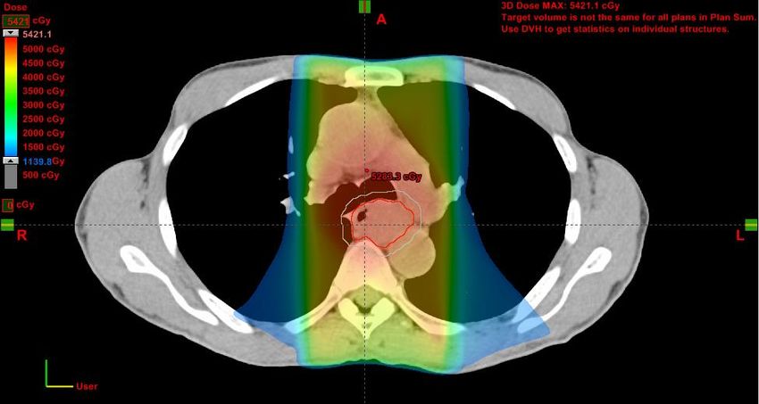
Case: Clinical Presentation
• 58 y/o male with 3 month history of dysphagia
  initially to solids, progressing to liquids
• Odynophagia
• Vague mid-chest discomfort
• 15 pound weight loss over the past 3 months
• Denies vomiting or regurgitation of food
• Denies cough/SOB
• KPS 80
Work-Up: Upper Endoscopy
• Large, friable, malignant-
  appearing mass noted
  spanning 25-31 cm from the
  incisors
• Occupying 50-60% of the
  lumen
• Remainder of endoscopic
  exam including stomach and
  duodenum were normal
• Biopsy was obtained,
  revealing moderately
  differentiated sqaumous cell
  carcinoma
Work-Up: PET/CT Scan

• Large area of FDG avid wall thickening seen in mid esophagus, with a
  maximal SUV of 12.7
• No FDG avid lymphadenopathy is identified to suggest metastatic disease
• The distribution of the FDG is otherwise within physiological limits
Work-Up: Endoscopic Ultrasound

• Hypoechoic lesion extending through the muscularis propria
• No abnormal lymphadenopathy was noted, confirming the lesion
  to be T3 N0
• No frank invasion into the surrounding structures was noted

Epidemiology
• Two distinct histopathologic types: squamous
  cell carcinoma and adenocarcinoma
• Relatively uncommon in the US
• Lifetime risk of being diagnosed with the disease
  is less than 1%
• 18,170 new cases in 2014
• 15,450 patients expected to die of the disease

3American   Cancer Society
Risk Factors
     •     Tylosis
     •     Plummer-Vinson Syndrome
     •     Caustic injury
     •     HPV (SCC)
     •     Tobacco
     •     Alcohol
            – 90% of SCC in Western Europe and North America can
              be attributed to tobacco and alcohol use
     • Obesity, GERD, Barrett’s Esophagus (adenoca)
     • Raw fruits and vegetables are protective
1DeVita, V. & Lawrence, T. & Rosenberg, S.
(2011). CANCER (9th). Philadelphia, PA;
Lippincott Williams & Wilkins.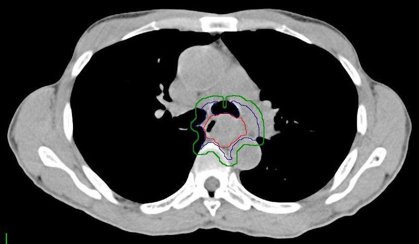
Anatomy
   • Cervical esophagus
           – Cricopharyngeus to the thoracic inlet
           – 15-18 cm from the incisors
   • Upper third
           – Thoracic inlet to the carina
           – 18-24 cm from the incisors
   • Middle third
           – Carina to the inferior pulmonary veins
           – 24-32 cm from the incisors
   • Lower third
           – Traversing the remaining distance to the GE junction
           – 32-40 cm from the incisors
1DeVita, V. & Lawrence, T. & Rosenberg, S.
(2011). CANCER (9th). Philadelphia, PA;
Lippincott Williams & Wilkins.
Lymphatic Drainage
     • Rich mucosal and submucosal lymphatic system
       which may extend long distances (reason why
       proximal/distal margins used for radiation planning
       have traditionally been a minimum of 5 cm)
     • Submucosal plexus drains into internal jugular,
       peritracheal, hilar, subcarinal, periesophageal,
       periaortic, and pericardial lesser curvature lymph
       nodes
     • Left gastric and celiac nodes for lower third lesions
2Minsky,Bruce D., MD,Goodman, Karyn, MD,
MS,Warren, Robert, MD - Leibel and Phillips
Textbook of Radiation Oncology, 772-787.
Histology
     • Squamous cell carcinomas
            – Majority of cases throughout the world
            – 40% of esophageal cancer in the US
            – 70% in the proximal and middle third
     • Adenocarcinoma
            – Frequently arise in the context of Barrett’s esophagus
            – Mainly occur in the distal third of the esophagus
            – Rate of adenocarcinoma rising in US (obesity & GERD)
     • No significant survival differences have been
       noted between various histologies
1DeVita, V. & Lawrence, T. & Rosenberg, S.
(2011). CANCER (9th). Philadelphia, PA;
Lippincott Williams & Wilkins.
Clinical Presentation
• Dysphagia
– Most common
– Initially to solids, then progressing to liquids
– Large impact on QOL
• Odynophagia
• Weight loss (Anorexia)
• Pain
• Cough/Hoarseness (Recurrent laryngeal nerve)
• Vomiting
2Minsky,Bruce D., MD,Goodman, Karyn, MD,
MS,Warren, Robert, MD - Leibel and Phillips
Textbook of Radiation Oncology, 772-787.Diagnosis/Work-Up
• Upper endoscopy - allows for biopsy and diagnosis
• Bronchoscopy in patients with tumors above the level of the
carina
• Barium esophagram (optional) – can identify a
tracheoesophageal fistula
• CT chest and abdomen – can identify extension beyond the
esophageal wall, enlarged lymph nodes, and visceral
metastases
• For cervical primaries, a neck CT should be performed to
evaluate for cervical lymph node involvement
• Endoscopic ultrasound – highly accurate in determining
depth of invasion as well as lymph node involvement
• FDG-PET scan for staging and response to pre-operative
treatment
2Minsky,Bruce D., MD,Goodman, Karyn, MD,
MS,Warren, Robert, MD - Leibel and Phillips
Textbook of Radiation Oncology, 772-787.TNM Staging, AJCC 7 th Edition
Primary Tumor Regional Lymph Nodes
TX Primary tumor cannot be assessed Nx Regional nodes not assessed
T0 No evidence of primary tumor N0 No regional lymph node metastasis
Tis High-grade dysplasia N1 Metastasis in 1-2 regional lymph
T1 Tumor invades lamina propria, muscularis nodes*
mucosae, or submucosa N2 Metastasis in 3-6 regional lymph
T1a Tumor invades lamina propria or nodes*
muscularis mucosae N3 Metastasis in 7 or more regional lymph
T1b Tumor invades submucosa nodes*
T2 Tumor invades muscularis propria
T3 Tumor invades adventitia Distant Metastasis
T4 Tumor invades adjacent structures MX Distant metastasis
cannot be assessed
T4a Resectable tumor invading pleura,
pericardium, or diaphragm M0 No distant metastasis
T4b Unresectable tumor invading other M1 Distant metastasis
adjacent structures, such as aorta, *Regional lymph nodes extend from cervical nodes to celiac nodes.
vertebral body, tracheaGroup Staging, AJCC 7th Edition
Adenocarcinoma Squamous Cell Carcinoma
Stage 0 Tis, N0, M0, grade 1 or X Stage 0 Tis, N0, M0, grade 1 or X, any location
Stage IA T1, N0, M0, grade 1-2 or X Stage IA T1, N0, M0, grade 1 or X, any location
Stage IB T1, N0, M0, grade 3 Stage IB T1, N0, M0, grade 2 or 3, any location
T2, N0, M0, grade 1-2 or X T2-3, N0, M0, grade 1 or X, lower esophagus or X
Stage IIA T2, N0, M0, grade 3 Stage IIA T2-3, N0, M0, grade 1 or X, upper and middle
esophagus
Stage IIB T3, N0, M0, any grade
T2-3, N0, M0, grade 2 or 3, lower esophagus or X
T1-2, N1, M0, any grade
Stage IIB T2-3, N0, M0, grade 2 or 3, upper and middle
Stage IIIA T1-2, N2, M0, any grade esophagus
T3, N1, M0, any grade T1-2, N1, M0, any grade, any location
T4a, N0, M0, any grade Stage IIIA T1-2, N2, M0, any grade, any location
Stage IIIB T3, N2, M0, any grade T3, N1, M0, any grade, any location
Stage IIIC T4a, N1-2, M0, any grade T4a, N0, M0, any grade, any location
T4b, any N, M0, any grade Stage IIIB T3, N2, M0, any grade, any location
Any T, N3, M0, any grade Stage IIIC T4a, N1-2, M0, any grade, any location
Stage IV Any T, any N, M1, any grade T4b, any N, M0, any grade, any location
Any T, N3, M0, any grade, any location
Stage IV Any T, any N, M1, any grade, any locationTreatment: T1 Disease
(Localized to the Mucosa)
• Little or no risk of lymph node metastases
• T1a (lamina propria or muscularis mucosa)
– Endoscopic mucosal resection followed by ablation
(preferred)
– Esophagectomy
• T1b (Invades submucosa)
– Esophagectomy
5NCCN. Esophageal and Esophagogastric
Junction Cancers (Version 1.2014)Treatment: Locally Advanced Disease
(Resectable)
• T1bN+, T2-T4aN0-N+
– Trimodality therapy with neoadjuvant
chemoradiotherapy (CRT) followed by surgical resection
• RT dose 41.4 - 50.4 Gy in 1.8 Gy daily fractions
– No utility in dose escalation
• RTOG 94-05 (Minsky et al) 50.4 v. 64.8 Gy (w/ cis/5-FU)
• Closed after interim analysis showed no probability of
superiority in the high-dose arm
• Multiagent chemotherapy with cisplatin and 5-FU or
paclitaxel and carboplatin typically used
5NCCN. Esophageal and Esophagogastric
Junction Cancers (Version 1.2014)CROSS Trial
• Preoperative Chemoradiotherapy for Esophageal
or Junctional Cancer
• 366 patients w/ T1N1 or T2-3N0 GE junction or
esophageal cancer
• Randomized
– Preoperative CRT (41.4 Gy & Carboplatin/Paclitaxel)
followed surgery
– Surgery aloneCROSS Trial Results • R0 resection – 92% in CRT v. 69% in surgery arm (p
Treatment Planning
     • CT Simulation
           – IV and/or esophageal contrast may be used to aid
             in target localization
           – Arms above head to maximize number of beam
             arrangements
           – Immobilization cradle
           – Consider 4D-CT for GE junction tumors

5NCCN.  Esophageal and Esophagogastric
Junction Cancers (Version 1.2014)
Target Volumes (RTOG 1010) • GTVp: primary tumor in the esophagus • GTVn: grossly involved regional lymph nodes • CTV = GTVp with a 4 cm expansion sup/inf along the length of the esophagus and gastric cardia and a 1.0- 1.5 cm radial expansion plus the GTVn with a 1.0-1.5 cm expansion in all dimensions • The celiac axis should be covered for tumors of the distal esophagus or GE junction • PTV (45Gy) expansion should be 0.5 to 1.0 cm and does not need to be uniform in all dimensions • Boost PTV (50.4Gy) = GTVp and GTVn with an expansion of 0.5 to 1.0 cm
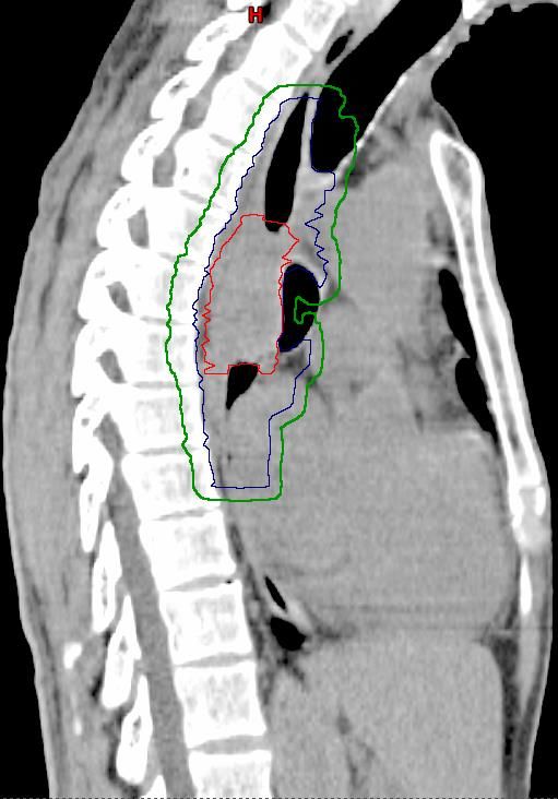
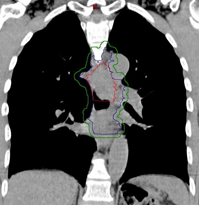
Target Volumes
• GTV
• CTV
– Cropped off anatomic
structures in which
invasion is not likely
(i.e. vertebrae,
trachea/bronchi, aorta,
lung)
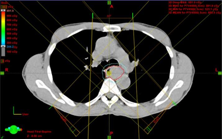
• PTVTarget Volumes • GTV • CTV • PTV (45Gy)
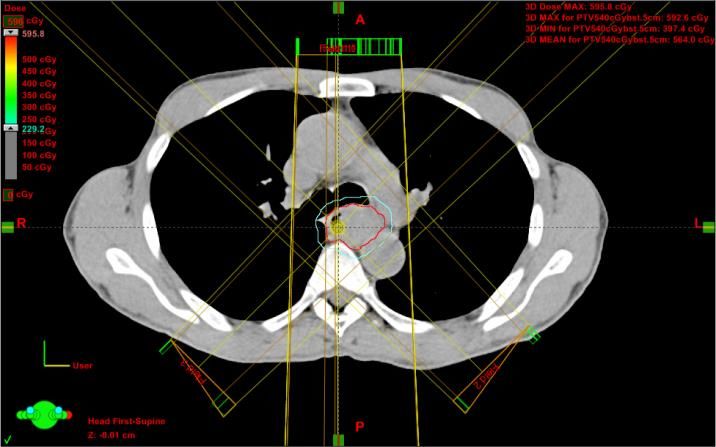
Boost Volumes
• Boost PTV (50.4Gy) = GTV with an
  expansion of 0.5 to 1.0 cm
Treatment Plan
•   3D-CRT with daily CBCT
•   AP/PA to 36 Gy followed by 3-field boost to 45 Gy
•   Additional cone down (Boost PTV) to 50.4 Gy
•   Concurrent chemotherapy with carbo/taxol
Plan Sum
Dose Constraints (RTOG 1010)
Cumulative DVH
Including dose to PTV1 and Boost PTV2
References
1.   DeVita, V. & Lawrence, T. & Rosenberg, S. (2011). CANCER (9th). Philadelphia, PA; Lippincott Williams &
     Wilkins.
2.   Minsky, Bruce D., MD,Goodman, Karyn, MD, MS,Warren, Robert, MD - Leibel and Phillips Textbook of
     Radiation Oncology, 772-787 © 2010 Copyright © 2010, 2004, 1998 by Saunders, an imprint of Elsevier
     Inc.
3.   American Cancer Society. http://www.cancer.org/cancer/esophaguscancer/detailedguide/esophagus-
     cancer-key-statistics . Accessed 9/22/2014
4.   AJCC cancer staging handbook, 7th ed. New York: Springer, 2010, published by Springer Science and
     Business Media LLC
5.   National Comprehensive Cancer Network. Esophageal and Esophagogastric Junction Cancers (Version
     1.2014). http://www.nccn.org/professionals/physician_gls/pdf/esophageal.pdf. Accessed 9/22/2014.
6.   INT 0123 (Radiation Therapy Oncology Group 94-05) phase III trial of combined-modality therapy for
     esophageal cancer: high-dose versus standard-dose radiation therapy. Minsky BD et al. J Clin Oncol. 2002
     Mar 1;20(5):1167-74.
7.   Preoperative Chemoradiotherapy for Esophageal or Junctional Cancer. Hagen et al.; NEJM 2012;366:2074-
     84.
8.   RTOG 1010: A Phase III Trial Evaluating the Addition of Trastuzumab to Trimodality Treatment of Her2-
     Overexpressing Esophageal Adenocarcinoma.
     http://www.rtog.org/ClinicalTrials/ProtocolTable/StudyDetails.aspx?action=openFile&FileID=6331.
     Accessed 9/22/2014.
You can also read