IHC PANEL MARKERS Pancreas Cancer Panel - BioGenex
←
→
Page content transcription
If your browser does not render page correctly, please read the page content below
IHC PANEL MARKERS
                               Pancreas Cancer Panel

BioGenex offers wide-ranging antibodies for several IHC panel for initial differentiation, tumor
origin, treatment methods, and prognosis. All BioGenex antibodies are validated on human
tissues to ensure sensitivity and specificity. BioGenex comprehensive IHC panels include a
range of mouse monoclonal, rabbit monoclonal, and polyclonal antibodies to choose from.

BioGenex offers a vast spectrum of high-quality antibodies for both diagnostic and reference
laboratories. BioGenex strives to support efforts in clinical diagnostics and drug discovery
development as we continue to expand our antibody product line offering in both
ready-to-use and concentrated formats for both manual and automation systems.

                            Antibodies for Pancreas Tumor
 Insulin, Chromogranin A, Glucagon, Synaptophysin, NSE, CEA, CA19.9, MUC4, MUC5AC, CK7,
 CK8, CK17, CK1, CK19
IHC PANEL MARKERS - Pancreas Cancer Panel
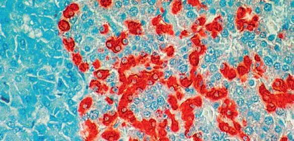
Insulin
Insulin is a hormone that regulates glucose homeostasis. It is
synthesized in the pancreas within the beta-cells of the islets of
Langerhans. Lack of insulin hormone gives rise to diabetes mellitus. This antibody
recognizes the A chain loop of human Insulin. Cross-reactivity with bovine,
rat and mouse Insulin has been observed. This antibody stains insulin in the
cytoplasm of beta cells in the pancreas. Insulin antibody is the most sensitive
and reliable means available for an accurate characterization of the function
of islet cell tumors.
Antibody Clone Localization Catalog Family
Insulin HB125 Cytoplasm AM029, AX029, MU029
Insulin EP125 Cytoplasm AN735, AY735, MU735
Chromoganin A
This antibody recognizes Chromogranin A (68 kD) and other related chro-
mogranin polypeptides from human, monkey, and pig. Chromogranin is widely
distributed has been demonstrated in several elements of the diffuse neuro-
endocrine system, including anterior pituitary, thyroid parafollicular C cells,
parathyroid chief cells, pancreatic islet cells, intestinal enteroendocrine cells,
and tumors derived from these cells. This antibody recognizes Chromogranin
A and other chromogranin polypeptides in cytoplasm of positive cells.
Antibody Clone Localization Catalog Family
Chromoganin A LK2H10 Cytoplasm AM126, AX126, MU126
Chromoganin A PHE-5 Cytoplasm AM356, AX356, MU356
Glucagon
Glucagon is a polypeptide of 29 amino acids produced by the pancreatic alpha
cells. In addition to its well-known effect of elevating blood glucose concen-
tration, glucagon functions to inhibit gastric and pancreatic secretions. Tumors
producing large amounts of glucagon are referred to as glucagonomas. This
antibody stains the cytoplasm in A cells of the endocrine pancreas and reacts
with glucagon in a number of mammalian species.
Antibody Clone Localization Catalog Family
Glucagon Polyclonal Cytoplasm AR039, AW039, PU039
Synaptophysin
Synaptophysin, a 38 kD glycoprotein, is the major integral membrane
protein of synaptic vesicles. It is a sensitive quantitative molecular marker of
synaptic density and a useful marker in the identification and characterization of
neuronal and neuroendocrine neoplasms of the adrenal medullary,
pituitary, thyroid and islet cell tumors, gastrointestinal, bronchial, thymic and
pancreatic carcinoid tumors. Immunohistochemistry of synaptophysin
has been used in the evaluation of functional bowel disorders, cortical
epileptogenesis, schizophrenia and amyotropic lateral sclerosis.
Antibody Clone Localization Catalog Family
Synaptophysin Snp88 Cytoplasm AM363, AX363, MU363
Synaptophysin EP158 Cytoplasm AN857, AY857, NU857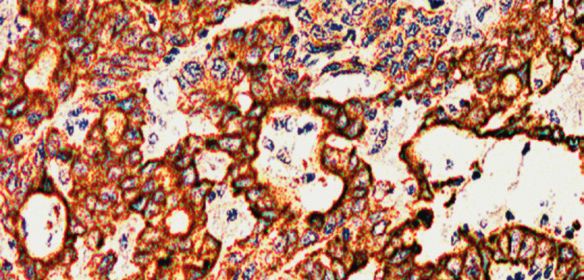
IHC PANEL MARKERS - Pancreas Cancer Panel
Neuron Specific Enolase (NSE)
NSE is a gene which encodes for a protein found in matured neurons and is
used in panels along with chromogranin, synaptophysin and neurofilament.
Elevated NSE concentrations are observed in patients with neuroblastoma,
pancreatic islet cell carcinoma, medullary thyroid carcinoma, pheochromocy-
toma, and other neuroendocrine tumors as well as certain benign conditions.
NSE is specific for such proteins, and aids in detection of neural and neuroen-
docrine lineage.
Antibody Clone Localization Catalog Family
NSE MIG-N3 Cytoplasm AM055, AX055, MU055
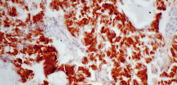
Carcinoembryonic Antigen
Carcinoembryonic Antigen (CEA) consists of a heterogeneous family of related
oncofetal 200 kD glycoproteins that is secreted into the glycocalyx surface
of gastrointestinal cells. Usually CEA is demonstrated as a linear labelling
of the apical poles of cells lining the glandular lumen and, occasionally, as
weak staining near the apex of colonic epithelial cells. Pancreatic carcinomas,
testicular tumor, gallbladder neoplasms and granular cell myoblastomas stain
positive, whereas malignant tumors of brain, prostate, skin, lymphoreticu-
lar tissues, hepatocellular carcinomas, esophageal squamous cell carcinomas,
and mesothelioma fail to stain for CEA. This antibody stains carcinoembryonic
antigen in the cytoplasm of positive cells.
Antibody Clone Localization Catalog Family
Carcinoembryonic Antigen B01-94-11M-P Cytoplasm AM009, AX009, MU009
Carcinoembryonic Antigen CEA88 Cytoplasm AM365, AX365, MU857
Carcinoembryonic Antigen Polyclonal Cytoplasm AR009, AW009, PU009
CA19.9
Carcinoma Antigen 19-9 (CA19-9) is a carbohydrate antigen that reacts
specifically with Sialyl Lewis-containing glycolipids and has been isolated and
characterized as the oligosaccharide sialylazed lacto-Nfucopentose II antigen.
This monoclonal antibody is directed against the CA19-9 antigen, which is
expressed in pancreatic carcinomas, and hepatobillary carcinomas, the tumor
cells of colorectal and gastric cancers. It can also be found in chronic pancre-
atitis and in healthy colonic mucosa of patients with colorectal cancer.
Antibody Clone Localization Catalog Family
CA19.9 C241:5:1:4 Cytoplasm AM424, AX424, MU424
MUC4
MUC4 is a membrane-associated protein of the mucin (MUC) gene family, en-
coded by a gene on chromosome 3q29 and produced by epithelial cells as a
heterodimer. The MUC4 protein is thought to play a protective role for vulner-
able epithelia, particularly in the airway, eye, female reproductive tract, and
mammary gland. Alterations in MUC4 expression have been observed in asso-
ciation with a variety of inflammatory and neoplastic states, reduction or loss
has been reported in non-small cell lung carcinoma, hyperplastic polyps of
the colon, and serrated colon adenomas, while overexpression of the MUC4/
Sialomucin complex (SMC) has been identified in malignant progression of
mammary tumors, and pancreas tumors in humans.
Antibody Clone Localization Catalog Family
MUC4 1G8 Cytoplasm AM455, AX455, MU455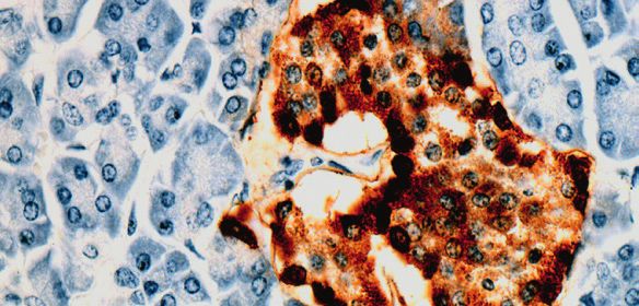
IHC PANEL MARKERS - Pancreas Cancer Panel
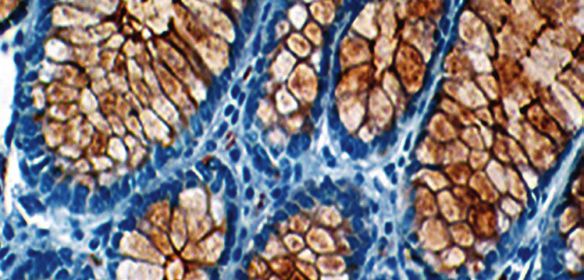
MUC5AC
Mucins are high molecular weight glycoproteins with 80% carbohydrates and
20% core protein. Gastric Mucin 5AC antigen is found in columnar mucus cells
of surface gastric epithelium and in goblet cells of the fetal and precancerous
colon but not in normal colon. Resurgence of gastric mucin during colonic
carcinogenesis is suggestive of either re-expression of the peptide core of
gastric mucin in the adult colon or due to changes in the glycosylation pattern
of mucin, which expose the hidden Mucin 5AC antigen.
Antibody Clone Localization Catalog Family
MUC5AC 45M1 Cytoplasm AM456, AX456, MU456
Cytokeratin 7
Cytokeratin 7 is a 54 kD marker of simple epithelium. Antibody to
Cytokeratin 7 strongly stains all cell layers of the urinary bladder transitional
epithelium. However, Cytokeratin 7 is absent from gastrointestinal epithelium,
hepatocytes, proximal and distal tubules of the kidney, and myoepithe-
lium, and also cannot be detected in the stratified epithelia of the skin,
tongue, esophagus, or cervix. Cytokeratin 7 recognizes specific subtypes of
adenocarcinomas and can be used to differentiate between Cytokeratin
7-positive tissues such as ovarian carcinomas and transitional cell carcinomas and
Cytokeratin 7-negative tissues such as carcinomas of the gastrointestinal tract
and prostate cancers.
Antibody Clone Localization Catalog Family
Cytokeratin 7 OV-TL12/30 Cytoplasm AM255, AX255, MU255
Cytokeratin 8
Cytokeratin 8 (52 kD) and 18 (45 kD) comprise a Cytokeratin pair as markers
for simple epithelia. In most situations, Cytokeratin 8 exists in tissues togeth-
er with Cytokeratin 18, but there are exceptions among some normal and
abnormal epithelial cells. Therefore, it is useful to use both Cytokeratin 8 and
Cytokeratin 18 in combination with other anti Cytokeratin antibody monoclo-
nals when studying cytokeratin expression patterns. Clone C-51 is designed
for the specific localization of Cytokeratin 8 and does not cross-react with
human cytokeratin numbers 7, 17, 18, or 19. This antibody stains Cytokeratin
8 in cytoplasm of positive epithelial cells.
Antibody Clone Localization Catalog Family
Cytokeratin 8 C51 Cytoplasm and Membrane AM142, AX142, MU142
Cytokeratin 17
Cytokeratin 17 is 46 kD intermediate filament found in simple epithelia some-
times in association with Cytokeratin 7. This antibody has been used to dis-
tinguish cervical immature squamous metaplasia from high grade cervical in-
traepithelial neoplasia (CIN III). Anti-CK17 also labels myoepithelial cells in
the benign breast tissue. CK17 labelling of breast carcinoma cells (so-called
basal phenotype) has been associated with a poor prognosis.
Antibody Clone Localization Catalog Family
Cytokeratin 17 E27 Cytoplasm AM572, AX572, MU572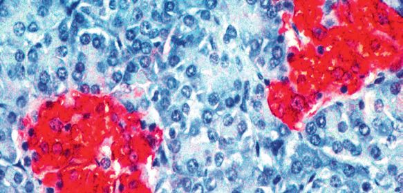
IHC PANEL MARKERS - Pancreas Cancer Panel

Cytokeratin 19
                 Cytokeratin 19 (molecular mass 40 kD) is a marker of simple epithe-
                 lia. Cytokeratin 19 has been found in mesothelial and mesothelioma
                 cells, ovarian cysts, cystadenomas, and ovarian carcinomas, in adeno-
                 carcinomas of the lung and in tumor cells of pulmonary metastases, in
                 the ductal cells of normal pancreas and in pancreatic cancers. It has
                 been shown to be present in the basal layer of non-keratinizing stratified
                 squamous epithelia such as the oral cavity and the ectocervix.

Antibody         Clone           Localization                Catalog Family
Cytokeratin 19   RCK108          Cytoplasm                   AM246, AX246, MU246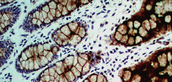
IHC PANEL MARKERS - Pancreas Cancer Panel

BioGenex Primary Antibody Format and Pack Size

BioGenex antibodies are optimized to provide a maximum signal with the minimum background
for immunohistochemical staining. All our antibodies are optimized and recommended for use
with all Super Sensitive™ Detection Systems to provide optimum staining.

BioGenex Ready-to-Use (RTU) antibodies are fully optimized for use with BioGenex Detection
Systems without the need for further dilution or titration. BioGenex concentrated antibodies
are provided with recommended dilutions for optimal use with BioGenex Detection Systems,
allowing rapid titration and testing.

 Prefix        Type                Species                            Suffix                      Volume and Format

 AM/AN         Monoclonal          AM-Mouse/AN-Rabbit                 -5M/5ME                     6 mL - Ready-to-use (manual)

 AM/AN         Monoclonal          AM-Mouse/AN-Rabbit                 -10M/10ME                   10 mL - Ready-to-use (i6000™)

                                                                      -YCD/YCDE and
 AX/AY         Monoclonal          AX-Mouse/AY-Rabbit                                             16 mL and 5 mL Ready-to-use (Xmatrx®)
                                                                      -50D/50DE

 AR            Polyclonal          Rabbit                             -5R/5RE                     6 mL - Ready-to-use (manual)

 AR            Polyclonal          Rabbit                             -10R/10RE                   10 mL - Ready-to-use (i6000™)

                                                                      -YCD/YCDE and
 AW            Polyclonal          Rabbit                                                         16 mL and 5 mL Ready-to-use (Xmatrx®)
                                                                      -50D/50DE
                                                                      -UC/UCE and
 MU/NU         Monoclonal          AM- Mouse/AN-Rabbit                                            1 mL and 0.5 mL Concentrate
                                                                      -5UC/5UCE
                                                                      -UC/UCE and
 PU            Polyclonal          Rabbit                                                         1 mL and 0.5 mL Concentrate
                                                                      -5UC/5UCE

                                                     Other Panel Markers from BioGenex
 Breast cancer panel                                                               Neuroendocrine tumor
 B&T cell Associated Lymphoma                                                      Liver cancer
 Cervical cancer                                                                   Kidney cancer
 Colorectal and stomach cancer                                                     Head & neck cancer
 Lung cancer                                                                       Bladder cancer
 Melanoma                                                                          Germ cell tumor
 Muscle cancer                                                                     Vascular tumor
 Ovarian cancer                                                                    Pituitary gland
 Prostate/Testicular cancer                                                        Esophagus cancer

For specific information on the individual antibody, please refer to the datasheets available
on www.biogenex.com or call BioGenex Technical Support at 1(800)421-4149 or write to
support@biogenex.com.

                                                                                            Customer Service
                                                                                             US:     customerservice@biogenex.com
In the U.S., call +1 (800) 421-4149                                                          India: indiacs@biogenex.com
Outside the U.S., call +91-40-27185500                       www.biogenex.com                Global: internationalcs@biogenex.com

© 2019 BioGenex Laboratories, Inc. All Rights Reserved, Doc No: 907-4110.0 Rev A
You can also read