Ballell, A., & Ferron, H. G. (2021). Biomechanical insights into the dentition of megatooth sharks (Lamniformes: Otodontidae). Scientific Reports ...
←
→
Page content transcription
If your browser does not render page correctly, please read the page content below
Ballell, A., & Ferron, H. G. (2021). Biomechanical insights into the
dentition of megatooth sharks (Lamniformes: Otodontidae). Scientific
Reports, 11, [1232]. https://doi.org/10.1038/s41598-020-80323-z

Publisher's PDF, also known as Version of record
License (if available):
CC BY
Link to published version (if available):
10.1038/s41598-020-80323-z

Link to publication record in Explore Bristol Research
PDF-document

This is the final published version of the article (version of record). It first appeared online via Nature Research at
https://doi.org/10.1038/s41598-020-80323-z . Please refer to any applicable terms of use of the publisher.

University of Bristol - Explore Bristol Research
General rights

This document is made available in accordance with publisher policies. Please cite only the
published version using the reference above. Full terms of use are available:
http://www.bristol.ac.uk/red/research-policy/pure/user-guides/ebr-terms/

www.nature.com/scientificreports
OPEN Biomechanical insights
into the dentition of megatooth
sharks (Lamniformes: Otodontidae)
Antonio Ballell & Humberto G. Ferrón*
The evolution of gigantism in extinct otodontid sharks was paralleled by a series of drastic
modifications in their dentition including widening of the crowns, loss of lateral cusplets, and
acquisition of serrated cutting edges. These traits have generally been interpreted as key functional
features that enabled the transition from piscivory to more energetic diets based on marine mammals,
ultimately leading to the evolution of titanic body sizes in the most recent forms (including the
emblematic Otodus megalodon). To investigate this hypothesis, we evaluate the biomechanics of
the anterior, lateral, and posterior teeth of five otodontid species under different loading conditions
by using two-dimensional finite element analysis. Stress distribution patterns are remarkably
similar among all models under puncture and draw (i.e., when subjected to vertical and lateral
forces, respectively). Contrary to expectation, higher average stress values are detected under both
loading scenarios in more recent species. Altogether, this suggests little correlation between tooth
morphology and key aspects of biomechanical behaviour in otodontids, making it difficult to frame
the morphological trend of their dentitions within an adaptive scenario. We propose that this pattern
most likely emerged as a non-functional by-product of heterochronic processes driven by selection
towards larger body sizes.
Otodontids, colloquially referred to as megatooth sharks, constitute a family of apex predatory selachians that
ranged from the Early Paleocene to the Pliocene1–3. This group experienced a trend towards gigantism throughout
the Cenozoic that culminated with Otodus megalodon, the largest macropredatory shark ever to e xist4. This spe-
cies overpassed 15 m in total length and likely weighed more than 50 t ons4–7. Historically, the evolution of such
titanic body sizes in otodontids has been related to the emergence of various marine mammal lineages during the
Paleogene (i.e., pinnipeds, sirenians, and cetaceans)5,8,9. Possessing thick layers of blubber, these taxa would have
represented ideal prey for large-sized mesotherms to meet the metabolic demands of their active lifestyles10–12.
Within this scenario, the earliest otodontids subsisted on comparatively small prey items, presumably fishes,
whereas the largest and more recent species, including O. megalodon, consumed larger marine m ammals8,9,13,14.
This dietary shift most likely required the acquisition of a series of anatomical innovations that enabled such
trophic specialisation.
The trend towards gigantism in otodontid sharks was paralleled by remarkable modifications in tooth mor-
phology, including an increase in crown width, the loss of lateral tooth cusplets, and the acquisition of ser-
rated cutting edges9,14,15. Collectively, these changes represent a shift from typical puncturing-tearing to cutting
dentitions16. These two dental types are usually associated with different ways of capturing and processing the
prey; accordingly, early otodontids were presumably adapted to prey upon small elusive animals, whereas the
most recent members of this family were likely adapted to tearing flesh from large prey or c arcasses17–20. However,
only few works have assessed morphofunctional questions about shark teeth from quantitative biomechanical
points of v iew21–26 and the most comprehensive studies in this regard did not find clear patterns between tooth
morphology and structural resistance (i.e., the ability to withstand the effect of forces and the deformation derived
from it) or puncture performance (i.e., efficiency to penetrate foodstuff). These findings called into question
the classical categorization of shark dentitions into functional t ypes27–29. As such, biomechanical testing of oto-
dontid teeth is crucial for clarifying the underlying mechanisms that promoted their presumed shift in dietary
preferences and better understanding the evolutionary factors that allowed them to reach the most gigantic sizes
among macropredatory s elachians1,16,30.
Here we evaluate the biomechanical behaviour of otodontid shark teeth by means of Finite Element Analysis
(FEA). Borrowed from engineering, FEA is one of the most commonly used computational methods in bio-
mechanics and functional p alaeobiology31. This technique reconstructs the mechanical behaviour of biological
Shool of Earth Sciences, University of Bristol, Bristol BS8 1RJ, UK. *email: humberto.ferron@bristol.ac.uk
Scientific Reports | (2021) 11:1232 | https://doi.org/10.1038/s41598-020-80323-z 1
Vol.:(0123456789)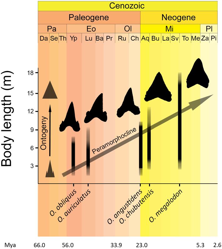
www.nature.com/scientificreports/
Figure 1. Von Mises stress distribution plots in the anterior (Ant.), lateral (Lat.), and posterior (Post.) teeth of
the five analysed otodontid species, simulating (a) puncture and (b) draw scenarios with scaled force magnitude.
Mesial is left, distal is right. Arrows indicate loading points. Grey areas represent von Mises stress values higher
than 5 GPa and 10 MPa in each of the scenarios, respectively.
structures, in terms of stress and strain, under simulated loads. To assess the functional significance of morpho-
logical trends in otodontid dentitions and test previous adaptive explanations, we analysed anterior, lateral and
posterior teeth of five otodontid chronospecies (Otodus obliquus, O. auriculatus, O. angustidens, O. chubutensis
and O. megalodon), thus capturing the diversity of dentitions exhibited by this lineage from the Palaeocene to the
Pliocene1. We tested loading scenarios of puncture, a vertical force acting on the tip of the crown; and unidirec-
tional draw, a lateral force acting along the distal cutting edge. Puncture was simulated under life-size absolute
force estimates (i.e., bite forces that each species would have exerted in life considering their estimated body
size) and scaled forces (i.e., force magnitudes scaled to maintain a constant force to surface area ratio across all
models to account for morphology o nly32); and draw was simulated under scaled forces only.
Results
Finite element models under puncture scenario with scaled forces show some similarities in von Mises stress dis-
tribution patterns (Fig. 1a). The region with the highest stress is located around the tip of the main tooth crown,
where the puncture force is acting, while the lowest stress is located in the root, where models are constrained.
The pattern of how stress dissipates from the loading point varies among models. Stress is distributed along the
center of the crown in teeth with crowns approximating an equilateral or isosceles triangular morphology, such
as the anterior teeth of O. chubutensis and O. megalodon. In crowns approaching a right triangular morphology,
as in the anterior teeth of O. obliquus, O. auriculatus, and O. angustidens, stress is mostly distributed along the
distal cutting edge. Teeth with recurved crowns, as in the lateral and posterior teeth of O. obliquus and the lateral
teeth of O. auriculatus and O. angustidens, show high stresses along the distal and (to a lesser extent) mesial
cutting edges while the center of the crown exhibits low stress, a pattern resembling the bending of a typical can-
tilever beam (i.e., a rigid structural element supported at one end and free at the other e nd33). When puncture is
simulated under estimated life-size bite force conditions for each species, same patterns of stress distribution are
obtained but stress values are higher due to the higher force magnitudes that are applied (Supplementary Fig. S1).
Comparing different tooth positions for the same taxa in this simulation, lateral and especially posterior teeth
Scientific Reports | (2021) 11:1232 | https://doi.org/10.1038/s41598-020-80323-z 2
Vol:.(1234567890)www.nature.com/scientificreports/
experience higher stresses than anterior teeth as a result of (1) experiencing higher forces because jaws behave
as third-class levers, in which output (bite) forces increase towards the jaw joint; and (2) having less surface area
due to their smaller size (see “Methods” section).
In the draw scenario with scaled force loadings, all models exhibit similar general distributions of stress
(Fig. 1b). The portions of the teeth exhibiting the lowest stresses are the root and the very apex of the crown. The
highest stress values are located along the distal cutting edge of the main crown, where the draw load is acting,
as well as along the mesial cutting edge, in resemblance to a cantilever-bending scenario. The centre of the tooth
crown between the cutting edges exhibits relatively lower stress, akin to the neutral axis of the beam. In taxa with
lateral cusplets, namely the four oldest species, these structures show moderate to low stresses, generally higher
in the distal cusplet than in the mesial, where the draw load is not acting directly.
Some general patterns can be extracted from comparing the von Mises stress mesh-weighted arithmetic means
(MWAM) across finite element models (Fig. 2). Under both puncture and draw scaled force loadings, relative
stresses decrease as tooth position becomes more distal, with the exception of the anterior and lateral teeth of O.
obliquus during puncture (Fig. 2a). When comparing different species, the teeth of older species display lower
stresses under both loading regimes than those of more recent taxa, although there are some exceptions (Fig. 2a).
For example, O. obliquus shows higher stress than O. auriculatus when comparing lateral and posterior teeth
during puncture, and anterior and lateral teeth during draw. Similarly, the anterior teeth of O. angustidens exhibit
higher stress than those of the younger species O. chubutensis, during both puncture and draw. In general, the
greatest differences in stress magnitude among taxa are seen in anterior and lateral teeth, while posterior teeth
show more similar stress values under both loading conditions. Correlation analyses support a general trend
towards increasing von Mises stress MWAM in time for all tooth positions and loading scenarios, apart from
posterior teeth under puncture where no trend is detected (Fig. 2b). These patterns remain broadly consistent
when von Mises stress MWAM is calculated considering only elements of the crown tooth, with the exception
of the teeth of O. obliquus which show higher stress values during both puncture and, to a lesser extent, draw
(Supplementary Fig. S2).
Discussion
Otodontid teeth show general patterns of stress distribution similar to those of extant e lasmobranchs29, with
high stresses concentrated around the crown apex and along the mesial and distal cutting edges during puncture
and draw, respectively (Fig. 1). FE models do not reveal structural weaknesses that could potentially lead to
failure under both loading scenarios in any of the considered teeth despite the high force magnitudes that were
simulated (Fig. 1 and Supplementary Fig. S1). Stress patterns during draw are consistent with cantilever beam
bending33, especially in anterior and lateral teeth that exhibit higher and straighter crowns (Fig. 1b), and similar
to those of extant species with elongate tooth crowns29. As such, despite covering a relatively diverse range of
shapes, with typical examples of distinct dental types1,9,15, the teeth of different otodontid species exhibit similar
patterns of stress distribution in both puncture and draw. This suggests that dental morphology is not a reliable
proxy for functional performance18,27–29, which undermines the traditional categorization of shark teeth into
morphofunctional classes (i.e., specific dental morphotypes presumably adapted to clutching, tearing, cutting,
crushing, or grinding) employed for decades to support dietary and ecological interpretations in both living
and extinct groups1,16,30.
Von Mises stress mesh-weighted arithmetic mean (MWAM) decreases towards more distal tooth positions
within otodontid species (Fig. 2a), indicating that the robust, shorter crowned posterior teeth are structurally
more resistant than the gracile anterior and lateral teeth. This suggests that heterodonty in the dentition of oto-
dontids could be a response to mechanical constraints where the morphology of more distal teeth is determined,
at least in part, by the need to resist higher bite forces. When comparing different taxa, the teeth of older species
exhibit, with few exceptions, lower von Mises stress values than those of more recent ones during both puncture
and draw (Fig. 2). However, this trend is not consistent with the mechanical properties presumed a priori for
the dental types found in taxa possessing extreme dental morphologies. Extant sharks with puncturing-tearing
dentitions, similar to that of O. obliquus, usually pierce and hold soft prey between their jaws before swallowing
them with little manipulation34; in contrast, species with cutting dentitions, similar to that of O. megalodon,
slice off large pieces of flesh through a combination of vertical bites and lateral head s haking35,36. Fossil evidence
supports that the latest otodontids (i.e., O chubutensis and O. megalodon) also possessed the ability to bite and
crush the bones of pinnipeds, sirenians, and cetaceans during hunting or s cavenging8,13,37–40. This implies that
the teeth of these species would impact hard mineralized endoskeletal tissues more often than those of their
earlier relatives that are presumed to have fed mostly on fish9,41. From this perspective, an optimization in draw
and, probably, puncture performances is expected through the evolution of otodontid dentitions in order to
support higher loads; an expectation not substantiated by our results. In any case, the evaluation of these aspects
should be conducted with caution given the complexity of feeding kinematics in sharks17–20 and the potential
effects of interspecific variation in the labio-lingual thickness and histology of the teeth. Nonetheless, planar
(two-dimensional) models have been established as a useful alternative to three-dimensional models for cap-
turing reaction forces and comparative patterns of stress and s train42. Our data support this approach given the
remarkable similarities between the general patterns of stress distribution recovered here for otodontid teeth
(Fig. 2), based on planar models with homogeneous material properties, and those previously reported for a
number of living sharks, based on more complex three dimensional models accounting for both the distribution
and properties of the different dental tissues29.
Our results reveal that the morphological trend recorded in otodontid dentitions is difficult to frame within
a functional c ontext9,14,15, thus calling into question its adaptive significance during the dietary transition of
this group and, ultimately, its causal impact on the evolution of gigantic body sizes in the most derived species.
Scientific Reports | (2021) 11:1232 | https://doi.org/10.1038/s41598-020-80323-z 3
Vol.:(0123456789)www.nature.com/scientificreports/
Figure 2. (a) Von Mises stress mesh-weighted arithmetic means (MWAM) calculated for anterior (Ant.),
lateral (Lat.), and posterior (Post.) teeth of the five analysed otodontid species, simulating puncture and draw
scenarios with scaled force magnitude. Data are shown in a temporal context (in million years ago, Mya)
where stratigraphic range of each taxa is represented by grey bars (stratigraphic ranges based on C appetta1 and
Diedrich9). (b) Density distributions of coefficients and p values derived from correlation analyses between von
Mises stress MWAM and species age (randomly selected within their chronostratigraphic range, n = 10,000).
Epoch: Pa, Paleocene; Eo, Eocene; Ol, Oligocene; Mi, Miocene; Pl, Pliocene; Age: Da, Danian; Se, Selandian; Th,
Thanetian; Yp, Ypresian; Lu, Lutetian; Ba, Bartonian; Pr, Priabonian; Ru, Rupelian; Ch, Chattian; Aq, Aquitanian;
Bu, Burdigalian; La, Langhian; Sv, Serravalian; To, Tortonian; Me, Messinian; Za, Zanclean; Pi, Piacenzian.
The presence of serrated edges (not captured in our FE models) is usually considered as a character related to
increased cutting efficiency18,22,30. Accordingly, the evolution of serrations in the tooth cutting edges of both
otodontids and the great white shark (i.e., Carcharodon carcharias)43–45 are interpreted as independent adapta-
tions to improve cutting performance triggered by the acquisition of comparable diets based mostly on marine
mammals9,46. However, the functional role of this feature has been challenged by recent biomechanical studies
on shark t eeth28 and the question of whether the acquisition of edge serrations in otodontids had some impact
on their ability to prey upon marine m ammals9 will remain unanswered until dynamic testing is conducted
specifically on these taxa26. Biomechanical testing of the cutting mechanics and efficiency of complete tooth
rows could also provide relevant functional insights in this context by revealing emergent functional properties
of the dentition as a whole. In analogy with the extant great white s hark46, the dentitions of more recent species
Scientific Reports | (2021) 11:1232 | https://doi.org/10.1038/s41598-020-80323-z 4
Vol:.(1234567890)www.nature.com/scientificreports/
Figure 3. Schematic representation of the trends in tooth morphology, body size and presumed heterochronic
phenomena through the evolution of otodontid sharks.
of otodontids might have comprised a continuous cutting edge spanning from one commissure through the
symphysis to the opposed commissure and provided with two orders of serrations (i.e., the teeth and the serrae
sensu stricto). Besides potential anatomical specializations, the dietary shift that occurred within Otodontidae
may have been an intrinsic consequence of body size increase4, allowing them to consume larger p rey47,48,
facilitated by the pre-existence of highly active metabolisms and mesothermy in smaller preceding f orms10,11.
In the absence of convincing functional evidence, other non-adaptive processes should be considered when
attempting to explain the morphological changes in the dentition of otodontids. Body size selection triggered by
heterochrony (i.e., changes to the timing or rate of developmental events, relative to an ancestor49) can produce
trends in traits that exhibit allometric variation (i.e., changes in morphology associated with size variation)50.
Heterochrony had a relevant role in the evolution of gigantism in o todontids51, where a general trend towards
the expansion of the somatic growth (i.e., peramorphosis) is recorded in the vertebral rings of successive taxa14.
This phenomenon appears to be a product of an increased rate of growth (i.e., acceleration) and a delayed offset
timing (i.e., hypermorphosis) in more recent species14. These heterochronic changes are mirrored in the denti-
tions of otodontids and are fundamental for understanding the ontogenetic and interspecific variation of tooth
morphology within the group52–54. When considered in the context of heterochrony, the evolution of otodontid
dentitions can be framed within a continuous morphological gradient where progressively larger and more
peramorphic species pass through more developmental stages during ontogeny, a trend that can be expressed as
a peramorphocline55,56 (Fig. 3). This may explain why the ontogenetic change in O. megalodon teeth mimics the
modifications that took place during their evolution within Otodontidae53,54,57,58. We propose that the morpho-
logical differences among otodontid dentitions may not be the result of selection acting on those traits but are
simple sequelae of size variation. Interestingly, a similar pattern is present within lamnid sharks (i.e., Carcharo-
don, Isurus and Lamna genera and extinct relatives), where size and similar aspects of tooth morphology (i.e.,
crown width and presence/absence of lateral cusplets) covary in comparable ways both along their o ntogeny59–62
44,63
and throughout phylogeny . In fact, heterochronic processes may have also been fundamental in the shaping
of dental morphological diversity of extinct and living lamnid s pecies63. Disentangling the causes that underly
these phenomena in different groups, and ascertaining whether they respond to common functional demands
and/or developmental mechanisms, might inform about the homology of key characters in these groups (e.g.,
lateral cusplets)60 and ultimately could provide new insights into their debated affinities5,8,9,43–45,53 by guiding
character selection in future phylogenetic analyses. From this perspective, and in agreement with the biome-
chanical evidence presented here, the long-term changes in the general morphology of otodontid teeth might be
better considered as a non-functional by-product of heterochronic phenomena, most likely driven by selection
on life history traits favouring the attainment of larger body sizes.
Methods
Model creation. Images of teeth in labial view were obtained for the otodontid species Otodus obliquus,
O. auriculatus, O. angustidens, O. chubutensis and O. megalodon from the literature and from specimens in
museum collections (Supplementary Table S1). We follow the taxonomic nomenclature of Cappetta1 and refer to
that study for a detailed discussion on alternative existing nomenclatures. For each species, we considered teeth
from the upper jaw with anterior, lateral and posterior positions in order to span the morphological diversity of
otodontid teeth related to heterodonty (i.e., anterior I–III, lateral III–IV, and posterior II–III, following the ter-
minology of Applegate & Espinosa-Arrubarrena53 and D iedrich9; see Supplementary Fig. S3). The images were
imported into ImageJ v. 1.51r64 and the outline of each tooth was drawn using the multipoint tool. The XY coor-
dinates of the outline were obtained using Microsoft Excel and imported into the CAD software Inventor Profes-
Scientific Reports | (2021) 11:1232 | https://doi.org/10.1038/s41598-020-80323-z 5
Vol.:(0123456789)www.nature.com/scientificreports/
sional 2016 (Autodesk). The outline was sketched from the XY coordinates and the planar models were exported
as STP files (available at the Open Science platform Figshare, https://figshare.com/s/e245548d6f31b226a7b0).
Bite force estimations. Anterior and posterior vertical bite forces were estimated for each species under
the assumption that bite force increases at 0.67 the power of body m ass65. Estimations were made presuming
isometry from values obtained in a jaw model of a 240 kg great white shark specimen (i.e., anterior and posterior
bite forces of 1602 N and 3131 N, respectively)66. The arithmetic average of anterior and posterior force values
was considered as the force exerted by the lateral region of the jaw. The body mass of each species was estimated
from exponential models established in living great white sharks5 using body length estimates reported in the
literature (Supplementary Table S1).
Finite element analysis. Two-dimensional FEA was performed in Abaqus v. 6.14-1 (Simulia). Tooth pla-
nar models were meshed prior to the analyses, using three-node linear triangular elements of type CPE3. The
optimal number of elements was determined in a convergence test, using the O. megalodon lateral tooth model
as a reference (Supplementary Fig. S4). Different element sizes were chosen for meshing in order to maintain
similar numbers of finite elements across models (from 29,323 to 42,922) (Supplementary Table S2).
Tooth models were assigned the elastic, isotropic, and homogeneous material properties of lamniform oste-
odentine, with Young’s modulus of 28.44 GPa67 and Poisson’s ratio of 0.368. Enameloid was not modelled as the
distribution and thickness of this tissue is virtually unknown for most otodontids69 and osteodentine represents
the vast majority of the tooth volume in lamniform sharks70. Boundary conditions were applied by constraining
all nodes within the tooth root in all three degrees of freedom ( U1, U2 and U R1). The loose attachment of teeth
to the dental ligament of shark jaws allows some degree of movement, especially in the labiolingual direction.
However, the mechanics of these movements are poorly understood and thus difficult to s imulate23,24,29. Addi-
tionally, our planar models do not capture the labiolingual axis, which is the main direction of tooth oscillation.
Thus, we assumed our models to be static in translation and rotation along the apicobasal and mesiodistal axes,
following previous a pproaches29.
FEA was performed under two loading scenarios: (1) puncture, simulating a vertical bite force applied to
the apex of the tooth crown; and (2) unidirectional draw, simulating a horizontal lateral force applied along the
distal cutting edge of the tooth crown. In the puncture simulation, the force was applied to a single node and
two sets of analyses were performed. The first one used bite forces taking into account size (see above), provid-
ing an estimate of the different absolute bite forces that each species would have experienced in different tooth
positions (Supplementary Table S3). A second analysis with scaled force magnitudes was performed to remove
the effect of size and compare shape differences only. The bite forces were scaled according to model surface
area (Supplementary Table S3), using the O. megalodon anterior tooth model (49,051 N) as a reference, so as to
keep the same F/SA ratio and allow shape c omparisons32. The draw load was applied to a set of nodes defining
the mesial edge of the tooth crowns. An arbitrary magnitude of 500 N, following previous works29, was used for
the O. megalodon anterior tooth model, and this force was scaled in the rest of the models to keep the same F/SA
ratio and account for shape only. The total draw force magnitude was divided by the number of nodes to which
the force was applied (Supplementary Table S3).
FEA results were summarised in field outputs including von Mises stress, a commonly used parameter in
palaeobiology71 which predicts failure under ductile fracture32,72. Areas of the models showing high stress values
indicate points of structural weakness which are more susceptible to failure. The von Mises stress mesh-weighted
arithmetic mean (MWAM) was calculated to account for element size differences within non-uniform m eshes73
considering finite elements from both the whole tooth and the tooth crown. Temporal trends in von Mises stress
MWAM were assessed with Pearson correlation analyses. Correlation between MWAM and species age was
evaluated accounting for the uncertainty associated to the duration of each taxon. For this purpose, repeated
correlation analyses (n = 10,000) were performed, where species ages were randomly subsampled within their
respective chronostratigraphic ranges. Derived correlation coefficients and p-values were displayed as violin
density plots generated using the package ‘ggplot2’74. All the analyses were performed in in R75 and resulting
scripts are available at the Open Science platform Figshare (https://figshare.com/s/e245548d6f31b226a7b0).
Data availability
The data set as well as the R syntax used for the analyses presented here are available at the Open Science platform
Figshare (https://figshare.com/s/e245548d6f31b226a7b0).
Received: 2 November 2020; Accepted: 17 December 2020
References
1. Cappetta, H. Chondrichthyes-Mesozoic and Cenozoic Elasmobranchii: Teeth (Verlag F, Pfeil, 2012).
2. Boessenecker, R. W. et al. The Early Pliocene extinction of the mega-toothed shark Otodus megalodon: A view from the eastern
North Pacific. PeerJ 7, e6088 (2019).
3. Pimiento, C. & Clements, C. F. When did Carcharocles megalodon become extinct? A new analysis of the fossil record. PLoS One
9, e111086 (2014).
4. Pimiento, C. & Balk, M. A. Body-size trends of the extinct giant shark Carcharocles megalodon: A deep-time perspective on marine
apex predators. Paleobiology 41, 479–490 (2015).
5. Gottfried, M. D., Compagno, L. J. V. & Bowman, S. C. Size and skeletal anatomy of the giant “megatooth” shark Carcharodon
megalodon. In Great White Sharks: The Biology of Carcharodon carcharias, Ch 7 (eds Klimley, A. P. & Ainley, D. G.) (Academic
Press, San Diego, 1996).
Scientific Reports | (2021) 11:1232 | https://doi.org/10.1038/s41598-020-80323-z 6
Vol:.(1234567890)www.nature.com/scientificreports/
6. Shimada, K. The size of the megatooth shark, Otodus megalodon (Lamniformes: Otodontidae), revisited. Hist. Biol. 20, 1–8 (2019).
7. Cooper, J. A., Pimiento, C., Ferrón, H. G. & Benton, M. J. Body dimensions of the extinct giant shark Otodus megalodon: A 2D
reconstruction. Sci. Rep. 10, 14596 (2020).
8. Purdy, R. W. Paleoecology of fossil white sharks. In Great White Sharks: The Biology of Carcharodon carcharias, Ch 8 (eds Klimley,
A. P. & Ainley, D. G.) (Academic Press, San Diego, 1996).
9. Diedrich, C. White and megatooth shark evolution and predation origin onto seals, sirenians and whales. Nat. Sci. 5, 1203–1218
(2013).
10. Pimiento, C., Cantalapiedra, J. L., Shimada, K., Field, D. J. & Smaers, J. B. Evolutionary pathways toward gigantism in sharks and
rays. Evolution 73, 588–599 (2019).
11. Ferrón, H. G. Regional endothermy as a trigger for gigantism in some extinct macropredatory sharks. PLoS One 12, e0185185
(2017).
12. Ferrón, H. G., Martínez-Pérez, C. & Botella, H. The evolution of gigantism in active marine predators. Hist. Biol. 30, 712–716
(2018).
13. Collareta, A. et al. Did the giant extinct shark Carcharocles megalodon target small prey? Bite marks on marine mammal remains
from the late Miocene of Peru. Palaeogeogr. Palaeoclimatol. Palaeoecol. 469, 84–91 (2017).
14. Ehret, D. J. Paleobiology and Taxonomy of Extinct Lamnid and Otodontid Sharks (Chondrichthyes, Elasmobranchii, Lamniformes)
(University of Florida, Florida, 2010).
15. Perez, V. J., Godfrey, S. J., Kent, B. W., Weems, R. E. & Nance, J. R. The transition between Carcharocles chubutensis and Carcharocles
megalodon (Otodontidae, Chondrichthyes): Lateral cusplet loss through time. J. Vertebr. Paleontol. 38, e1546732 (2018).
16. Cappetta, H. Types dentaires adaptatifs chez les sélaciens actuels et post-paléozoïques. Palaeovertebrata 16, 57–76 (1986).
17. Motta, P. J. & Wilga, C. D. Advances in the study of feeding behaviors, mechanisms, and mechanics of sharks. Environ. Biol. Fishes
60, 131–156 (2001).
18. Motta, P. J. & Huber, D. R. Prey capture behavior and feeding mechanics of elasmobranchs. In Biology of Sharks and Their Relatives
(eds Carrier, J. C. et al.) (CRC Press, London, 2004).
19. Wilga, C. A. & Ferry, L. A. Functional anatomy and biomechanics of feeding in elasmobranchs. Fish Physiol. 34, 153–187 (2015).
20. Huber, D. et al. Feeding in cartilaginous fishes: An interdisciplinary synthesis. In Feeding in Vertebrates (eds Bels, V. & Whishaw,
I.) (Springer, Berlin, 2019).
21. Nobiling, G. Die Biomechanik des Kiefferapparates beim Stierkopfhai (Heterodontus portusjacksoni = Heterodontus philippi). Adv.
Anat. Embryol. Cell Biol. 52, 3–52 (1977).
22. Frazzetta, T. H. The mechanics of cutting and the form of shark teeth (Chondrichthyes, Elasmobranchii). Zoomorphology 108,
93–107 (1988).
23. Powlik, J. J. On the geometry and mechanics of tooth position in the white shark Carcharodon carcharias. J. Morphol. 226, 277–288
(1995).
24. Ramsay, J. B. & Wilga, C. D. Morphology and mechanics of the teeth and jaws of white-spotted bamboo sharks (Chiloscyllium
plagiosum). J. Morphol. 268, 664–682 (2007).
25. Dean, M. N., Ramsay, J. B. & Schaefer, J. T. Tooth reorientation affects tooth function during prey processing and tooth ontogeny
in the lesser electric ray, Narcine brasiliensis. Zoology 111, 123–134 (2008).
26. Corn, K. A., Farina, S. C., Brash, J. & Summers, A. P. Modelling tooth–prey interactions in sharks: The importance of dynamic
testing. R. Soc. Open Sci. 3, 160141 (2016).
27. Whitenack, L. B. The Biomechanics and Evolution of Shark Teeth (University of South Florida, South Florida, 2008).
28. Whitenack, L. B. & Motta, P. J. Performance of shark teeth during puncture and draw: Implications for the mechanics of cutting.
Biol. J. Linn. Soc. 100, 271–286 (2010).
29. Whitenack, L. B., Simkins, D. C. Jr. & Motta, P. J. Biology meets engineering: The structural mechanics of fossil and extant shark
teeth. J. Morphol. 272, 169–179 (2011).
30. Cappetta, H. Chondrichthyes II Mesozoic and Cenozoic Elasmobranchii (Verlag F, Pfeil, 1987).
31. Bright, J. A. A review of paleontological finite element models and their validity. J. Paleontol. 88, 760–769 (2014).
32. Dumont, E. R., Grosse, I. R. & Slater, G. J. Requirements for comparing the performance of finite element models of biological
structures. J. Theor. Biol. 256, 96–103 (2009).
33. Gere, J. M. Mechanics of Materials (Thomson Learning, Belmont, 2004).
34. Moyer, J. K., Shannon, S. F. & Irschick, D. J. Bite performance and feeding behaviour of the sand tiger shark Carcharias taurus. J.
Fish Biol. 95, 881–892 (2019).
35. Tricas, T. C. Feeding ethology of the white shark, Carcharodon carcharias. Mem. South. Calif. Acad. Sci. 9, 81–91 (1985).
36. Tucker, J. P., Vercoe, B., Santos, I. R., Dujmovic, M. & Butcher, P. A. Whale carcass scavenging by sharks. GECCO 19, e00655 (2019).
37. Godfrey, S. J. & Altman, J. A Miocene cetacean vertebra showing a partially healed compression fracture, the result of convulsions
or failed predation by the Giant White Shark, Carcharodon megalodon. Jeffersoniana 16, 1–12 (2005).
38. Aguilera, O. A., García, L. & Cozzuol, M. A. Giant-toothed white sharks and cetacean trophic interaction from the Pliocene Carib-
bean Paraguaná Formation. Palaontol. Z. 82, 204–208 (2008).
39. Carrillo-Briceño, J. D. et al. An early Neogene elasmobranch fauna from the southern Caribbean (western Venezuela). Palaeontol.
Electron. 20, 1–31 (2016).
40. Godfrey, S. J., Ellwood, M., Groff, S. & Verdin, M. S. Carcharocles-bitten odontocete caudal vertebrae from the Coastal Eastern
United States. Acta Palaeontol. Pol. 63, 20 (2018).
41. Diedric, C. G. & Felker, H. Middle Eocene shark coprolites from shallow marine and deltaic coasts of the pre-North Sea Basin in
central Europe. In Vertebrate Coprolites (eds Hunt, A. P. et al.) (New Mexico Museum of Natural History and Science, Albuquerque,
2012).
42. Morales-García, N. M., Burgess, T. D., Hill, J. J., Gill, P. G. & Rayfield, E. J. The use of extruded finite-element models as a novel
alternative to tomography-based models: A case study using early mammal jaws. J. R. Soc. Interface 16, 20190674 (2019).
43. Nyberg, K. G., Ciampaglio, C. N. & Wray, G. A. Tracing the ancestry of the great white shark, Carcharodon carcharias, using
morphometric analyses of fossil teeth. J. Vertebr. Paleontol 26, 806–814 (2006).
44. Ehret, D. J., Hubbell, G. & MacFadden, B. J. Exceptional preservation of the white shark Carcharodon (Lamniformes, Lamnidae)
from the early Pliocene of Peru. J. Vertebr. Paleontol 29, 1–13 (2009).
45. Ehret, D. J. et al. Origin of the white shark Carcharodon (Lamniformes: Lamnidae) based on recalibration of the Upper Neogene
Pisco Formation of Peru. Palaeontology 55, 1139–1153 (2012).
46. Martin, R. A., Hammerschlag, N., Collier, R. S. & Fallows, C. Predatory behaviour of white sharks (Carcharodon carcharias) at
Seal Island, South Africa. J. Mar. Biol. Assoc. UK 85, 1121–1136 (2005).
47. Lucifora, L. O., García, V. B., Menni, R. C., Escalante, A. H. & Hozbor, N. M. Effects of body size, age and maturity stage on diet
in a large shark: Ecological and applied implications. Ecol. Res. 24, 109–118 (2009).
48. Estrada, J. A., Rice, A. N., Natanson, L. J. & Skomal, G. B. Use of isotopic analysis of vertebrae in reconstructing ontogenetic feeding
ecology in white sharks. Ecology 87, 829–834 (2006).
49. Klingenberg, C. P. Heterochrony and allometry: The analysis of evolutionary change in ontogeny. Biol. Rev. 73, 79–123 (1998).
50. McKinney, M. L. Allometry and heterochrony in an Eocene echinoid lineage: Morphological change as a by-product of size selec-
tion. Paleobiology 10, 407–419 (1984).
Scientific Reports | (2021) 11:1232 | https://doi.org/10.1038/s41598-020-80323-z 7
Vol.:(0123456789)www.nature.com/scientificreports/
51. Renz, M. Megalodon: Hunting the Hunter (Paleo Press, Fort Myers, 2002).
52. Menesini, E. Ittiodontoliti delle formazioni terziarie dell‘archipelago maltese. Palaeontogr. Ital. 68, 121–162 (1974).
53. Applegate, S. P. & Espinosa-Arrubarrena, L. The fossil history of Carcharodon and its possible ancestor, Cretolamna: A study in tooth
identification. In Great White Sharks: The Biology of Carcharodon carcharias, Ch 7 (eds Klimley, A. P. & Ainley, D. G.) (Academic
Press, San Diego, 1996).
54. Ward, D. J. & Bonavia, G. C. Additions to, and a review of, the Miocene shark and ray fauna of Malta. Cent. Mediterr. Nat. 3,
131–146 (2001).
55. McNamara, K. J. Heterochrony and phylogenetic trends. Paleobiology 20, 130–142 (1982).
56. McNamara, K. J. Heterochrony: The evolution of development. Evol. Educ. Outreach 5, 203–218 (2012).
57. Pimiento, C., Ehret, D. J., MacFadden, B. J. & Hubbell, G. Ancient nursery area for the extinct giant shark Megalodon from the
Miocene of Panama. PLoS One 5, e10552 (2010).
58. Herraiz, J. L., Ribé, J., Botella, H., Martínez-Pérez, C. & Ferrón, H. G. Use of nursery areas by the extinct megatooth shark Otodus
megalodon (Chondrichthyes: Lamniformes). Biol. Lett. 16, 20200746 (2020).
59. Shimada, K. Teeth of embryos in lamniform sharks (Chondrichthyes: Elasmobranchii). Environ. Biol. Fishes 63, 309–319 (2002).
60. Bemis, W. E., Moyer, J. K. & Riccio, M. L. Homology of lateral cusplets in the teeth of lamnid sharks (Lamniformes: Lamnidae).
Copeia 103, 961–972 (2015).
61. Tomita, T. et al. Dental ontogeny of a white shark embryo. J. Morphol. 278, 215–227 (2017).
62. Collareta, A. et al. A well preserved skeleton of the fossil shark Cosmopolitodus hastalis from the late Miocene of Peru, featuring
fish remains as fossilized stomach contents. Riv. Ital. Paleontol. Stratigr. 123, 11–22 (2017).
63. Kriwet, J., Mewis, H. & Hampe, O. A partial skeleton of a new lamniform mackerel shark from the Miocene of Europe. Acta
Palaeontol. Pol. 60, 857–875 (2014).
64. Schneider, C. A., Rasband, W. S. & Eliceiri, K. W. NIH Image to ImageJ: 25 years of image analysis. Nat. Methods. 9, 671–675 (2012).
65. Huber, D. R., Eason, T. G., Hueter, R. E. & Motta, P. J. Analysis of the bite force and mechanical design of the feeding mechanism
of the durophagous horn shark Heterodontus francisci. J. Exp. Biol. 208, 3553–3571 (2005).
66. Wroe, S. et al. Three-dimensional computer analysis of white shark jaw mechanics: How hard can a great white bite?. J. Zool. 276,
336–342 (2008).
67. Whitenack, L. B., Simkins, D. C. Jr., Motta, P. J., Hirai, M. & Kumar, A. Young’s modulus and hardness of shark tooth biomaterials.
Arch. Oral Biol. 55, 203–209 (2010).
68. Waters, N. E. Some mechanical and physical properties of teeth. Symp. Soc. Exp. Biol. 34, 99–134 (1980).
69. Bendix-Almgreen, S. E. Carcharodon megalodon from the Upper Miocene of Denmark, with comments on elasmobranch tooth
enameloid: Coronoïn. B Geol. Soc. Denmark 32, 1–32 (1983).
70. Jambura, P. L. et al. Micro-computed tomography imaging reveals the development of a unique tooth mineralization pattern in
mackerel sharks (Chondrichthyes; Lamniformes) in deep time. Sci. Rep. 9, 1–13 (2019).
71. Rayfield, E. J. Finite element analysis and understanding the biomechanics and evolution of living and fossil organisms. Annu.
Rev. Earth Planet. Sci. 35, 541–576 (2007).
72. Dumont, E. R., Piccirillo, J. & Grosse, I. R. Finite-element analysis of biting behavior and bone stress in the facial skeletons of bats.
Anat. Rec. 283A, 319–330 (2005).
73. Marcé Nogué, J., de Esteban-Trivigno, S., Escrig Pérez, C. & Gil Espert, L. Accounting for differences in element size and homo-
geneity when comparing finite element models: Armadillos as a case study. Palaeontol. Electron. 19, 1–22 (2016).
74. Wickham, H. ggplot2: Elegant Graphics for Data Analysis (Springer, Houston, 2016).
75. R Development Core Team. R: A Language and Environment for Statistical Computing (R Foundation for Statistical Computing,
Geneva, 2020).
Acknowledgements
We thank Dr. Alberto Collareta and two anonymous reviewers for their helpful comments on the manu-
script. We also thank Jim Bourdon for providing images of Otodus obliquus and O. megalodon teeth from
the Gordon Hubbell collection (Gainesville, Florida, US), Melisa Morales García (University of Bristol, UK)
for advising on FEA and Thomas Smith (University of Bristol, UK) for proofreading the manuscript. A.B.
is supported by a NERC GW4+ Doctoral Training Partnership studentship from the Natural Environment
Research Council (NE/L002434/1). H.G.F. is a recipient of a Marie Skłodowska-Curie Individual Fellowship
(H2020-MSCA-IF-2018-839636).
Author contributions
A.B. and H.G.F. designed the research; A.B. and H.G.F. gathered the data; A.B. carried out finite element analyses;
H.G.F. wrote the R script. A.B. and H.G.F. wrote the manuscript.
Competing interests
The authors declare no competing interests.
Additional information
Supplementary Information The online version contains supplementary material available at https://doi.
org/10.1038/s41598-020-80323-z.
Correspondence and requests for materials should be addressed to H.G.F.
Reprints and permissions information is available at www.nature.com/reprints.
Publisher’s note Springer Nature remains neutral with regard to jurisdictional claims in published maps and
institutional affiliations.
Scientific Reports | (2021) 11:1232 | https://doi.org/10.1038/s41598-020-80323-z 8
Vol:.(1234567890)www.nature.com/scientificreports/
Open Access This article is licensed under a Creative Commons Attribution 4.0 International
License, which permits use, sharing, adaptation, distribution and reproduction in any medium or
format, as long as you give appropriate credit to the original author(s) and the source, provide a link to the
Creative Commons licence, and indicate if changes were made. The images or other third party material in this
article are included in the article’s Creative Commons licence, unless indicated otherwise in a credit line to the
material. If material is not included in the article’s Creative Commons licence and your intended use is not
permitted by statutory regulation or exceeds the permitted use, you will need to obtain permission directly from
the copyright holder. To view a copy of this licence, visit http://creativecommons.org/licenses/by/4.0/.
© The Author(s) 2021
Scientific Reports | (2021) 11:1232 | https://doi.org/10.1038/s41598-020-80323-z 9
Vol.:(0123456789)You can also read