Short review of dental microstructure and dental microwear - Pfeil Verlag
←
→
Page content transcription
If your browser does not render page correctly, please read the page content below
231

 Short review of dental microstructure and dental microwear
                     in xenarthran teeth
                                                            Daniela C. Kalthoff

                                                            Introduction
Dentin is one of three tooth tissues occurring in mammals,                 Also using SEM technique, the most comprehensive and
the other two being the hypermineralized tooth enamel                      detailed histological study to date is from Kalthoff (2011),
and tooth cementum. In respect to volume, dentin usu-                      comprising teeth from about 50 xenarthran taxa dating
ally makes up the largest part of the tooth. Interestingly,                from late Eocene to Holocene.
a number of mammal taxa have reduced the resistant                              Dental microwear analysis is the study of microscopic
layer of tooth enamel during their evolutionary history,                   scars on the chewing surface of cheek teeth. Last consumed
thus focusing on dentin as the main tooth building tissue.                 food items (and potentially also exogenous grit; Hoffman
Enamelless teeth can be found in various extant and ex-                    et al. 2015) leave these scars, mainly different kinds of
tinct taxa of odontocete whales (Odontoceti), aardvarks                    pits and scratches, during mastication and different food
(Tubulidentata), armadillos, glyptodonts and pampatheres                   categories (leaves, grass, seeds, fruits, meat, insects,
(Cingulata), sloths (Folivora), walruses (Odobenidae), sea                 etc.) produce different kinds of scars. Extant species with
cows (Dugongidae), and elephants (Proboscidea).                            known diets are used to calibrate microwear scars. From
     This review will give an overview of recent results on                the beginning of dental microwear analysis, the highly
histology as well as dietary adaptations using the distinctly              mineralized enamel had been the target tissue, and this
built dentin teeth in armadillos and sloths and their fossil               technique has been employed to various mammal clades
relatives (in the following referred to with the informal term             (e. g., Walker 1976, Walker et al. 1978, Rensberger 1978,
“xenarthran(s)”). Toothed xenarthrans show an astonishing                  Teaford 1991, Semprebon et al. 2004), but has also been
diversity regarding tooth shapes (e. g., complex to simpli-                used to recognize dietary adaptations in other vertebrates
fied; Fig. 12.1) and number of teeth (severely reduced to                  such as reptiles and fish (e. g., Purnell et al. 2012, Winkler et
abundant supernumerary teeth).                                             al. 2019). The much softer dentin was thought not suitable
     Afrotheria, Laurasiatheria, Euarchontoglires, and Xe-                 to reliably record microwear features; however, this as-
narthra are the four major clades within placental mammals                 sumption was challenged and proven incorrect. Beginning
(Murphy et al. 2001, O’Leary et al. 2013). Showing unique                  with Oliveira (2001), several studies successfully applying
characters in e. g., skeletal morphology and life history traits,          dental microwear on dentin have been published, leading
both extinct and extant xenarthrans are well investigated                  to new and exciting insights in xenarthran paleobiology
and even have experienced increased scientific attention                   (e. g., Green 2009a,b, Green & Resar 2012, Haupt et al.
in the last two decades (e. g., Fariña et al. 2003, 2013,                  2013, Resar et al. 2013, Green & Kalthoff 2015, Kalthoff
Vizcaíno & Loughry 2008, Bargo & Nyakatura 2018). On                       & Green 2018). Thus, the analysis of dentin has been
the other hand, research on dental histology in this group                 proven feasible and very interesting as independent proxy
is rather meagre. Historically, studies on xenarthran dentin               for feeding ecology in taxa with enamelless teeth.
date back to the first half of the 19th century (Retzius 1837,                  In the following, established knowledge and recent
Owen 1840-1845, Owen 1842). Only 80 years later, some                      results on dentin microstructure analysis as well as dentin
more papers were published (Schmidt 1924, Arsuffi 1938,                    microwear analysis will be reviewed. If not mentioned differ-
Schmidt & Keil 1958, Keil & Venema 1963), all focusing on                  ently, they are based on Kalthoff (2011), Green & Kalthoff
one or few taxa and not revealing dentin microstructure in                 (2015), and Kalthoff & Green (2018). Appendices 1 and 2
necessary detail, mainly because all these studies relied on               provide an overview about specimen details and their most
light microscopy. Ferigolo (1985) was the first investigating              conspicuous characters in respect to dentin microstructure
a larger number of both fossil and extant species and also                 and/or dental microwear.
the first one applying scanning electron microscopy (SEM).

                                                Preparation techniques
In the fossil record, dentin microstructures – similar as those            are not affected. This means that dentin of extinct and
of enamel – are usually preserved without alterations. As                  extant taxa can be analyzed with the same techniques.
a general phenomenon of taphonomic processes, a tooth                      However, if alterations occur, they are obvious and easy
can be impregnated with different minerals (e. g., Mn, Fe)                 to recognize, regardless whether they occurred at an early
and thus change color, but the internal structures usually                 (Kalthoff et al. 2011) or later post-mortem stage.

T. Martin & W. v. Koenigswald (eds.): Mammalian Teeth – Form and Function. Pp. 231-241, 3 figs.
© 2020 by Verlag Dr. Friedrich Pfeil, München, Germany – ISBN 978-3-89937-266-3
DOI: http://doi.org/10.23788/mammteeth.12 Published 22 December 2020
232
Ost
Vd
C
B
Ostd
Ord
HOrd C Ord Vd
A C D
Fig. 12.1. Tooth morphology in extant and extinct xenarthrans. A, Left maxilla fragment of Glyptodon clavipes (Glyptodontidae)
in occlusal view with three complete, “Christmas tree-shaped” cheek teeth (NHMD-ZMK 70/1885). The slightly darker, ramify-
ing, and elevated material in the tooth center is osteodentin (Ostd), outward follows orthodentin (Ord), then a thin, slightly
darker and elevated layer of hard orthodentin (HOrd). Rostral is up. B, Fragment of right mandible of Lestodon armatus
(Mylodontidae) in oblique view from lateral with three complete molariform teeth (NHMD-ZMK 52/1888). The center layer is
vasodentin (Vd), the hazelnut-brown layer orthodentin (Ord), the outer, gray-brown later is tooth cementum (C). Rostral is to
the right. C, Megatherium americanum (Megatheriidae), undetermined cheek tooth (NHMD-ZMK 56/1885) in occlusal view.
The thin layer between the arrows is orthodentin (Ord), forming sharp cutting edges. Outward is a thick layer of softer tooth
cementum (C), the inner part is made of softer vasodentin (Vd). D, Left lateral view of dentition of the living six-banded ar-
madillo Euphractus sexcinctus (NHMD L62). The peg-like teeth show anterior- and posterior-facing facets and interlocking of
maxillary and mandibular teeth. NHMD, Natural History Museum of Denmark, Copenhagen, Denmark; NHMD-ZMK, Zoologisk
Museum Kvartærzoologi (Quaternary Zoology collection), Natural History Museum of Denmark, Copenhagen. Scale bars
equal 1 cm.
Dentin needs “special attention” when it comes to for thin sectioning, it is even advised to use the resin
preparation in order to expose the full set of microstructural for repeated vacuum impregnation during the different
details. Dentin microstructures can be analyzed with trans- preparation steps. For SEM preparation, it has proven most
mitted light microscopy using thin sections or by scanning advantageous to etch the trimmed surfaces with Mutvei’s
electron microscopy (SEM) using pseudo-3D surfaces solution (Schöne et al. 2005). Mutvei’s solution comprises
produced through etching. In contrast to enamel, dentin (1) acetic acid for gentle and slow etching, (2) glutardial-
is a friable tissue and easily fractured under preparation dehyde as fixation agent, and (3) alcian blue for staining.
stresses. Therefore, tooth samples should be completely A detailed description of preparation procedures can be
embedded and thus stabilized in translucent epoxy resin; found in Kalthoff (2011).
Different types of dentinal tissues in xenarthran cheek teeth
In mammalian teeth, dentin (Fig. 12.2) occurs in three weight of collagenous proteins (Carlson 1990) and is not
different varieties, namely orthodentin, vasodentin, and vascularized. Its microstructure is characterized by sub-
osteodentin. Orthodentin is the most common variety (often parallel, slightly S-curved dentinal tubules raising radially
simply referred to as “dentin”) and occurs in all mamma- in direction of the tooth crown in a rather homogeneous
lian teeth (regardless whether enameled or enamelless), dentin matrix. Each dentinal tubule accommodates a thin
whereas vasodentin and osteodentin are restricted to xe- cytoplasmic process, the so-called odontoblastic process
narthrans. Compared to the almost completely mineralized or Tomes’ process, and dentinal fluid. From the outer wall
tooth enamel, the moderately porous orthodentin is much of the odontoblastic process protrude even thinner exten-
softer and elastic making it an ideal counter bearing tissue sions, which normally are short and occasionally bifurcate
in enameled teeth. Orthodentin contains about 20 % by (Fig. 12.2E). The odontoblastic process and its extensions
T. Martin & W. v. Koenigswald: Mammalian Teeth – Form and Function. – München (Pfeil) 2020 – ISBN 978-3-89937-266-3233
are commonly preserved as calcified tubes (Fig. 12.2A,E). it forms an outer rim surrounding the tooth and thus be-
Orthodentin can further be split into two subtypes ing more wear resistant than the neighbouring ‘regular
of different biomechanical properties: firstly, the organic orthodentin’ (Figs. 1A, 2F, G). Microstructurally, the ‘hard
matter-rich intertubular dentin, and secondly, the almost orthodentin’ is undistinguishable from the regular type
inorganic peritubular dentin (Ten Cate 1998) (Fig. 12.2C,D). but µCT images reveal that the ‘hard orthodentin’ is opti-
Intertubular dentin makes up the dentin matrix being a cally more dense, which can be translated to being higher
network of collagenous fibers with embedded mineral mineralized (Fig. 12.2G). The fact that the hard orthodentin
crystals. In contrast, peritubular dentin is a collagen-free type only occurs in fossil cingulate teeth makes it difficult
and highly mineralized dentinal tissue (Habelitz et al. 2007), to reveal the reasons for its increased hardness. Analysis
and in this respect resembles tooth enamel. However, in of mineral composition and hardness tests by indentation
contrast to the dense enamel, peritubular dentin is rather seem appropriate techniques to approach this question;
porous. Peritubular dentin is located at the inner periphery however, taphonomic processes have to be taken into
of the individual tubules (Fig. 12.2C) and assumed to be account when interpreting the results. Still, the increased
precipitated during dentinogenesis (Ten Cate 1998). hardness is very likely to be primary because the prominent
Orthodentin occurs in all xenarthrans and is the hardest outer rim is a consistent feature.
dental tissue in sloths (Fig. 12.2B). Xenarthran orthodentin The second dentinal tissue, true osteodentin, occurs
is microstructurally different from that of other mammals. within Mammalia exclusively in extinct cingulates (Glyp-
Firstly, dentinal tubules are arranged in close proximity todontidae, Pampatheriidae, Proeutatus, Eutatus). This
throughout the entire tooth height, herewith differing from dentin variety consists of densely packed primary osteons
e. g., human teeth in which tubule density is wider at the and is permeated with blood vessels and nerves, both
enamel-dentin junction (Mjör & Nordahl 1996). Secondly, of which are often arranged in rows. True osteodentin is
individual dentinal tubules are on average wider as in other always located in the center of the tooth, followed toward
mammals, measuring up to 7 µm in, e. g., the extant three- the outer tooth surface by orthodentin and hard orthodentin
toed sloth Bradypus. Both SEM and synchrotron radiation (Fig. 12.2F,G). In glyptodontids and pampatheres, the area
X-ray tomographic microscopy results suggest that large occupied by osteodentin is elongated and shows branch-
parts of the lumen in the wider dentinal tubules are occupied ing toward the convexities (Fig. 12.1A). The osteodentin
by the highly mineralized peritubular dentin (Fig. 12.2D), area always forms the highs in the tooth relief, suggesting
thus suggesting that xenarthran orthodentin is biomechani- it being hard and resistant to abrasion. The teeth of the
cally more resistant than that of other mammals. Thirdly, in Miocene horned armadillo Peltephilus, the Pliocene arma-
xenarthran orthodentin and here especially in sloths, the dillo Macroeuphractus, and, unexpectedly, also the extant
small extensions protruding from the odontoblastic process three-banded armadillo Tolypeutes, the hairy armadillo
are very long, regularly reaching and piercing through the Chaetophractus, and the pichi Zaedyus, show a swelled,
tubule wall and thereby entering the neighboring tubule. In central cloud-shaped region encircling a tissue, harder
addition, extensions show frequent branching and even are than the surrounding orthodentin. Although being devoid
connected to each other and, as a consequence, form a of vascularization and identifiable primary osteons, the
dense but irregular meshwork (Fig. 12.2E). In other mam- microstructural appearance reminds of osteodentin, which
mals, side-extensions are either absent (e. g., diprotodont is why this central tissue is interpreted as ‘degenerated
marsupials) or fewer and shorter without forming networks osteodentin’.
(many placental mammals). The third dentin variety is vasodentin (Fig. 12.2B). As
The reason behind the differing orthodentin architecture the term suggests, vasodentin is vascularized and shows
in xenarthran teeth is far from being understood and needs relatively wide vascular canals (20-25 µm on average) and
further investigation. As mentioned above, one possible an overall porous appearance. Vasodentin lacks dentinal
explanation lies in the improvement of the biomechanical tubules and is relatively soft, compared to orthodentin
properties of the tooth, which evolved to counterbalance and osteodentin. It occurs as central tissue in the teeth of
the lack of a resistant enamel layer. Alternatively, the extant and extinct sloths (Fig. 12.1B,C) and some living
biochemical regulating transport system of the tooth is armadillos such as Dasypus. Vasodentin is absent in fossil
enhanced, in which peritubular dentin plays an important cingulates and it has never been proven to be present in
role (Gotliv & Veis 2007, 2008). Both explanations can be any other mammal.
true, of course. The so-called “globular dentin” of the walrus Odobenus
A conspicuous type of orthodentin exclusively occurs rosmarus (Kastelein 2009) is currently under study by the
in extinct cingulates (Glyptodontidae, Pampatheriidae, author.
Proeutatus, Eutatus). It is termed ‘hard orthodentin’ for
Form and function of xenarthran cheek teeth
Xenarthran teeth are unusual in several aspects. Above, it (Martin 1916). The reasons for xenarthran teeth becoming
has already been mentioned that they generally are devoid enamelless are still not solved, however, the fossil record
of enamel; despite the great systematic diversity of fossil in armadillos shows that this happened synchronous with
xenarthrans, only two Eocene armadillo species show teeth becoming rootless and continuously growing. To
remnants of enamel (Simpson 1932; Ciancio et al. 2014), date, no fossil sloth teeth with an outer enamel coating
likewise do the milk teeth of the extant genus Dasypus have been discovered.
Chapter 12. D. C. Kalthoff: Dental microstructure and dental microwear in xenarthran teeth234
Genetic studies suggest that reduction and loss of crushing-grinding dentitions show a forward directed jaw
enamel occurred independently in cingulates and folivores movement and gently sloped tooth relief (e. g., Glyptodon:
(Meredith et al. 2009). This result makes sense in the light Fig. 12.1A; Lestodon: Fig. 12.1B), contrasting strongly with
of their different strategies of dentin tissue organization: xenarthrans with shear-cutting dentitions (e. g., Euphractus:
in sloths and their fossil relatives (Folivora), we see a Fig. 12.1D) showing a simpler vertical (open-close) jaw
layered composition of a central vasodentin core, which movement but pronounced tooth relief with cusps, valleys,
is surrounded by a thick orthodentin layer, followed by an and cutting edges. The latter occur as self-sharpening
outer collar of cementum (Figs. 1B, 2B). In this make-up, cutting blades, e. g., in the very large-bodied fossil ground
orthodentin is the most wear-resistant (= hard) layer form- sloths Eremotherium and Megatherium (Fig. 12.1C). The
ing the highest rims of the tooth, while vasodentin and self-sharpening effect is achieved by considerable thinning
cementum are comparably less wear-resistant (= soft). of the orthodentin and orientation of the cutting blade per-
By contrast, fossil cingulates combine an inner layer of pendicular to the jaw movement. In addition, cheek teeth
osteodentin (or ‘degenerated osteodentin’ in some taxa, of Megatherium and Eremotherium show an especially
see above), surrounded by orthodentin as middle layer dense meshwork of the odontoblastic process extensions.
and followed by an outer layer of ‘hardened orthodentin’ Another peculiarity can be seen in species of the giant
(Figs. 1A, 2F,G). In this make-up, both osteodentin types ground sloths Scelidotherium, Lestodon, Glossotherium,
and the ‘hardened orthodentin’ are the most wear resist- Paramylodon, and Mylodon. Microstructurally, the outer
ant, rim-forming tissues (= very hard) while the regular orthodentin portion shows fusion of five to fifteen individual
orthodentin is comparably less wear-resistant (= less hard). dentinal tubules to cone-shaped bundles, each bundle rais-
Living armadillos may show divergent tissue combinations, ing slightly above the chewing surface. Raised parts are
i. e. they lack true osteodentin, never show ‘hardened ortho radially orientated and perpendicular to the outer margin
dentin’, might have an outer collar of cellular (Dasypus) or of the tooth, thus having a washboard-like appearance.
acellular cementum (Priodontes, Cabassous, Zaedyus), or Although a functional interpretation does not suggest
have an inner vasodentin core (Dasypus). itself, the structure might have a positive effect on food
The combination of dentin varieties (and cementum) processing. Morphologically similar structures are known
of differing biomechanical properties, mainly correlated from enamel in rhinoceros cheek teeth, here caused by a
with hardness, allows xenarthrans to develop a proper special microstructural arrangement of the enamel prisms
tooth relief depicting food choice and mastication move- (vertical Hunter-Schreger bands; Koenigswald 1997).
ments (Fig. 12.1). Regarding function, xenarthrans with
Dental microwear
Dental microwear depicts the relationship of the scaring dillos and sloths in four dietary classes, namely folivore,
of the chewing surface and the animal’s last food items. folivore-frugivore, insectivore, and carnivore-omnivore, and
Different techniques have been successfully employed evaluated in a follow-up study (Green 2009b) inter-tooth
to explore dietary adaptations in xenarthrans: scanning variations of microwear signals depending on tooth position
electron microscopy, stereomicroscopy, and confocal mi- and facet analyzed. Since then and with these prerequisites
croscopy, each of them being different from the others in at hand, various fossil ground sloths have been analyzed
regard to magnification, area and parameters analyzed, in respect to their feeding ecology.
and illumination (see references in Kalthoff & Green 2018), Two of the stratigraphically earliest definite sloths,
but all of them using orthodentin as target tissue. Green Octodontotherium grande and Orophodon hapaloides,
(2009a) used stereomicroscopy to categorize living arma- are known from the Late Oligocene of Patagonia, and
Fig. 12.2. Dentin microstructures in xenarthran cheek teeth. A, Glyptodontidae indet. (KOE 2879), longitudinal section, scan- /
ning electron micrograph. The subparallel oriented, calcified odontoblastic processes are densely packed and show short but
strong side extensions. B, Hapalops sp. (KOE 3012), longitudinal section, scanning electron micrograph. In sloths, cheek teeth
show an outer collar of tooth cementum (C), followed by a layer of orthodentin (Ord), and a layer of vasodentin (Vd) in the
tooths’ center. A crack in the vasodentin is filled by epoxy resin, appearing dark grey in the image. C, Nothrotheriops texanus
(KOE 3053), transverse section, scanning electron micrograph. Detail of the orthodentin layer showing dentinal tubules with
odontoblastic processes (OP); the orthodentin matrix consists of intertubular dentin (ITD), dentinal tubules are lined with the
highly mineralized peritubular dentin (PTD). D, Bradypus tridactylus (KOE 3070), unprepared cheek tooth, three dimensional
synchrotron radiation X-ray tomographic micrograph. Light grey color represent the densely spaced, peritubular dentin (PTD)
filled dentinal tubules with the odontoblastic process (OP, black spot). Darker grey color surrounding the tubules represent
intertubular dentin (ITD). E, Megatheriidae indet. (KOE 3003), longitudinal section, scanning electron micrograph. The calcified
tubes of the odontoblastic processes show very long extensions, the latter forming a dense network. F, Proeutatus sp. (KOE
3425), transverse section, scanning electron micrograph. In a number of fossil cingulates, cheek teeth show a central, cloud-
shaped core of osteodentin (Ostd), followed by a thick layer of orthodentin (Ord), and an outer rim of hardened orthodentin
(HOrd). The orthodentin layer displays well preserved incremental lines. G, Glyptodon clavipes (KOE 3060), transverse section,
µ-CT image. The tooth fragment shows the same built-up as described in (F), the hardened orthodentin (HOrd) is light-grey
colored and thus optically more dense. KOE, Enamel collection, established by Wighart von Koenigswald, housed in the In-
stitute of Geosciences, Section Paleontology, Rheinische Friedrich-Wilhelms-Universität Bonn.
T. Martin & W. v. Koenigswald: Mammalian Teeth – Form and Function. – München (Pfeil) 2020 – ISBN 978-3-89937-266-3235
C Ord Vd
A 25 µm B 1 mm
PTD
ITD
ITD
OP
PTD
OP
C 20 µm D 100 µm
Ostd
Ord HOrd
E 50 µm
Ostd
Ord
HOrd
F 1 mm G 50.5
mmcm
Chapter 12. D. C. Kalthoff: Dental microstructure and dental microwear in xenarthran teeth236
A 50
50 µm
µm
A 50
50 µm
µm B 50
50 µm
µm
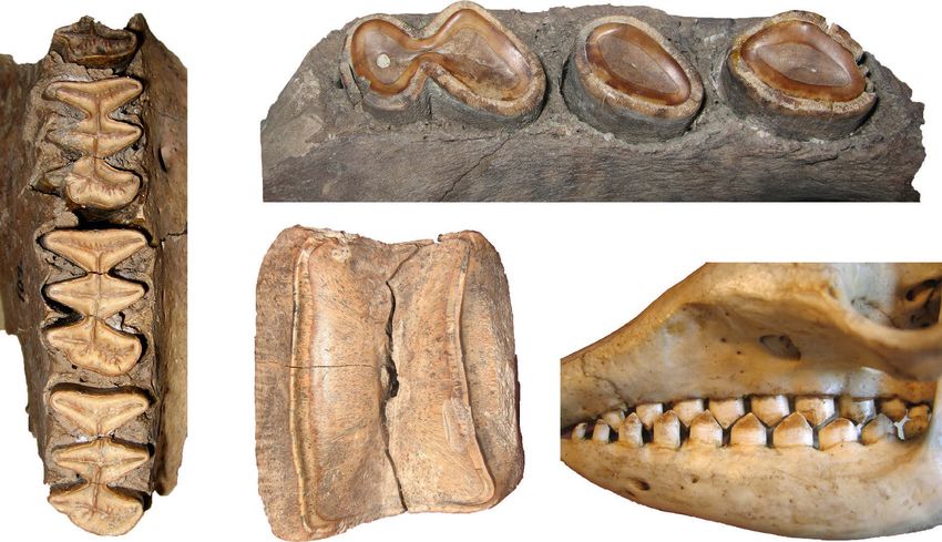
Fig. 12.3. Orthodentin surface of the stratigraphically earliest definite sloths showing representative microscopic scars.
A, Orophodon hapaloides (MNHN.F.DES 267), right lower cheek tooth, scanning electron micrograph. B, Octodontotherium
grande (MNHN.F.DES 238), right upper cheek tooth, scanning electron micrograph. Both wear surfaces display fine (i. e. nar-
row), coarse (i. e. wide), and hypercoarse (i. e. very wide scratches). Small (i. e. shallow) and large (i. e. deep) pits are scattered
over the surfaces but not so easy to discern. MNHN: Muséum national d’Histoire naturelle, Paris.
systematically belong to the Mylodontoidea. Both species and the all folivore Bradypus. A browsing behavior was
show microwear signals (Fig. 12.3), which are very differ- also identified in the North American Shasta ground sloth
ent from all other analyzed sloths to date, both fossil and Nothrotheriops shastensis, being in its food choice most
living. The results in Kalthoff & Green (2018) suggest a similar to extant Bradypus. In contrast, the North American
generalized herbivorous diet consisting of softer (foliage, mylodontid Thinobadistes segnis and the giant ground
fruit pulp) to harder (fruit, seeds, twigs) food items that sloth Megatherium americanum are characterized by
were taken both from higher vegetation levels as well scratch-dominated microwear signals, suggesting a more
B 50
50 µm
µm
as from ground level. On the base of their wide muzzle, abrasive herbivorous diet comprising mature leafs, woody
Octodontotherium and Orophodon are interpreted as so- plants, and fruit. In both species, high scratching might
called bulk feeders, meaning that the morphology and/or be related to contamination of food items with grit, which
dimensions of their feeding apparatus prevents selective is in accordance with habitat reconstructions suggesting
foraging; this is supported by their unspecialized, varied a dryer, more open landscape, at least for Megatherium
menu. In the early days of their evolution, sloths clearly (Bargo 2001). These results fit well with the idea that large
were herbivorous but still unspecific in their food choice megatheriid sloths ecologically represented the “giraffe
distinguishing them from both stratigraphically younger niche” (McDonald 2005) feeding in the higher levels of the
sloths and the living species, all of which are much more canopy, in which dust particle accumulation might occur
specialized in respect to their diet. (Ungar et al. 2005).
A number of Pleistocene ground sloths have been The above results clearly show a dietary niche partition-
analyzed and compared to the living two- and three-toed ing in Pleistocene ground sloths. However, only a fraction
sloths, i. e. the frugivore-folivore Choloepus and the folivore of their diversity has been analyzed to date, not to mention
Bradypus (Green 2009a, Green & Resar 2012, Resar fossil cingulates, i. e. fossil Dasypodidae, Glyptodontidae,
et al. 2013, Green & Kalthoff 2015). The Antillean sloth Pampatheriidae), for which microwear studies are currently
Acratocnus odontrigonus and the North American Megal- unavailable. Much work remains to be done, and we have
onyx wheatleyi have pit-dominated microwear signals with only opened a small window into the feeding ecology of
A. odontrigonus falling in the dietary ecospace together fossil xenarthrans.
with Choloepus, while M. wheatleyi falls between the latter
Summary
The monophyletic Xenarthra show a large number of unu- against abrasion. These specific microstructural make-ups
sual morphological synapomorphies distinguishing them lead to diverse tooth reliefs, ranging from dentitions with
from all other placental clades. Within toothed xenarthrans lobed, rather shallow teeth having a predominant grinding
(sloths, armadillos, and their fossil relatives), their ever- function (e. g., Mylodontidae) to dentitions with bilopho-
growing, enamelless teeth with a layered composition of dont teeth having a predominant shear-cutting function
dentin varieties are unique. Each of these dentin varieties (Megatheriidae). This result implies that the loss of the
is identified by a specific microstructural make-up, repre- hard outer enamel layer has never been a drawback in the
senting different biomechanical properties from weaker evolution and diversification of xenarthrans, probably due
(vasodentin) to strong (orthodentin) to very strong (hard to hypselodonty and also relaxed selection on the dentition.
orthodentin and osteodentin) capability of resistance Recent studies on the feeding ecology of extant and extinct
T. Martin & W. v. Koenigswald: Mammalian Teeth – Form and Function. – München (Pfeil) 2020 – ISBN 978-3-89937-266-3237
ground sloths revealed a variety of dietary adaptations toughness. They allow exciting insights into the biology of
along a line of specialized leaf browsing to more generalized this clade and independent evidence for testing existing
browsing comprising also food items with a higher intrinsic assumption on life history traits of fossil xenarthrans.
Acknowledgments
I deeply thank Wighart von Koenigswald and Thomas Martin and Olaf Dülfer (both Institute of Geosciences, Section Pale-
(both Institute of Geosciences, Section Paleontology, Rheinische ontology, Rheinische Friedrich-Wilhelms-Universität Bonn) for
Friedrich-Wilhelms-Universität Bonn) as speakers of the DFG Re- professional help with photography and specimen preparation.
search Unit FOR 771, for excellent guiding of our group through I would like to express my sincere thanks to the two reviewers
six years of funding. I am also grateful to all members of FOR Helder Gomes-Rodrigues (Muséum national d’Histoire naturelle,
771 for inspiring discussions and fruitful collaborations. I thank all Paris) and Ryan Haupt (Smithonian Institution, Washington) for
curators and collection managers of a large number of South and their insightful comments.
North American as well as European museums for their generos- This research was funded by grants KA 1556/4-1 and 5-1 of
ity to make material available for studies on microstructure and the Deutsche Forschungsgemeinschaft (DFG, German Research
microwear by DCK. Thanks also go to Jeremy Green (Kent State Foundation) Research Unit 771 “Function and performance
University at Tuscarawas) for an excellent collaboration on two enhancement in the mammalian dentition – phylogenetic and
microwear projects. I am deeply indebted to Georg Oleschinski ontogenetic impact on the masticatory apparatus”.
References
* indicates publications that originated from the DFG Research Green, J. L. & Resar, N. A. (2012): The link between dental mi-
Unit 771. crowear and feeding ecology in tree sloths and armadillos.
Biological Journal of the Linnean Society 107: 277-294.
Arsuffi, E. (1938): Beiträge zur Kenntnis des Vasodentins. Zeitschrift Habelitz, S., Rodriguez, B. J., Marshall, S. J., Marshall Jr, G. W.,
für Anatomie und Entwicklungsgeschichte 108: 749-760. Kalinin, S. V. & Gruverman, A. (2007): Peritubular dentin lacks
Bargo, M. S. (2001): The ground sloth Megatherium americanum: piezoelectricity. Journal of Dental Research 86: 908-911.
skull shape, bite forces and diet. Acta Palaeontologica Polonica Haupt, R. J., DeSantis, L. R. G., Green, J. L. & Ungar, P. S. (2013):
46: 173-192. Dental microwear texture as a proxy for diet in xenarthrans.
Bargo, M. S. & Nyakatura, J. A. (eds.) (2018): Special Issue: Mor- Journal of Mammalogy 94: 856-866.
phology and Evolution of Xenarthra. Journal of Mammalian Hoffman, J. M., Fraser, D. & Clementz, M. T. (2015): Controlled
Evolution 25: 445-588. feeding trials with ungulates: a new application of in vivo dental
Carlson, S. J. (1990): Vertebrate dental structures. In: Carter, J. molding to assess the abrasive factors of microwear. Journal
G. (ed.). Skeletal Biomineralisation: Patterns, Processes and of Experimental Biology 218: 1538-1547.
Evolutionary Trends. Vol. 1. Van Nostrand Reinhold, New *Kalthoff, D. C. (2011): Microstructure of dental hard tissues in fossil
York: 531-556. and Recent xenarthrans (Mammalia: Folivora and Cingulata).
Ciancio, M. R., Vieytes, E. C. & Carlini, A. A. (2014): When Journal of Morphology 272: 641-661.
xenarthrans had enamel: insights on the evolution of their *Kalthoff, D. C. & Green, J. L. (2018): Feeding ecology in Oligocene
hypsodonty and paleontological support for independent mylodontidoid sloths (Mammalia, Xenarthra) as revealed
evolution in armadillos. Naturwissenschaften 101: 715-725. by orthodentine microwear analysis. Journal of Mammalian
Fariña, R. A, Vizcaíno, S. F. & Iuliis, G. de (2013): Megafauna. Gi- Evolution 25: 551-564.
ant Beasts of Pleistocene South America. Indiana University *Kalthoff, D. C., Rose, K. D. & Koenigswald, W. v. (2011): Dental mi-
Press, Bloomington. crostructure in Palaeanodon and Tubulodon (Palaeanodonta)
Fariña, R. A, Vizcaíno, S. F. & Storch, G. (eds.) (2003): Morpho- and bioerosional tunneling as a widespread phenomenon in
logical Studies in Fossil and Extant Xenarthra (Mammalia). fossil mammal teeth. Journal of Vertebrate Paleontology 31:
Senckenbergiana Biologica 83: 1-101. 1303-1313.
Ferigolo, J. (1985): Evolutionary trends in the histological pattern Kastelein, R. A. (2009): Walrus: Odobenus rosmarus. In: Perrin,
in the teeth of Edentata (Xenarthra). Archives of Oral Biol- W. F., Würsig, B. & Thewissen, J. G. M. (eds.). Encyclopedia
ogy 30: 71-82. of Marine Mammals (Second Edition). Academic Press in
Gotliv, B. A. & Veis, A. (2007): Peritubular dentin, a vertebrate Elsevier, Burlington, San Diego, London: 1212-1217.
apatitic mineralized tissue without collagen: Role of a phos- Keil, A. & Venema, B. (1963): Struktur und Mikrohärteuntersu-
pholopid-proteolipid complex. Calcified Tissue International chungen an Zähnen von Gürteltieren. Zoologische Beiträge
81: 191-205. 9: 173-195.
Gotliv, B. A. & Veis, A. (2008): The composition of bovine peritubular Koenigswald, W. v. (1997): Evolutionary trends in the differentiation
dentin: Matching TOF-SIMS, scanning electron microscopy of mammalian enamel ultrastructure. In: Koenigswald, W. v. &
and biochemical component distributions. Cells Tissues and Sander, P. M. (eds.). Tooth Enamel Microstructure. Balkema,
Organs 189: 12-19. Rotterdam: 203-235.
Green, J. L. (2009a): Dental microwear in the orthodentine of Martin, B. E. (1916): Tooth replacement in Dasypus novemcinctus.
the Xenarthra (Mammalia) and its use in reconstructing the Journal of Morphology 27: 647-682.
paleodiet of extinct taxa: the case study of Nothrotheriops McDonald, H. G. (2005): Paleoecology of extinct xenarthrans and
shastensis (Xenarthra, Tardigrada, Nothrotheriidae). Zoologi- the Great American Biotic Interchange. Bulletin of the Florida
cal Journal of the Linnean Society 156: 201-222. Museum of Natural History 45: 313-333.
Green, J. L. (2009b): Intertooth variation of orthodentine microwear Meredith, R. W., Gatesy, J., Murphy, W. J., Ryder, O. A. & Springer,
in armadillos (Cingulata) and tree sloths (Pilosa). Journal of M. S. (2009): Molecular decay of the tooth gene enamelin
Mammalogy 90: 768-778. (ENAM) mirrors the loss of enamel in the fossil record of
*Green, J. L. & Kalthoff, D. C. (2015): Xenarthran tooth archi- placental mammals. PLoS Genetics 5: 1-12.
tecture and dietary adaptations from analyses of dental Mjör, I. A. & Nordahl, I. (1996): The density and branching of dentinal
microstructure and microwear, with new data for the giant tubules in human teeth. Archive of Oral Biology 41: 401-412.
sloth Megatherium americanum (Megatheriidae). Journal of Murphy, W. J., Eizirik, E., Johnson, W. E., Zhang, Y. P., Ryder, O. A.
Mammalogy 96: 645-657. & O’Brien, S. J. (2001): Molecular phylogenies and the origin
of placental mammals. Nature 409: 614-618.
Chapter 12. D. C. Kalthoff: Dental microstructure and dental microwear in xenarthran teeth238
O’Leary, M. A., Bloch, J. I., Flynn, J. J., Gaudin, T. J., Giallombardo, Appendix 1. Specimen details of the extinct and extant sloths
A., Giannini, N. P., Goldberg, S. L., Kraatz, B. P., Luo, Z.-X., mentioned in the text. Dietary adaptations follow Green (2009a),
Meng, J., Ni, X., Novacek, M. J., Perini, F. A., Randall, Z. S., Resar et al. (2013), Green and Kalthoff (2015), and Kalthoff and
Rougier, G. W., Sargis, E. J., Silcox, M. T., Simmons, N. B., Green (2018). Country codes refer to ISO 3166-1 alpha-2. Abbre-
Spaulding, M., Velazco, P. M., Weksler, M., Wible, J. R. & viations: LMA, Land Mammal Age; m, lower molariform; M, upper
Cirranello, A. L. (2013): The placental mammal ancestor and molariform. For Acronyms of collections, see Appendix 2.
the post-K-Pg radiation of placentals. Science 339: 662-667.
Oliveira, E. V. (2001): Micro-desgaste dentario em alguns Dasy- Taxon Stratigraphical age / LMA
podidae (Mammalia, Xenarthra) [Dental microwear in some
Dasypodidae]. Acta Biologica Leopoldensia 23: 83-91.
Owen, R. (1840-1845): Odontography; or, a Treatise on the Mylodontidae
Comparative Anatomy of the Teeth; their Physiological Rela- Scelidotherium sp. Pleistocene (“Pampeano”)
tions, Mode of Development, and Microscopic Structure, in
Vertebrate Animals. Hippolyte Bailliere, London. Scelidotherium Early Pleistocene
Owen, R. (1842): Description of the Skeleton of an Extinct Gi- (Ensenadense)
gantic Sloth, Mylodon robustus, Owen, with Observations on Mylodontidae indet. No data
the Osteology, Natural Affinities, and Probable Habits of the
Megatheroid Quadruped in General. Taylor, London. Mylodontidae indet. Late Miocene-Pliocene
Purnell, M., Seehausen, O. & Galis, F. (2012): Quantitative three- (Mesopotamian)
dimensional microtextural analyses of tooth wear as a tool for Mylodon sp. No data
dietary discrimination in fishes. Journal of the Royal Society
Interface 9: 2225-2233. Thinobadistes segnis Miocene
Rensberger, J. M. (1978): Scanning electron microscopy of wear
and occlusal events in some small herbivores. In: Butler, P. M.
& Joysey, K. A. (eds.). Development, Function, and Evolution Glossotherium robustus Late Miocene/Early Pliocene
of Teeth. Academic Press, New York: 415-438 (‘Mesopotamiense’)
Resar, N. A., Green, J. L. & McAfee, R. K. (2013): Reconstructing Paramylodon harlani Late Pleistocene
paleodiet in ground sloths (Mammalia, Xenarthra) using dental (Rancholabrean)
microwear analysis. Kirtlandia 58: 61-72. Paramylodon harlani Late Pleistocene
Retzius, A. (1837): Bemerkungen über den inneren Bau der Zähne (Rancholabrean)
mit besonderer Rücksicht auf den in Zahnknochen vorkom- Lestodon armatus Early Pleistocene
menden Röhrenbau. Archiv für pathologische Anatomie und (Pampas inferior)
Physiologie und für klinische Medizin 1837: 486-566. Octodontotherium sp. ?Late Oligocene (Deseadan)
Schmidt, W. J. (1924): Über das Dentin von Bradypus tridactylus.
Anatomischer Anzeiger 58: 97-107. Octodontotherium grande Late Oligocene (Deseadan)
Schmidt, W. J. & Keil, A. (1958): Die gesunden und die erkrankten
Zahngewebe des Menschen und der Wirbeltiere im Polarisa-
tionsmikroskop. Hanser, München. Orophodon hapaloides Late Oligocene (Deseadan)
Schöne, B. R., Dunca, E., Fiebig, J. & Pfeiffer, M. (2005): Mutvei’s
solution: An ideal agent for resolving microgrowth structures Megatheriidae
of biogenic carbonates. Palaeogeography, Palaeoclimatology, Eremotherium eoimigrans Early Pleistocene
Palaeoecology 228: 149-166.
Semprebon, G. M., Godfrey, L. R., Solounias, N., Sutherland, M. Eremotherium laurillardi Late Pleistocene
R. & Jungers, W. L. (2004): Can low-magnification stereomicro
scopy reveal diet? Journal of Human Evolution 47: 115-144. Megatheriidae indet., small Late Miocene-Pliocene
Simpson, G. G. (1932): Enamel on the teeth of an Eocene edentate. (Promegatherium vel Pliomegatherium
American Museum Novitates 567: 1-4. vel Pyramiodontherium)
Teaford, M. F. (1991): Dental microwear: what can it tell us about diet Megatheriidae indet. Pleistocene
and dental function. In: Kelley, M. A. & Larsen, C. S. (eds.). Ad-
vances in Dental Anthropology. Wiley-Liss, New York: 341-356. Megatheriidae indet. No data
Ten Cate, A. R. (1998): Oral Histology, 5th ed. Mosby, St. Louis.
Ungar, P. S., Teaford, M. F., Glander, K. E. & Pastor, R. F. (1995): Megatherium americanum Pleistocene
Dust accumulation in the canopy: a potential cause of dental
microwear in primates. American Journal of Physical Anthro-
pology 97: 93-99.
Vizcaíno, S. F. & Loughry, W. L. (eds.) (2008): The Biology of the Nothrotheriidae
Xenarthra. University Press of Florida, Gainesville. Nothrotheriops shastensis Late Pleistocene
Walker, P. L. (1976): Wear striations on the incisors of cercopithe- (Rancholabrean)
coid monkeys as an index of diet and habitat preference. Nothrotheriops texanus Early Pleistocene
American Journal of Physical Anthropology 45: 299-308. (Irvingtonian)
Walker, A., Hoeck, H. N. & Perez, L. (1978): Microwear of mam- Megatheroidea indet.
malian teeth as an indicator of diet. Science 201: 908-910. Hapalops sp. Late Early Miocene
Winkler, D. E., Schulz-Kornas, E., Kaiser, T. M. & Tütken, T. (2019): (Santacrucian)
Dental microwear texture reflects dietary tendencies in extant Megalonychidae
Lepidosauria despite their limited use of oral food processing. Choloepus sp. Recent
Proceedings of the Royal Society B 286. https://doi.10.1098/ Acratocnus odontrigonus Pleistocene
rspb.2019.0544 Megalonyx wheatleyi Pleistocene
Bradypodidae
Bradypus torquatus Recent
Bradypus tridactylus Recent
T. Martin & W. v. Koenigswald: Mammalian Teeth – Form and Function. – München (Pfeil) 2020 – ISBN 978-3-89937-266-3239
Sample provenance Sample no. Original coll. no. Tooth position Special dentin character Diet based on dentin
microwear
No data KOE 3577 MLP 50-VIII-1-1 Probably M2 Cone-shaped bundles
of dentinal tubules
Entre Pto. de Olivos y Pta. KOE 3433 MLP 54-VI-19-8 Molariform Cone-shaped bundles
Anchore, prov. Buenos Aires, AR of dentinal tubules
No data KOE 3004 AMNH 11282 Molariform Cone-shaped bundles
of dentinal tubules
Paraná, prov. Entre Rios, AR KOE 3432 MLP 41-XII-13-2080 Molariform Cone-shaped bundles
of dentinal tubules
No data KOE 3000 AMNH 132682 Molariform Cone-shaped bundles
of dentinal tubules
Mixon’s Bone Bed, Florida, US – AMNH FAM (2 teeth); molariforms (5) Plants with low to moderate
FMNH (3 teeth) (2 MF, 3 mf) intrinsic toughness;
possible influence of grit/dust
No data KOE 3541 AMNH 11273 Molariform Cone-shaped bundles
of dentinal tubules
Rancho La Brea, California, US KOE 3569 LACMHC 130 757 Left M2 Cone-shaped bundles
of dentinal tubules
Rancho La Brea, California, US KOE 3573 LACMHC 130 761 Right m2 Cone-shaped bundles
of dentinal tubules
Rio Arrecifes, AR –
NHMD-ZMK 52/1888 Right m1-3 Cone-shaped bundles
(former ZMUC 154) of dentinal tubules
Ez. Co. Alto. zona del Rio KOE 3434 MLP 52-XI-3-15, Molariform Small amount of central Generalized herbivorous diet;
Deseado, Prov. Santa Cruz, AR diverse numbers vasodentin bulk feeding
La Flecha, Patagonia, AR – MNHN.F.DES molariforms (9) Small amount of central Generalized herbivorous diet;
(5 MF, 3 mf, 1 vasodentin bulk feeding
MF/mf)
La Flecha, Patagonia, AR – MNHN.F.DES molariforms (5) Small amount of central Generalized herbivorous diet;
diverse numbers (2 MF, 3 mf) vasodentin bulk feeding
AL033: Haile 16A, Alachua Co., KOE 3044 UF 46361 Molariform Strong thinning of
Florida, US orthodentin layer
LV028: Waccassassa River, KOE 3045 UF 16416 Molariform Strong thinning of
Levy Co., Florida, US orthodentin layer
Paraná, Prov. Entre Rios, AR KOE 3431 MLP 325 Molariform Strong thinning of
orthodentin layer
Quebrada de Agua, ?AR KOE 3003 AMNH 123683 Molariform Strong thinning of
orthodentin layer
No data KOE 3578 YPM 25011 PU Molariform Strong thinning of
orthodentin layer
“Plata-Landene” – NHMD-ZMK 56/1885 Molariform Strong thinning Plants with low to moderate
(territory of today’s Argentina, (former ZMUC 19) of orthodentin layer intrinsic toughness;
Paraguay, Uruguay, possible influence of grit/dust
and parts of Bolivia)
Potter Creek Cave, – UCMP 8141, 8336, molariforms (4) Thinning of orthodentin Herbivorous diet
California, US 8704, 8715 (all M2/M3) layer
HI007: Leisey Shell pit 1A, KOE 3053 UF 87136 Molariform Thinning of orthodentin
Hillsborough Co., Florida, US layer
Estancia La Costa, layer ?3, KOE 3012 IGPB uncat. Molariform
Prov. Santa Cruz, AR
No data KOE 3590 SMF 35.784 Molariform Frugivore - folivore
Puerto Rico, PR – AMNH molariforms (3) Frugivore - folivore
Smith Pit, Florida, US – AMNH molariforms (6) Folivore
No data KOE 3018 ZSM 1903/9534 Molariform Folivore
“Vivarium”, supposedly from KOE 3070 MNHN CG 1954-268 Molariform Folivore
Ménagerie du Jardin
des Plantes, Paris, FR
Chapter 12. D. C. Kalthoff: Dental microstructure and dental microwear in xenarthran teeth240
Appendix 2. Specimen details of the extinct and extant cingulates mentioned in the text. Dietary categories follow Green (2009a).
Country codes refer to ISO 3166-1 alpha-2.
Abbreviations: LMA, Land Mammal Age; M, upper molariform. Acronyms to museum collections: AMNH, American Museum of
Natural History, New York, USA; FMNH, Field Museum of Natural History, Chicago, USA; IGPB, Institute of Geosciences, Section
Paleontology, Rheinische Friedrich-Wilhelms-Universität Bonn, Bonn, Germany; KOE, Enamel collection, established by Wighart von
Koenigswald, Institute of Geosciences, Section Paleontology, Rheinische Friedrich-Wilhelms-Universität Bonn, Germany; LACMHC,
Los Angeles County Museum Hancock Collection (Page Museum), Los Angeles, USA; MACN, Museo Argentino de Ciencias Na-
turales “Bernardino Rivadavia”, Buenos Aires, Argentina; MLP, Museo de La Plata, La Plata, Argentina; MNHN, Muséum national
d’Histoire naturelle, Paris, France; NRM, Swedish Museum of Natural History, Stockholm, Sweden; NHMD, Natural History Museum
of Denmark, Copenhagen, Denmark; NHMD-ZMK, Natural History Museum of Denmark: Quaternary Zoology collection, Copenhagen,
Denmark; SMF, Senckenberg Forschungsinstitut und Naturmuseum Frankfurt, Germany; UCMP, University of California Museum of
Paleontology, Berkeley, USA; UF, Florida Museum of Natural History, University of Florida, Gainesville, USA; YPM, Peabody Muse-
um of Natural History, Yale University, New Haven, USA; ZMB, Museum für Naturkunde (Zoologisches Museum), Berlin, Germany;
ZSM, Zoologische Staatssammlung München, Germany. Uncat.: uncataloged.
Taxon Stratigraphical age / LMA Sample provenance Sample no.
Cingulata: Dasypodinae
Dasypus hybridus Recent No data KOE 3010
Dasypus novemcinctus Recent No data KOE 3442
Dasypus novemcinctus Recent Golf Course near Friesenhahn Ranch, Texas, US KOE 3534
Euphractinae
Chaetophractus villosus Recent No data KOE 3046
Chaetophractus villosus Recent No data KOE 3441
Chlamyphorus truncatus Recent No data KOE 3021
Euphractus sexcinctus Recent No data KOE 3594
Euphractus sexcinctus Recent Lagoa Santa, Minas Gerais, BR –
Eutatus seguini Pleistocene (“Pampeano”) No data KOE 3424
Macroeuphractus sp. Pliocene No data KOE 3588
Proeutatus sp. Late Early Miocene (Santacrucian) Monte León, Santa Cruz, AR KOE 3425
Zaedyus pichiy Recent Puerto Jenkins, Santa Cruz, AR KOE 3022
Tolypeutinae
Cabassous unicinctus Recent Ipitingua, Rio Acara, BR KOE 3019
Priodontes maximus Recent El Cerro, Chiguitos, BO KOE 3015
Tolypeutes matacus Recent La Urbana, Tapikiolé, lower Rio Pilcomayo, PY KOE 3020
Peltephilidae
Peltephilus ferox Late Early Miocene (Santacrucian) No data KOE 3429
Pampatheriidae
Pampatherium sp. No data No data KOE 3002
Pampatherium sp. Pleistocene (“Pampeano”) Algarrobo, Ptdo C. Casares, Buenos Aires province, AR KOE 3426
Pampatherium Early Pleistocene (Irvingtonian) HI007: Leisey Shell pit 1A, Hillsborough County, Florida, US KOE 3047
(= Holmesina) floridanus
Vassallia minuta Pliocene (“Araucanense”) Catamarca province, AR KOE 3428
Glyptodontidae
Propalaehoplophorinae indet. No data Santa Cruz province, AR KOE 3436
Propalaehoplophorus australis Late Early Miocene (Santacrucian) Santa Cruz province, AR KOE 3538
Glyptodontidae indet. Early Pleistocene (Ensenadan) Buenos Aires province, AR KOE 2879
Glyptodontidae indet. No data No data KOE 3430
Glyptodon sp. No data No data KOE 3001
Glyptodon sp. No data South America KOE 3535
Glyptodon reticulatus Pleistocene Tarija, BO KOE 3026
Glyptodon clavipes Pleistocene Arroyo del medio, Pampa intermedio, AR KOE 3060
Hoplophorus ornatus Pleistocene Pergamino, Province Buenos Aires, AR KOE 3061
T. Martin & W. v. Koenigswald: Mammalian Teeth – Form and Function. – München (Pfeil) 2020 – ISBN 978-3-89937-266-3241
Original coll. no. Tooth position Special dentin character Dietary catagory based
on dentin microwear
MLP uncat. Molariform Milk tooth generaton with enamelled teeth;
?central vasodentin
– Molariform Milk tooth generaton with enamelled teeth; Insectivore
central vasodentin
IGPB M-6357 Molariform Milk tooth generaton with enamelled teeth; Insectivore
central vasodentin
MLP uncat. Molariform ‘Degenerated osteodentin’
MLP uncat. Molariform ‘Degenerated osteodentin’
ZSM AM/1125 Right M7 and M8 Insectivore
SMF 908 Molariform Carnivore - omnivore
NHMD L62 (former ZMUC L62) Skull and mandibles Carnivore - omnivore
MLP 16-146 Molariform Hard orthodentin; osteodentin
FMNH P 14493 Molariform ‘Degenerated osteodentin’
MLP 67-X-31-1 Molariform Hard orthodentin; osteodentin
ZMB 48626 Molariform ‘Degenerated osteodentin’ Carnivore - omnivore
ZSM 1910/252 Molariform Insectivore
ZSM 1926/359 Right M3
ZSM 1925/595 Molariform ‘Degenerated osteodentin’ Insectivore
MLP 69-VIII-13-3 Molariform ‘Degenerated osteodentin’
– Molariform Hard orthodentin; osteodentin
MLP 70-III-10-1 Molariform Hard orthodentin; osteodentin
UF 65890 Molariform Hard orthodentin; osteodentin
MLP 29-X-10-71 Molariform Hard orthodentin; osteodentin
MACN A-11229 Upper molariform Hard orthodentin; osteodentin
AMNH 132681 Molariform Hard orthodentin; osteodentin
– Molariform Hard orthodentin; osteodentin
MLP s/n Molariform Hard orthodentin; osteodentin
– Molariform Hard orthodentin; osteodentin
AMNH 11301 Molariform Hard orthodentin; osteodentin
NRM-PZ M2519 Molariform Hard orthodentin; osteodentin
NHMD-ZMK 70/1885 (former ZMUC 109) Molariform Hard orthodentin; osteodentin
NHMD-ZMK 77/1888 (former ZMUC 170) Molariform Hard orthodentin; osteodentin
Chapter 12. D. C. Kalthoff: Dental microstructure and dental microwear in xenarthran teethYou can also read