Cardiac anatomy in the 'Dreyfus Madonna' by Leonardo da Vinci - Oxford Academic Journals
←
→
Page content transcription
If your browser does not render page correctly, please read the page content below
Interactive CardioVascular and Thoracic Surgery 32 (2021) 582–584 BRIEF COMMUNICATION
doi:10.1093/icvts/ivaa314 Advance Access publication 9 December 2020
Cite this article as: Keshelava G. Cardiac anatomy in the ‘Dreyfus Madonna’ by Leonardo da Vinci. Interact CardioVasc Thorac Surg 2021;32:582–4.
Cardiac anatomy in the ‘Dreyfus Madonna’ by Leonardo da Vinci
Grigol Keshelava*
Department of Vascular Surgery, Clinic “Helsicore”, Tbilisi, Georgia
* Corresponding author: Department of Vascular Surgery, Clinic “Helsicore”, Tevdore Mgvdeli St. 13, Tbilisi 0121, Georgia. Tel: +995-99424832;
e-mail: gagakeshelava@gmail.com (G. Keshelava).
Received 13 July 2020; received in revised form 15 October 2020; accepted 9 November 2020
Downloaded from https://academic.oup.com/icvts/article/32/4/582/6132061 by guest on 09 August 2021
Abstract
Leonardo’s findings on the anatomy and physiology of the heart, preserved in his private sketches and notes, reveal a remarkable period of
transition in the early history of cardiology. The object of this research is a ‘Dreyfus Madonna’. Through the programme Paint X, we moved
1 detail (blue marked) in the direction of the green arrow and we get an image of the heart with branches of the aortic arch. If we compare
the image of the heart obtained by us with the anatomical sketch of Leonardo (RL 19073-74v; K/P 1 66v), we will see a sharp resemblance
to the branches of the aortic arch.
Keywords: Cardiac and aortic arch anatomy • Leonardo da Vinci
LEONARDO DA VINCI’S ANATOMY OF THE Hidden description of anatomical elements in the painting has
HEART also been made by other artists of the Renaissance era.
Researchers have found that the pomegranate in Botticelli’s
Leonardo da Vinci (1452–1519) conducted many anatomical ‘Madonna of the Pomegranate’ corresponds to the appearance of
studies during the second half of his life and his drawings not the heart [10].
only demonstrate his artistic genius but also prove that he was a
great scientist. Today almost the complete set of these anatomi-
cal drawings and comments is owned by the British Crown and is ANALYSIS OF THE ‘DREYFUS MADONNA’ BY
in the Royal Library at Windsor Castle, UK [1]. Leonardo’s findings LEONARDO DA VINCI
on the anatomy and physiology of the heart, preserved in his pri-
vate sketches and notes, reveal a remarkable period of transition It was done by Leonardo da Vinci between 1469 and 1471. The
in the early history of cardiology. At the time of Leonardo’s birth, painting features a young woman with a naked baby standing
the prevailing understanding of the structures and functions of her laps. The young lady is believed to be Virgin Mary and the in-
the heart in Europe derived from ancient authorities [2]. At the fant is baby Jesus. The woman is carrying pomegranate, a symbol
same time, the heart had an almost spiritual role [3]. The vast ma- of fertility. Leonardo wants to show his audience that the light
jority of Leonardo’s studies on the heart are reported in drawings display hope for humanity as Christ is born through some awk-
and notes he produced in Milan from 1508 to 1513, though af- ward circumstances.
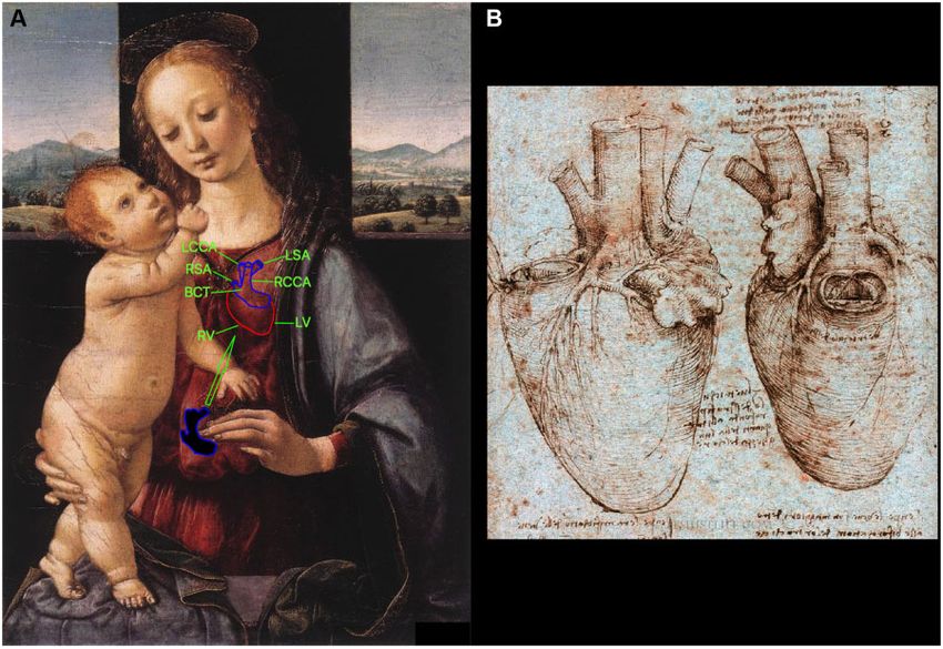
terwards he continued to work on in Rome [4]. To explore the The object of this research is a ‘Dreyfus Madonna’ (Fig. 1).
working of the heart, Leonardo injected warm wax into a bull’s Through the program Paint X, we moved 1 detail (blue
heart to examine the valves’ forms [5]. He then created a glass marked) in the direction of the green arrow to the breast of Mary
model of the heart to study the hydraulic characteristics of the and we get an image of the heart with branches of the aortic
blood flux and the function of valves [6]. arch (Fig. 2), (Video 1). The moving detail is circled along the faint
In this period, anatomical knowledge in Europe was largely contour by Leonardo da Vinci himself and is located near the left
based on manuscripts from classical Greece and medieval Italy, hand cluster of Mary. One gets the impression that Mary’s fingers
the dissection of animals and the intermittent dissection of the are pointing at this detail. The received heart imaging has an ex-
condemned criminal [7, 8]. Despite the fact that cadaver dissec- act anatomical location and inclination. It reflects the brachio-
tion was illegal in this epoch, physicians still managed to deepen cephalic trunk, right subclavian artery, right common carotid ar-
knowledge in human anatomy. Leonardo fundamentally studied tery, left common carotid artery, left subclavian artery, left ventri-
the anatomy and used this knowledge in the art. Leonardo filled cle and right ventricle (Fig. 2).
notebooks with carefully drawn two-dimensional representations The harmonious combination of the colours of the heart and
of the organs, tissues and skeletal formations uncovered during aortic arch with the colours of neighbouring details is remarkable
his dissections [9]. (Fig. 1).
C The Author(s) 2020. Published by Oxford University Press on behalf of the European Association for Cardio-Thoracic Surgery. All rights reserved.
V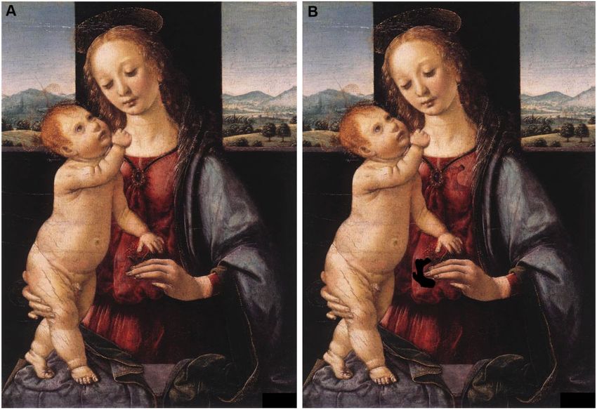
G. Keshelava / Interactive CardioVascular and Thoracic Surgery                                                   583

                                                                                                                                                                           Downloaded from https://academic.oup.com/icvts/article/32/4/582/6132061 by guest on 09 August 2021
                                                                                                                                                                           VASCULAR
Figure 1: (A) ‘Dreyfus Madonna’ by Leonardo da Vinci (1469–71); (B) the image obtained after moving the detail.

Figure 2: (A) The blue marked detail moves in the direction of the green arrow. (B) Anatomical drawing by Leonardo da Vinci (RL 19073-74v; K/P 1 66v). BCT: bra-
chio-cephalic trunk; LCCA: left common carotid artery; LSA: left subclavian artery; LV: left ventricle; RCCA: right common carotid artery; RSA: right subclavian artery;
RV: right ventricle.
584                                          G. Keshelava / Interactive CardioVascular and Thoracic Surgery

                                                                               Reviewer information
                                                                               Interactive CardioVascular and Thoracic Surgery thanks Eugenio Neri and the
                                                                               other, anonymous reviewer(s) for their contribution to the peer review pro-
                                                                               cess of this article.

                                                                               REFERENCES
                                                                                 [1] Schultheiss D, Laurenza D, Gotte B, Jonas U. The Weimar anatomical
                                                                                     sheet of Leonardo da Vinci (1452–1519): an illustration of the genitouri-
                                                                                     nary tract. BJU Int 1999;84:595–600.
                                                                                 [2] Cambiaghi M, Hausse H. Leonardo da Vinci and his study of the heart.
                                                                                     Eur Heart J 2019;40:1823–31.
                                                                                 [3] Heater W. The Medieval Heart. New Haven, London: Yale University
                                                                                     Press, 2010.
                                                                                 [4] Shoja MM, Agutter PS, Loukas M, Benninger B, Shokouhi G, Namdar H

                                                                                                                                                                 Downloaded from https://academic.oup.com/icvts/article/32/4/582/6132061 by guest on 09 August 2021
                                                                                     et al. Leonardo da Vinci’s studies of the heart. Int J Cardiol 2013;167:
Video 1: Move the detail on the painting ‘Dreyfus Madonna’ by Leonardo da            1126–33.
Vinci.                                                                           [5] Boon B. Leonardo da Vinci on atherosclerosis and function of the
                                                                                     sinuses of Valsalva. Neth Heart J 2009;17:496–9.
                                                                                 [6] Sterpetti AV. Cardiovascular research by Leonardo da Vinci (1452–1519).
                                                                                     Circ Res 2019;124:189–91.
  If we compare the image of the heart obtained by us with the                   [7] Park K. The criminal and the saintly body: autopsy and dissection in
                                                                                     Renaissance Italy. Renaiss Q 1994;47:1–33.
anatomical sketch of Leonardo (RL 19073-74v; K/P 1 66v) (Fig. 2),                [8] Olry R. Medieval neuroanatomy: the text of Mondino dei Luzzi and the
we will see a sharp resemblance to the branches of the aortic                        plates of Guido da Vigevano. J Hist Neurosci 1997;6:113–23.
arch.                                                                            [9] Cothern AM. The perfect machine: the reason behind the anatomical
                                                                                     studies of Leonardo da Vinci. Kaleidoscope 2008;7:19–29.
                                                                               [10] Lazzeri D, Al-Mousawi A, Nicoli F. Sandro Botticelli’s Madonna of the
                                                                                     Pomegranate: the hidden cardiac anatomy. Interact CardioVasc Thorac
Conflict of interest: none declared.                                                 Surg 2019;28:619–21.
You can also read