Case Report Encephalopathy Associated with Autoimmune Thyroid Disease: A Potentially Reversible Condition - Core
←
→
Page content transcription
If your browser does not render page correctly, please read the page content below
Hindawi Publishing Corporation
Case Reports in Medicine
Volume 2016, Article ID 9183979, 6 pages
http://dx.doi.org/10.1155/2016/9183979

Case Report
Encephalopathy Associated with Autoimmune Thyroid Disease:
A Potentially Reversible Condition

 Inês Correia,1 Inês B. Marques,1 Rogério Ferreira,2 and Lívia Sousa1
 1
 Department of Neurology, Centro Hospitalar e Universitário de Coimbra, Praceta Professor Mota Pinto,
 3000-075 Coimbra, Portugal
 2
 Department of Internal Medicine, Centro Hospitalar e Universitário de Coimbra, Praceta Professor Mota Pinto,
 3000-075 Coimbra, Portugal

 Correspondence should be addressed to Inês Correia; mcorreia.ines@gmail.com

 Received 23 December 2015; Revised 13 March 2016; Accepted 22 March 2016

 Academic Editor: Di Lazzaro Vincenzo

 Copyright © 2016 Inês Correia et al. This is an open access article distributed under the Creative Commons Attribution License,
 which permits unrestricted use, distribution, and reproduction in any medium, provided the original work is properly cited.

 Autoimmune thyroid disease may occasionally associate with unspecific neurological symptoms, which are more commonly
 insidious, include cognitive or behavioural symptoms, and may associate with tremor, myoclonus, or ataxia. We report a 61-year-old
 female patient who presented with chronic headache, insidious mood, and cognitive disturbance which evolved in a few months
 to dementia associated with exuberant limb myoclonus. Diagnostic workup revealed high anti-thyroid peroxidase antibody titers
 and an inflammatory CSF profile, and it was negative for other possible etiologies. Treatment with steroids induced significant
 improvement. The diagnosis of encephalopathy associated with autoimmune thyroid disease is still controversial given the fact
 that the clinical presentation and diagnostic workup are unspecific, the pathophysiology is still undetermined, and the diagnosis is
 mostly of exclusion. No direct correlation is found between anti-thyroid antibody titers and clinical presentation, and it is currently
 speculated that other still unrecognized antibodies may be responsible for this clinical entity. It is extremely important to recognize
 this entity because it is potentially treatable with immunotherapies. It is also increasingly recognized that clinical improvement with
 first-line treatment with steroids may be absent or incomplete, and other immunotherapies as immunosuppressants, intravenous
 immunoglobulin, or plasma exchange must be attempted in the clinical suspicion of EEAT.

1. Introduction We report a 61-year-old female patient who presented
 with chronic headache, insidious mood, and cognitive distur-
Encephalopathy associated with autoimmune thyroid dis- bance evolving to rapidly progressive dementia with exuber-
ease (EAAT) is a rare clinical entity, which presents with ant limb myoclonus. Diagnostic workup identified high anti-
unspecific neurological symptoms. The clinical presentation thyroid antibody titers and excluded other causes, leading to
is more frequently insidious, with cognitive and behavioural diagnosis of EAAT. Significant improvement was achieved
disturbance that may associate with tremor, myoclonus, or with steroid treatment.
ataxia. More rarely, clinical onset may be acute as stroke-like With this paper we underline the importance of con-
episodes, epilepsy, or psychosis [1–6]. sidering EAAT when approaching patients with rapidly
 Diagnostic investigation is usually unspecific and there progressive neurological or psychiatric symptoms, since it is
is no direct correlation between thyroid hormone levels or a potentially reversible condition with appropriate treatment.
anti-thyroid antibody titers and the clinical presentation [1–
3]. The current diagnostic criteria are based on the association 2. Case Report
of neurological or psychiatric symptoms, presence of anti-
thyroid antibodies, exclusion of other possible causes, and We report a 61-year-old Caucasian woman who first pre-
significant improvement with immunotherapies [7], which sented to our Neurology Emergency Department in March
make this entity mostly a diagnosis of exclusion. 2012 complaining of severe chronic daily headache. The
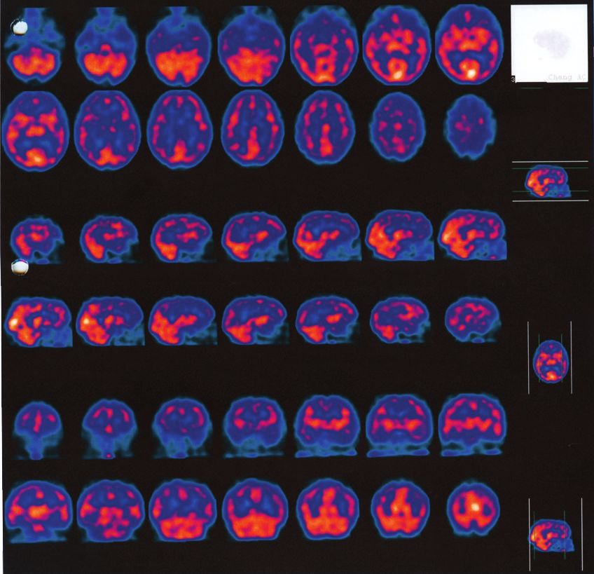
2 Case Reports in Medicine

headache was started 9 months before and was described Cerebrospinal fluid (CSF) analysis revealed slightly
as bilateral, with pressing-type quality, without associated increased proteins (72 mg/dL) and lymphocytic pleocy-
symptoms such as nausea, photophobia, or phonophobia, tosis with 17 cells/mm3 . CSF direct microscopy, cultures,
and did not worsen with recumbency, exercise, or Valsalva and serologies (Herpes simplex 1 and 2, Cytomegalovirus,
manoeuvres. The patient also presented apathy with progres- Epstein-Barr virus, Treponema pallidum, Borrelia burgdorferi,
sive loss of interest in life for 18 months and had already and Brucella spp.) were negative. Oligoclonal bands were
been evaluated by a Psychiatrist who diagnosed depressive absent and CSF dementia biomarkers (beta-amyloid peptide,
syndrome given that these features immediately followed the tau protein, and phosphorylated tau protein) were normal.
return to her country after 40 years living abroad, leaving Electroencephalogram (EEG) revealed rhythmic slow
behind her children and grandchildren. However, despite activity in both temporal regions, with normal background
antidepressive treatment, she presented gradual worsening rhythm and without paroxysmal activity.
and became unable to perform her usual activities of daily Brain Magnetic Resonance Imaging (MRI) was unre-
living without supervision (such as cooking, using the tele- markable and brain perfusion single-photon emission com-
phone, handling money, and taking her medication) and she puted tomography (SPECT) imaging revealed hypoperfusion
spent most days in bed in the last three months. The patient in frontal, temporal, and parietal regions with left predomi-
also complained of upper limb tremor with left predominance nance (Figure 1).
for the same period. The rapidly progressive neurological and psychiatric
 She had history of arterial hypertension, dyslipidemia, symptoms presented by the patient were unspecific and it
and hypothyroidism due to Hashimoto’s thyroiditis chron- could be the presentation of several different conditions
ically treated with levothyroxine. There was no history of including metabolic or toxic encephalopathy, CNS infection,
recent or chronic infections or toxic exposure. Familial cerebrovascular disease, CNS tumor, and CNS inflamma-
medical history was unremarkable. tory conditions, such as cerebral vasculitis, autoimmune
 The general physical examination was normal. The neu- encephalitis, or paraneoplastic syndromes, or, more remotely,
rological examination revealed cognitive impairment with a rapidly progressive presentation of a degenerative dementia.
Mini Mental State Examination of 23 points (3 years of The clinical history and diagnostic investigations excluded
education). She presented frontal functions impairment with most of these causes and given that the only relevant find-
low verbal fluency, perseveration, impairment of abstract ings were an inflammatory CSF profile and an increased
thinking, and signs of frontal release, namely, glabellar reflex. anti-TPO antibodies titer, encephalopathy associated with
Visuospatial impairment was also observed with inability autoimmune thyroid disease was diagnosed and treatment
to copy a drawing or perform the clock-drawing test. An with intravenous methylprednisolone (1 g/day for 5 days)
upper limb rest and postural tremor with left predominance was performed. A significant improvement occurred after
was identified, without other focal signs in the neurological five days of therapy with complete resolution of mood and
examination. cognitive disturbance (MMSE = 29) and disappearance of the
 A brain computerized tomography (CT) and blood anal- myoclonic movements. EEG was repeated with normal result.
ysis were performed in the Emergency Department with No steroid side effects occurred and, after improvement, the
normal results. On discharge a follow-up appointment was patient was discharged with oral prednisolone (1 mg/kg/day).
planned for dementia study. In the next weeks, a rapidly One month after discharge, steroid dosage reduction
progressive neurological deterioration occurred; the patient was attempted, but neurocognitive symptoms and distal
became unable to walk and totally dependent and presented limb myoclonus rapidly returned (anti-TPO antibodies =
exuberant myoclonus in the distal upper limbs, so she was 217 UI/mL). Improvement was promptly seen after pred-
admitted for more investigations. nisolone reincrease to 1 mg/kg/day; however, steroid side
 Extensive blood workup including full blood count, effects developed, including Diabetes and Cushing Syn-
coagulation study, liver function test, creatinine, erythrocyte drome, so azathioprine was added as a steroid sparing agent.
sedimentation rate, c-reactive protein, protein electrophore- Although steroid gradual withdrawal was possible after 6
sis, vitamin B12, folic acid, thyroid function, and serologies months without symptoms recurrence, the patient developed
(Treponema pallidum, Brucella spp., Borrelia burgdorferi, toxic hepatitis in relation to azathioprine and the drug
Coxiella burnetii, Rickettsia conorii, HIV, and hepatitis B and was stopped. Neurological and psychiatric symptoms and
C) was normal. Study of systemic autoimmunity, includ- myoclonic movements returned and were rapidly controlled
ing antinuclear antibodies, anti-ds-DNA, anti-SSA, anti-SSB, with low prednisolone dosage (10 mg/day) without associated
anti-RNP, anti-Scl70, anti-Jo1, anti-neutrophil cytoplasmic side effects, and other therapies such as plasma exchange,
antibodies, and anti-thyroid antibodies, revealed only high intravenous immunoglobulins, or other immunosuppressive
titers of anti-thyroid peroxidase antibodies (anti-TPO anti- drugs were not necessary. Three years later the patient was
bodies) equal to 1008 UI/mL (normal
Case Reports in Medicine 3

 3 Change AC
 6 7 8 9 10 11 12 Vol Rendered

 13 14 15 16 17 18 19
 Feet to Head Transversal Slice thickness 6.63 mm
 1

 9 10 11 12 13
 8
 7

 14 15 16 17 18 19 20
 Right to Left Sagittal Slice thickness 6.63 mm
 1

 6 7 8 9 10 11 12

 17 18 19
 13 14 15 16
 Anterior to Posterior Coronal Slice thickness 8.84 mm 1

Figure 1: Brain perfusion SPECT. Reduced 99mTc-HMPAO uptake in parietal, temporal, and frontal lobes. Hypoperfusion is more severe
on the left hemisphere.

 3. Discussion
 Anti-TPO antibody titer

 1200
 1000
 800 Encephalopathy associated with autoimmune thyroid dis-
 600 ease, also called Hashimoto’s encephalopathy or Steroid-
 400 Responsive Encephalopathy associated with Autoimmune
 200 thyroid disease, is a controversial entity as its pathophysi-
 0 ology is not yet well defined and it is usually a diagnosis
 0 4 8 12 16 20 24 28
 of exclusion. It is known to be associated with clinical or
 Time (months)
 subclinical autoimmune thyroid disease, most commonly
 Clinical exacerbation
 Hashimoto’s thyroiditis, but there are also some reports of
 Anti-TPO
 association with Graves Disease, a clinical entity without true
 thyroiditis [8–13].
Figure 2: Evolution of anti-thyroid peroxidase antibodies (anti- Although autoimmune thyroiditis and the presence of
TPO antibodies) titers in time and their relationship with clinical anti-thyroid antibodies are relatively common in the popula-
exacerbation. tion, with an estimated prevalence of Hashimoto thyroiditis
 of 0.3 to 2% [14–16] and detection of anti-thyroid antibodies
 in up to 10% healthy population [17–19], encephalopathy
antibody titer is increased, there is no direct relationship associated with thyroiditis or anti-thyroid antibodies is
with clinical presentation, including asymptomatic periods very uncommon, with an estimated prevalence of 2.1 per
associated with elevated antibodies. 100.000 habitants [20]. It occurs more commonly in females
4 Case Reports in Medicine

 EEG abnormalities are usually present, occurring in 90
 Thyroid hormones levels

 4
 to 98% of patients, usually with unspecific findings [22]. The
 3 most common presentations are diffuse background slowing
 2 and frontal intermittent rhythmic delta activity (FIRDA)
 [1, 6], but other EEG patterns have been described such as
 1
 periodic lateralized epileptiform discharges (PLEDs) [23] and
 0 temporal epileptiform activity [24, 25]. There is usually a cor-
 0 4 8 12 16 20 24 28
 relation between the slowing severity and the encephalopathy
 Time (months)
 severity [6, 24, 26] and an EEG normalization occurs with
 Clinical exacerbation successful treatment, which may also be used to support the
 TSH
 diagnosis [3, 27].
 Free T4 Brain MRI may be normal or, in up to 50% of cases,
 present unspecific anomalies, and it is very important to
Figure 3: Evolution of thyroid hormone levels in time and their exclude other etiologies [1, 6]. The abnormalities that may
relationship with clinical exacerbation. be seen are cerebral atrophy, or less frequently, focal cortical
 abnormalities or unspecific focal or diffuse subcortical white
 matter hyperintensities which may be reversible with treat-
(4 : 1 ratio), and, although there are cases reported from ment [1, 3].
childhood through the eighth decade of life, the mean age of SPECT scan is not routinely used, but it was performed
onset is in the fourth decade [1, 2]. in some previous reports, with unspecific patterns of reduced
 Since the first description of Hashimoto’s encephalopathy perfusion, which can be diffuse or patchy and may involve
in 1966 [21], the clinical spectrum has been widened and dif- multiple different regions [3, 28–30]. Positron emission
ferent possible presentations are now recognized, including tomography (PET) is also unspecific and usually shows
acute-onset presentations as stroke-like episodes, epilepsy, widespread multifocal hypometabolism [31, 32]. The findings
or psychosis, and a slowly progressive presentation, which in SPECT and PET do not appear to be correlated with the
appears to be more common, characterized by cognitive and clinical presentation, EEG, or neuroradiological findings and
behavioral disturbances which may be associate with tremor, usually improve after successful treatment.
myoclonus, or ataxia [1–4]. The pathogenesis underlying the encephalopathy asso-
 The diagnostic criteria of EAAT include the association of ciated with autoimmune thyroid disease is still unknown.
neurological or psychiatric manifestations, high titers of anti- Although anti-thyroid antibodies have no established
thyroid antibodies, exclusion of other possible causes with pathogenic role, as there is no direct correlation between
complementary exams, and a good response to immuno- antibody titers and clinical severity [1–3], the high prevalence
suppressive therapy [3, 7]. However, these vague diagnostic of coexistence of thyroid autoimmune diseases and other
criteria may contribute to masquerade of other autoimmune autoimmune diseases is well described [33, 34]. Therefore,
encephalopathies instead. the presence of anti-thyroid antibodies may be related to an
 In fact, blood workup is usually normal, except for autoimmune predisposition and the presence of other, still
the presence of increased anti-thyroid antibodies, more unrecognized, antibodies responsible for the encephalopathy
commonly against thyroid peroxidase (86%), but anti- may be speculated.
thyroglobulin antibodies may be present in some cases (48%) Antibodies against alpha-enolase, an antigen present in
[1–3]. Despite this association, there is no direct correlation thyroid and also diffusely in the brain, have been described
between antibody titers and the clinical severity, with some in some patients [35, 36]; however, their involvement in the
asymptomatic patients having high antibody titers and some pathogenesis has not been yet documented. The presence
patients with severe encephalopathy having only mildly of antibodies against neuronal antigens is also suggested
increased antibody titers. The absence of direct correlation although it remains to be proven [37].
between clinical presentation and anti-thyroid antibodies Given that EAAT is considered an inflammatory con-
levels complicates the diagnosis and therefore the evidence of dition, the current treatment is based on immunotherapy.
CNS inflammation, assessed in the cerebrospinal fluid (CSF), The most commonly used treatment is intravenous methyl-
so the exclusion of other causes is essential to the assumption prednisolone (500–1000 g/day, for 3 to 5 days) followed by
of this diagnosis. Thyroid hormones levels are also not related oral prednisone (1-2 mg/kg/day), which is gradually tapered
to the course of the disease with the majority of patients being with clinical improvement. Immunosuppressants, more com-
euthyroid (18–45%) or hypothyroid (clinical in 25–35% and monly azathioprine, may be used as steroid sparing agents
subclinical in 17–20%) and less commonly hyperthyroidism [3].
(7%) [1–3]. In fact thyroid hormone levels are usually normal A prompt response to steroids occurs in most patients,
or only mildly abnormal not to explain the psychiatric or usually with a favourable prognosis [3]. However, there is
neurological symptoms. only partial benefit in some patients, no response to steroids
 CSF analysis usually reveals mild and nonspecific inflam- is seen in a few cases, and, even after successful treatment,
mation that is mild mononuclear pleocytosis or slightly some patients may have relapses, usually during treatment
increased proteins [2, 13]; oligoclonal bands have been withdrawal [3, 13, 25, 37–40]. However, the absence of
reported [6]. response to steroids should not be regarded as a factor against
Case Reports in Medicine 5

this diagnosis, and other immunotherapies must be tried References
in cases of strong clinical suspicion. In cases of suboptimal
or absent response to first-line treatment with steroids, [1] J. Y. Chong, L. P. Rowland, and R. D. Utiger, “Hashimoto
 encephalopathy: syndrome or myth?” Archives of Neurology,
good results have been reported with immunosuppressants
 vol. 60, no. 2, pp. 164–171, 2003.
(methotrexate, azathioprine, and cyclophosphamide), peri-
odic intravenous immunoglobulin [41, 42], and plasma [2] F. Ferracci and A. Carnevale, “The neurological disorder associ-
 ated with thyroid autoimmunity,” Journal of Neurology, vol. 253,
exchange [25]. More recently [43] a role for levetiracetam in
 no. 8, pp. 975–984, 2006.
patients ineligible to steroid treatment has been suggested, as
having, in addition to its antiepileptic effects, a possible anti- [3] N. C. P. de Holanda, D. D. de Lima, T. B. Cavalcanti, C. S.
 Lucena, and F. Bandeira, “Hashimoto’s encephalopathy: system-
inflammatory effect mediated through interleukin-1 beta and
 atic review of the literature and an additional case,” Journal of
transforming growth factor beta 1. Neuropsychiatry and Clinical Neurosciences, vol. 23, no. 4, pp.
 In our case, both clinical presentation and response to 384–390, 2011.
treatment are in favour of an autoimmune encephalopathy [4] A. Sánchez Contreras, S. A. Rojas, A. Manosalva et al.,
and criteria to EAAT are met. However, both the absence “Hashimoto encephalopathy (autoimmune encephalitis),” Jour-
of CSF oligoclonal bands and the correlation between blood nal of Clinical Rheumatology, vol. 10, no. 6, pp. 339–343, 2004.
antibodies and clinical exacerbation may favour the hypoth- [5] J. Payer, T. Petrovic, L. Lisy, and P. Langer, “Hashimoto
esis of an autoimmune encephalopathy due to unknown encephalopathy: a rare intricate syndrome,” International Jour-
antibodies, where anti-thyroid antibodies are just indicative nal of Endocrinology and Metabolism, vol. 10, no. 2, pp. 506–514,
of autoimmune predisposition. 2012.
 In conclusion, encephalopathy associated with autoim- [6] I. Kothbauer-Margreiter, M. Sturzenegger, J. Komor, R. Baum-
mune thyroid disease is a diagnostic challenge as the clinical gartner, and C. W. Hess, “Encephalopathy associated with
presentation and complementary exams are unspecific and Hashimoto thyroiditis: diagnosis and treatment,” Journal of
no diagnostic markers are currently available. This condition Neurology, vol. 243, no. 8, pp. 585–593, 1996.
is probably underdiagnosed in our clinical practice and, [7] G. Tamagno, G. Federspil, and G. Murialdo, “Clinical and diag-
therefore, a high clinical suspicion is required. With this clini- nostic aspects of encephalopathy associated with autoimmune
cal case we underline the importance of making the diagnosis thyroid disease (or Hashimoto’s encephalopathy),” Internal and
of this entity, since it can be treated with immunotherapy and Emergency Medicine, vol. 1, no. 1, pp. 15–23, 2006.
patient prognosis can be significantly improved. [8] A. Cantón, O. de Fàbregas, M. Tintoré, J. Mesa, A. Codina, and
 R. Simó, “Encephalopathy associated to autoimmune thyroid
 disease: a more appropriate term for an underestimated con-
Abbreviations dition?” Journal of the Neurological Sciences, vol. 176, no. 1, pp.
EAAT: Encephalopathy associated with autoimmune 65–69, 2000.
 thyroid disease. [9] S. W. Seo, B. I. Lee, J. D. Lee et al., “Thyrotoxic autoimmune
 encephalopathy: a repeat positron emission tomography study,”
 Journal of Neurology, Neurosurgery & Psychiatry, vol. 74, no. 4,
Additional Points pp. 504–506, 2003.
 [10] U. Utku, T. Asil, Y. Çelik, and D. Tucer, “Reversible MR
 angiographic findings in a patient with autoimmune Graves
 (i) Clinical presentation of EAAT is variable and unspe- disease,” American Journal of Neuroradiology, vol. 25, no. 9, pp.
 cific. 1541–1543, 2004.
 (ii) Complementary exams results are unspecific and no [11] M. Dihné, F. J. Schuier, M. Schuier et al., “Hashimoto
 diagnostic biomarker is available. encephalopathy following iodine 131 (131I) radiotherapy of
 Graves disease,” Archives of Neurology, vol. 65, no. 2, pp. 282–
 (iii) There is no direct correlation between anti-thyroid 293, 2008.
 antibody titers and the clinical presentation. [12] G. Gelosa, J. C. DiFrancesco, L. Tremolizzo et al., “Autoimmune
 encephalopathy in Graves’ disease: remission after total thy-
 (iv) The diagnosis consists mostly of exclusion of other
 roidectomy,” Journal of Neurology, Neurosurgery and Psychiatry,
 possible causes. vol. 80, no. 6, pp. 698–699, 2009.
 (v) EAAT recognition is extremely important as it usually [13] G. Tamagno, Y. Celik, R. Simó et al., “Encephalopathy associ-
 improves with immunotherapies. ated with autoimmune thyroid disease in patients with Graves’
 disease: clinical manifestations, follow-up, and outcomes,” BMC
 Neurology, vol. 10, article 27, 2010.
Consent [14] A. Staii, S. Mirocha, K. Todorova-Koteva, S. Glinberg, and J. C.
 Jaume, “Hashimoto thyroiditis is more frequent than expected
A signed release from the patient authorizing publication has
 when diagnosed by cytology which uncovers a pre-clinical
been obtained. state,” Thyroid Research, vol. 3, no. 1, article 11, 2010.
 [15] M. P. J. Vanderpump, W. M. G. Tunbridge, J. M. French et
Competing Interests al., “The incidence of thyroid disorders in the community:
 a twenty-year follow-up of the Whickham Survey,” Clinical
All authors report that they have no conflict of interests. Endocrinology, vol. 43, no. 1, pp. 55–68, 1995.
6 Case Reports in Medicine

[16] C. Wang and L. M. Crapo, “The epidemiology of thyroid disease [32] E. Pari, F. Rinaldi, E. Premi et al., “A follow-up 18 F-FDG brain
 and implications for screening,” Endocrinology and Metabolism PET study in a case of Hashimoto’s encephalopathy causing
 Clinics of North America, vol. 26, no. 1, pp. 189–218, 1997. drug-resistant status epilepticus treated with plasmapheresis,”
[17] M. Afshari, Z. S. Afshari, and S. U. Schuele, “Pearls & oy- Journal of Neurology, vol. 261, no. 4, pp. 663–667, 2014.
 sters: Hashimoto encephalopathy,” Neurology, vol. 78, no. 22, pp. [33] K. Boelaert, P. R. Newby, M. J. Simmonds et al., “Prevalence
 e134–e137, 2012. and relative risk of other autoimmune diseases in subjects
[18] L. Punzi and C. Betterle, “Chronic autoimmune thyroiditis and with autoimmune thyroid disease,” The American Journal of
 rheumatic manifestations,” Joint Bone Spine, vol. 71, no. 4, pp. Medicine, vol. 123, no. 2, pp. 183.e1–183.e9, 2010.
 275–283, 2004. [34] P. E. Knapp, “Risk of other autoimmune diseases increased in
[19] C. P. Mavragani, S. Danielides, E. Zintzaras, P. G. Vla- people with Graves’ disease or Hashimoto’s thyroiditis relative
 choyiannopoulos, and H. M. Moutsopoulos, “Antithyroid anti- to the general UK population,” Evidence-Based Medicine, vol.
 bodies in antiphospholipid syndrome: prevalence and clinical 15, no. 5, pp. 158–159, 2010.
 associations,” Lupus, vol. 18, no. 12, pp. 1096–1099, 2009. [35] A. Fujii, M. Yoneda, T. Ito et al., “Autoantibodies against the
[20] F. Ferracci, G. Bertiato, and G. Moretto, “Hashimoto’s enceph- amino terminal of -enolase are a useful diagnostic marker of
 alopathy: epidemiologic data and pathogenetic considerations,” Hashimoto’s encephalopathy,” Journal of Neuroimmunology, vol.
 Journal of the Neurological Sciences, vol. 217, no. 2, pp. 165–168, 162, no. 1-2, pp. 130–136, 2005.
 2004. [36] H. Ochi, I. Horiuchi, N. Araki et al., “Proteomic analysis of
[21] L. Brain, E. H. Jellinek, and K. Ball, “Hashimoto’s disease and human brain identifies -enolase as a novel autoantigen in
 encephalopathy,” The Lancet, vol. 2, no. 7462, pp. 512–514, 1966. Hashimoto’s encephalopathy,” FEBS Letters, vol. 528, no. 1–3, pp.
 197–202, 2002.
[22] R. Henchey, J. Cibula, W. Helveston, J. Malone, and R. L.
 Gilmore, “Electroencephalographic findings in Hashimoto’s [37] T. Oide, T. Tokuda, M. Yazaki et al., “Anti-neuronal
 encephalopathy,” Neurology, vol. 45, no. 5, pp. 977–981, 1995. autoantibody in Hashimoto’s encephalopathy: neuropath-
 ological, immunohistochemical, and biochemical analysis of
[23] C. P. Doherty, M. Schlossmacher, N. Torres, E. Bromfield,
 two patients,” Journal of the Neurological Sciences, vol. 217, no.
 and M. A. Samuels, “Hashimoto’s encephalopathy mimicking
 1, pp. 7–12, 2004.
 Creutzfeldt-Jakob disease: brain biopsy findings,” Journal of
 Neurology Neurosurgery and Psychiatry, vol. 73, no. 5, pp. 601– [38] P. Castillo, B. Woodruff, R. Caselli et al., “Steroid-responsive
 602, 2002. encephalopathy associated with autoimmune thyroiditis,”
 Archives of Neurology, vol. 63, no. 2, pp. 197–202, 2006.
[24] B. Schäuble, P. R. Castillo, B. F. Boeve, and B. F. Westmoreland,
 “EEG findings in steroid-responsive encephalopathy associated [39] J. Lopez-Giovaneli, O. Moreaud, P. Faure, I. Debaty, O. Chabre,
 with autoimmune thyroiditis,” Clinical Neurophysiology, vol. 114, and S. Halimi, “Cortico-responsive encephalopathy associ-
 no. 1, pp. 32–37, 2003. ated with autoimmune thyroiditis (SREAT): about two case
 reports characterized by a gap between the diagnosis of
[25] T. Nagpal and S. Pande, “Hashimoto’s encephalopathy: response
 autoimmune thyroiditis and neurological disorders,” Annales
 to plasma exchange,” Neurology India, vol. 52, no. 2, pp. 245–247,
 d’Endocrinologie, vol. 68, no. 2-3, pp. 173–176, 2007.
 2004.
 [40] R. Mocellin, D. I. Lubman, J. Lloyd, E. B. Tomlinson, and D.
[26] A. J. Rodriguez, G. A. Jicha, T. D. L. Steeves, E. E. Benarroch,
 Velakoulis, “Reversible dementia with psychosis: Hashimoto’s
 and B. F. Westmoreland, “EEG changes in a patient with steroid-
 encephalopathy,” Psychiatry and Clinical Neurosciences, vol. 60,
 responsive encephalopathy associated with antibodies to thy-
 no. 6, pp. 761–763, 2006.
 roperoxidase (SREAT, Hashimoto’s encephalopathy),” Journal of
 Clinical Neurophysiology, vol. 23, no. 4, pp. 371–373, 2006. [41] R. Cornejo, P. Venegas, D. Goñi, A. Salas, and C. Romero,
 “Successful response to intravenous immunoglobulin as rescue
[27] M. Mijajlovic, M. Mirkovic, J. Dackovic, J. Zidverc-Trajkovic,
 therapy in a patient with Hashimoto’s encephalopathy,” BMJ
 and N. Sternic, “Clinical manifestations, diagnostic criteria and
 Case Reports, vol. 2010, 2010.
 therapy of Hashimoto’s encephalopathy: report of two cases,”
 Journal of the Neurological Sciences, vol. 288, no. 1-2, pp. 194– [42] S. Jacob and Y. A. Rajabally, “Hashimoto’s encephalopathy:
 196, 2010. steroid resistance and response to intravenous immunoglobu-
 lins,” Journal of Neurology, Neurosurgery and Psychiatry, vol. 76,
[28] C. M. Forchetti, G. Katsamakis, and D. C. Garron, “Autoim-
 no. 3, pp. 455–456, 2005.
 mune thyroiditis and a rapidly progressive dementia: global
 hypoperfusion on SPECT scanning suggests a possible mech- [43] L. C. Wong, J. D. Freeburg, G. D. Montouris, and A. D.
 anism,” Neurology, vol. 49, no. 2, pp. 623–626, 1997. Hohler, “Two patients with Hashimoto’s encephalopathy and
 uncontrolled diabetes successfully treated with levetiracetam,”
[29] A. Bocchetta, G. Tamburini, P. Cavolina, A. Serra, A. Loviselli,
 Journal of the Neurological Sciences, vol. 348, no. 1-2, pp. 251–
 and M. Piga, “Affective psychosis, Hashimoto’s thyroiditis, and
 252, 2015.
 brain perfusion abnormalities: case report,” Clinical Practice
 and Epidemiology in Mental Health, vol. 3, article 31, 2007.
[30] F. H. Mahmud, A. N. Lteif, D. L. Renaud, A. M. Reed, and C.
 K. Brands, “Steroid-responsive encephalopathy associated with
 Hashimoto’s thyroiditis in an adolescent with chronic halluci-
 nations and depression: case report and review,” Pediatrics, vol.
 112, no. 3, pp. 686–690, 2003.
 [31] K.-I. Kaida, K. Takeda, N. Nagata, and K. Kamakura,
 “Alzheimer’s disease with asymmetric parietal lobe atrophy: a
 case report,” Journal of the Neurological Sciences, vol. 160, no. 1,
 pp. 96–99, 1998.
You can also read