Revista Agrária Acadêmica - Agrarian Academic Journal
←
→
Page content transcription
If your browser does not render page correctly, please read the page content below
Rev. Agr. Acad., v.3, n.5, Set/Out (2020)

                      Revista Agrária Acadêmica
                                 Agrarian Academic Journal
                      Volume 3 – Número 5 – Set/Out (2020)
________________________________________________________________________________
                                                                    doi: 10.32406/v3n52020/5-13/agrariacad

Visceral Botryomycosis by Pseudomonas aeruginosa in a bovine - case report. Botriomicose
visceral por Pseudomonas aeruginosa em um bovino - relato de caso.

Thatyane Carla de Lima 1, Raquel Ribeiro Colares1, Antônio Flávio Medeiros Dantas2, José Wilton
Pinheiro Junior3, Raylson Pereira de Oliveira4, Carla Lopes de Mendonça 5, José Augusto Bastos Afonso 5,
Jobson Filipe de Paula Cajueiro5, Rodolfo José Cavalcanti Souto5

1-
   Programa de Residência em Sanidade de Ruminantes, Clínica de Bovinos de Garanhuns, Universidade Federal Rural
de Pernambuco - UFRPE, Av. Bom Pastor, s/n, box 152, Boa Vista, 55292-272, Garanhuns - PE- Brasil. E-mail:
thatyane.carla@gmail.com, raquelcolaresvet@gmail.com
2-
   Centro de Saúde e Tecnologia Rural (CSTR), Hospital Veterinário, Universidade Federal de Campina Grande -
UFCG, Patos - PB - Brasil.
3-
   Departamento de Medicina Veterinária, Universidade Federal Rural de Pernambuco - UFRPE, Recife - PE- Brasil.
4-
   Programa de Pós Graduação em Biociência Animal, Universidade Federal Rural de Pernambuco - UFRPE, Recife –
PE - Brasil.
5-
   Clínica de Bovinos de Garanhuns, Universidade Federal Rural de Pernambuco - UFRPE, Garanhuns - PE – Brasil.

________________________________________________________________________________
Abstract
Botriomycosis is a rare and chronic granulomatous disease, whose main etiologic agents are Staphylococcus aureus and
Pseudomonas aeruginosa. Animed to describe a case of visceral botryomycosis in a bovine attended at the Garanhuns
Cattle Clinic, Campus of the Federal Rural University of Pernambuco (CBG), with clinical signs of chronic evolution.
Rectal palpation dispalyed nodular structures of firm consistency in organs of abdomen. Ultrasonography and blood
count revealed changes. The anatomopathological findings revelead nodular lesions in the gastrointestinal tract.
Histopathology showes the presence of the Splendori-Hoeppli phenomenon. Microbiological analysis isolated
Pseudomonas aeruginosa. It was concluded that it is necessary to include this disease as differential diagnosis of
granulomatous lesions in ruminants.
Keywords: Digestive system. Cow. Splendore-Hoeppli. Intestinal granuloma.

Resumo
A botriomicose é uma doença granulomatosa crônica e rara, cujos principais agentes etiológicos são Staphylococcus
aureus e Pseudomonas aeruginosa. Objetivou-se descrever um caso de botriomicose visceral em um bovino atendido
na Clínica de Bovinos de Garanhuns, Campus da Universidade Federal Rural de Pernambuco (CBG), com sinas clínicos
de evolução crônica. Na palpação retal constatou-se estruturas nodulares de consistência firme em órgãos do abdômen.
A ultrassonografia e o hemograma revelaram alterações. Os achados anatomopatológicos evidenciaram lesões nodulares
no trato gastrointestinal. A histopatologia revelou a presença do fenômeno de Splendori-Hoeppli. A análise
microbiológica isolou Pseudomonas aeruginosa. Concluiu-se que é necessário incluir essa enfermidade como
diagnóstico diferencial das lesões granulomatosas em ruminantes.
Palavras-chave: Sistema digestório. Vaca. Splendori-Hoeppli. Granuloma intestino.

________________________________________________________________________________

                                                                                                                       5
Rev. Agr. Acad., v.3, n.5, Set/Out (2020)

Introduction

        Botryomycosis is a suppurative granulomatous bacterial disease, with a chronic and rare
character. It has as synonyms staphylococcal actinophytosis, bacterial pseudomycosis, or granular
bacteriosis 12. The term botryomycosis (from Greek - botrys: grape clusters, mycosis: of fungal
origin) was coined by Rivolta in 1984 due to the clinical and histological similarity with
granulomatous fungal lesions (BONIFAZ; CARRASCO, 1996; RIVOLTA, 1884; SCOTT, 2007).
        Botryomycosis can be caused by anaerobic or aerobic bacteria, of which Staphylococcus
aureus is the most frequently isolated, accounting for 42.9% of cases, followed by Pseudomonas
aeruginosa which corresponds to 19.9% of cases, and, to a lesser extent, the disease can be caused
by other agents such as bacteria of the genera Proteus, Microccus, and Escherichia, among others
(BONIFAZ; CARRASCO, 1996).
        The first reported case of botryomycosis was in an equine after a post-orchiectomy
complication (BOLLINGER, 1870). Only 43 years later, in 1913, was the first case of visceral
human botryomycosis reported in the United States, by three American researchers: Lignieres, Spitz
and Archbald (WINSLOW, 1959).
        This disease, in humans, occurs in cutaneous, affecting muscles and bones, and visceral
forms, affecting mainly lung, liver and gastrointestinal tract (BONIFAZ; CARRASCO, 1996). The
lesions may be single or multiple with the presence of ulcers or micro abscesses that can fistulate
and suppurate white-yellow purulent material (CUDMORE, 2012; SCOTT, 1988). Histopathology
shows lesions containing non-filamentous granules and bacterial colonies surrounded by
eosinophilic material, denominated the Splendore-Hoeppli phenomenon (BONIFAZ; CARRASCO,
1996; THOMPSON et al., 2001). This phenomenon is able to prevent phagocytosis and intracellular
bacterial destruction, functioning as a defense mechanism of the microorganism (BERSOFF-
MATCHA et al. 1998; HUSSEIN, 2008; PADILLA-DESGARENNES, 2012). Although most cases
report visceral involvement, it is believed that the cutaneous form of the disease is much more
frequent (HACKER, 1983).
        In addition to humans, botryomycosis can affect several species of animals without
predisposition in relation to race, age, or sex (SCOTT, 1988). The cutaneous form of the disease has
been reported in guinea pigs (BOSTROM et al., 1969), dogs (SCOTT, 2007; WALTON; SCOTT;
MANNING, 1982), cats (WALTON; SCOTT; MANNING, 1982), hamsters (GROSSET et al.,
2014), horses (SCOTT, 1988) and zebra (PANDEY, 1998). The visceral form is rarer and has been
described involving hamster lungs (BOSTROM et al., 1969), dog pericardium (CASAMIÁN-
SORROSAL, 2008), and the mammary glands, uterus, cerebellum, and lungs of horses
(ELLENBERGER; SCHOON; SCHOON, 2006; JOHNS et al., 2014; MOREIRA, 2018; SMIET et
al., 2012).
        In bovines, there are descriptions of the two forms of the disease, but the literature data are
very scarce. There are reports of the cutaneous form (DONOVAN; GROSS, 1984; SPAGNOLI et
al., 2011), and the visceral form in this species has been described by Thompson et al. (2001),
Miller et al. (2001), and Sartelet et al. (2015), describing a case of botryomycosis in the
nasopharynx of a cow, in the lungs of a bullock, and in the uterus of cows post-surgical caesarean
section, respectively (MILLER, 2001; SARTELET, 2015; THOMPSON et al., 2001).
        To date, the pathogenesis of the disease has not been fully elucidated. However, for
infection to occur, a high infective dose, low agent virulence, and decrease in host immunity is
required. The predisposing factors for the occurrence include trauma or solution of continuity for

                                                                                                          6
Rev. Agr. Acad., v.3, n.5, Set/Out (2020)

the cutaneous form, and postoperative complications for the visceral cases (BONIFAZ;
CARRASCO, 1996).
             Considering that there are few reports in the literature on the occurrence of
botryomycosis, this article aimed to report a case of visceral botryomycosis in bovines caused by
Pseudomonas aeruginosa.

Materials and methods

        The findings were obtained from the clinical care of a bovine, female, Dutch half-breed,
seven years old and weighing 500 kg was attended at the Bovine Clinic of Garanhuns, Campus of
the Federal Rural University of Pernambuco (CBG). The owner reported that for about a month the
animal had shown signs of apathy, hyporexia, and reduced milk production. The owner suspected
that the bovine was affected by hemoparasitosis and had therefore used oxytetracycline and
diminazene diaceturate based drugs, but no improvement was reported to the institution.
        The animal was examined clinically following the recommendations of Dirksen et al.
(1993). The internal organs of the abdominal and thoracic cavities were evaluated through
transabdominal and transrectal techniques with convex transducers, at 5MHz, and the transrectal
linear method, at the frequency of 7.5 MHz, respectively. The device used was the Z6 Vet (Mindray
Bio-Medical Eletronics Co. Ltd., Shenzhen China). Blood samples were collected for complete
blood count, which was performed following the recommendations of Jain et al. (1993)
        Due to the suspicion of incurable granulomatous disease, the animal was euthanized using a
protocol with 2% xylazine hydrochloride (0.05 mg / kg), 10% ketamine hydrochloride (2mg / kg)
and potassium chloride (1ml / kg). Was submitted to necropsy and, during the procedure, the
material was collected from the mesentery, great omentum and liver, some fragments were fixed in
10% buffered formalin and later stained with hematoxylin and eosin and others kept cooled to 2 ° C
for subsequently sent to the laboratory for histopathological and microbiological analysis.
        In the laboratory microbilogic, the biological sample was seeded on a base agar plate with
addition of 7% (v/v) of defibrinated sheep blood and incubated in a microbiological oven at 37ºC,
with readings after 24, 48, and 72 hours to observe the morphological characteristics of the
colonies. The microscopy showed the arrangement of the cells and the morphotintorial
characteristics through the Gram staining technique (CARTER et al., 1991). An inoculum was
prepared for identification of the bacterial species in the VITEK®2Compact™ automated system
according to the manufacturer's instructions for reading and interpretation.

Results
        In the physical examination of the animal, the clinical signs that should be emphasized are,
behavior in the period, with anterior kyphosis, abducted thoracic limbs, enlarged left precrural
lymph nodes, a temperature of 39.1°C, body score II, and hyporexia. The nostrils were dilated,
presenting tachypnea respiration with polypnea intensity with expiratory dyspnea. Abdominal
tension was slightly increased, with the sound of liquid present in the ballottement test; in the pain
test, the percussion test was positive. The feces of the animal were diarrheal, overly digested, and
brownish in color. When rectal palpation was performed, the presence of several nodular structures
of firm consistency were found, located on the medial aspect of the rumen, peritoneum, dorsal
aspect of the left kidney, and lateral aspect of the uterus. The hemogram revealed neutrophilic
leukocytosis (Table 1).

                                                                                                         7
Rev. Agr. Acad., v.3, n.5, Set/Out (2020)

Table 1 - Hemogram of a bovine attended at the Bovine Clinic of Garanhuns, Campus of the Federal Rural
University of Pernambuco (CBG)
He: 6.32 x 10⁶ /µl                                         Leukocytes: 16,300/µl
Ht: 30%                                              Lymphocytes 34% ----- 5,542/µl
Hb: 9.45 g/dL                                         Segmented 66% ----- 10,758/µl
MCV: 47.46 fL
CMCH: 31.5%
TPP: 8.4 g/dL
PF: 600 mg/dL
He: red blood cells; Ht: hematocrit; Hb: hemoglobin; MCV: mean corpuscular volume; CMCH:
concentration of mean corpuscular hemoglobin; TPP: total plasma protein; and PF: plasma fibrinogen

        In ultrasonographic examination alterations were found in the reticulum, liver, rumen,
greater omentum, and mesentery. In the reticulum, a reduction in the number and intensity of
reticular contractions was observed (2 biphasic contractions in 3 minutes with maximum
displacement of 14cm). The liver had a parenchyma with a heterogeneous echotexture, presenting
nodular structures in large numbers, with sizes varying from 1 to 6cm in diameter in the ventral
region (right lobe) at the height of the 9th and 10th intercostal spaces of the right antimere. The
content of these nodules had an echotexture a little heterogeneous, but more echogenic than the
normal hepatic parenchyma, and although suggestive of abscesses, these were not delimited by a
well-defined capsule (Figure 1). Similar nodules were found in the greater omentum (Figure 2A),
mesentery (Figure 2B), and adhered to the ruminal serosa.

    A                                                 B

   Figure 1 - Bovine botryomycosis. A- Liver presenting nodular structures (white arrows) with greater
   echotexture than the normal hepatic parenchyma, multifocal and sizes varying between 1 and 2 cm.
   B- Liver presenting nodular structures (white arrow), similar to those visualized in Figure 1A, but
   measuring 5x5.9cm.

        Due to the clinical condition of the patient, the extent of the lesions found, and the
echotextural characteristics of the nodulations, which suggested abscess lesions and, therefore, an
unfavorable prognosis, euthanasia of the animal was authorized by the owner in order to arrive at a
definitive diagnosis through anatomopathological examination.

                                                                                                         8
Rev. Agr. Acad., v.3, n.5, Set/Out (2020)

                Figure 2 - Bovine botryomycosis. Greater omentum with nodular
                structure (white ar-row) in a heterogeneous e chotexture measuring
                1.1x1.3cm suggestive of abscess.

       In the macroscopic examination of the abdominal cavity, the most prominent features were;
in the greater omentum and lymphatic chain of the mesentery, nodular structures of hardened
consistency, multifocal and pleomorphic, which drained contents of caseous consistency and a
yellowish color, characterized as abscesses (Figures 3A and 3B). There were, also, adhesions of the
great omentum to the peritoneum and the serosa of the rumen. In the reticulum, a metallic foreign
body (screw, approximately 10 cm) was found lodged in the mucosa, with adhesions between the
inner surface of the left lobe of the liver, as well as the presence of abscesses on the surface of this
organ that extended to the parenchyma. The bladder was empty and adherent to the intestine
segment. The kidneys presented capsules adhered to and on the surface of the scar formation area,
in addition to clear points distributed, mainly in the left kidney. The lungs were congested, with
mild alveolar emphysema. The nodular structures present in the mesentery and greater omentum, in
addition to the liver fragment, were collected for microbiological and histopathological analysis.

  A                                                  B

 Figure 3 - Bovine botryomycosis. A- Greater omentum with the presence of nodular structures of
 hardened consistency, multifocal, and of differentiated sizes, which when cut drained content of
 yellowish color and creamy consistency (abscess), adhesions to the peritoneum. B- Mesentery: lymphatic
 chain with the presence of several distributed nodular structures of differentiated sizes, which, when cut
 drained yellowish caseous content (abscess).
                                                                                                              9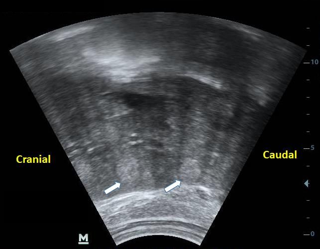
Rev. Agr. Acad., v.3, n.5, Set/Out (2020)
The microbiological examination of the nodular structures, through microscopy, confirmed
the presence of Gram-negative bacteria, where the isolated species was Pseudomonas aeruginosa.
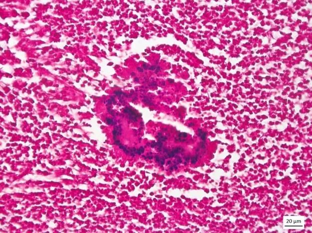
In the histopathology, a lesion was observed with the characteristic of a pyogranulomatous
process, associated with the presence of neutrophils and multinucleated giant cells surrounding
bacteria, forming the phenomenon known as Splendore-Hoeppli (Figures 4A and 4B).
A B
Figure 4 - Bovine botryomycosis. A- A marked inflammatory infiltrate of intact and
degenerate neutrophils (asterisk) can be observed, associated with myriads of bacteria
bordered by Splendore-Hoeppli material (black arrow). HE. Obj.20x. B- Gram staining
demonstrating a myriad of Gram-positive coccoid bacteria amid the Splendore-Hoeppli
material. Obj. 40x.
Discussion
Several species of bacteria are included as causes of botryomycosis, among them
Staphylococcus aureus, which corresponds to the most commonly isolated agent, however in the
present case Pseudomonas aeruginosa was isolated as the agent responsible for the lesion. This
finding corroborates with reports that described the involvement of this pathogen affecting other
organs in cattle (DONAVAN; GROSS, 1984, THOMPSON et al. 2001, MILLER et al. 2001,
SARTELET et al. 2015). Some fewer common pathogens such as Escherichia coli, Proteus
vulgaris, Streptococcus spp., Biberstenia trealosi may be involved (WINSLOW et al, 1959,
SPAGNOLI et al, 2011).
The etiopathogenesis of this disease is not well understood (SCOTT, 2007), but it is known
that factors such as immunodepression of the host, low virulence of the pathogen, and a large
number of inoculums are important to establish the infection (BONIFAZ & CARRASCO, 1996).
Botryomycosis is defined as a chronic suppurative piogranulomatous inflammation caused by
bacteria, which can occur in both cutaneous and visceral forms (WINSLOW, 1959). The latter form
can be divided into primary disease, where the infection starts in the affected organ, or secondary,
when the infection originates from an injury in one organ and spreads to others (PADILLA-
DESGARENNES et al, 2012).
Predisposing factors that include trauma, presence of foreign bodies and postoperative
complications may be involved (HACKER, 1983; CONSTABLE et al, 2017). In this study, the
10
Rev. Agr. Acad., v.3, n.5, Set/Out (2020)

condition of visceral botriomycosis diagnosed in the cow, probably originated from reticulitis
traumatic peritonitis caused by the foreign body
        This could justify the colonization by opportunistic bacteria of the species Pseudomonas
aeruginosa and subsequent systemic spread, mainly affecting organs of the gastrointestinal tract,
resulting in a framework of secondary visceral botryomycosis in the animal.
        In the clinical examination, in addition to changes in the white blood cell, the animal in this
report presented alterations indicative of a severe process, such as apathy, reduced milk production,
impaired gastrointestinal tract function, and including slightly increased abdominal tension,
diarrhea, and the presence of nodular structures on the surface of the rumen, kidneys, peritoneum
and mesentery, which were evidenced by rectal palpation. These structures can be observed in some
bovine diseases, among them, intestinal tuberculosis, enzootic bovine leukosis (lymphosarcoma),
and less frequent actinobacillosis and actinomycosis (SILVA FILHO et al., 2011; CONSTABLE et
al., 2017).
        In the macroscopy, the nodular structures found, more frequently in the greater omentum, of
hardened, multifocal consistency and of varying sizes, draining yellowish contents with a caseous
consistency, resembled lesions commonly found in cases of intestinal tuberculosis (RIET-CORREA
& GARCIA, 2007; PAES & FRANCO, 2016). However, to a lesser extent, similar lesions have
been found in cases of subcutaneous, pulmonary botryomycosis, in the nasopharynx and uterus of
cattle (MILLER, et al., 2001; THOMPSON et al, 2001; SPAGNOLI et al, 2011; SARTELET et al,
2015). Botryomycosis had not been raised as a possible diagnosis for the disease of this animal,
since this disease presents nonspecific clinical signs and there are few descriptions in the literature,
there being no reported cases of visceral botryomycosis visceral with involvement of the omentum,
mesentery lymphonodes and liver of cow.
        Histopathological examination showed a lesion characteristic of the disease, termed the
Splendore-Hoeppli phenomenon, which corresponds to a pyogranulomatous reaction characterized
by an eosinophilic matrix formed by an antigen-antibody complex, cellular debris, and fibrin
disposed between bacterial granules. This phenomenon has frequently been described in cases of
visceral and cutaneous botryomycosis (MILLER et al. 2001, THOMPSON et al. 2001, HUSSEIN,
2008).
        The diagnosis of botryomycosis is performed through the association of cytological and
histopathological findings (SCOTT, 2007; HUSSEIN, 2008), and isolation of the agent, the latter
being considered the gold standard for diagnosis of the disease (PADILLA- DESGARENNES et al,
2012). In the present work, anatomopathological and histopathological findings associated with the
isolation of the etiological agent were fundamental to confirm the diagnosis of this disease.

Conclusion

        Botryomycosis is a chronic granulomatous disease, which can be confused with other
diseases, especially due to the scarcity of studies on the disease in production animals in veterinary
medicine. The present report has great relevance in the literature because it is the first record of the
disease affecting the gastrointestinal tract of bovines, which demonstrates the need to include this
disease as a differential diagnosis of granulomatous lesions in this specie, in addition to alerting the
scientific community to the underreporting of this disease due to lack of diagnosis.

                                                                                                           11
Rev. Agr. Acad., v.3, n.5, Set/Out (2020)
References
BERSOFF-MATCHA, S.J.; ROPER, C.C.; LIAPIS, H.; LITTLE, J.R. Primary pulmonary botryomycosis:
cases report and review. Clinical Infectious Diseases, v. 26, p. 620-624, 1998.
BOLLINGER, O. Mycosen der Lunge beim Pferde. Virchows Archiv Pathological Anatomy, v. 49, p. 583-
586, 1870.
BONIFAZ, A.M.B.; CARRASCO, E.M.D. Botryomycosis. International Journal of Dermatology, v. 35,
n. 6, p. 381-386, 1996.
BOSTROM, R.E.; HUCKINS, J.G.; KROE, D.J.; LAWSON, N.S.; MARTIN, J.E.; FERREL, J.F.;
WHITNEY, R.A. Atypical fatal pulmonary botryomycosis in two guinea pigs dues to Pseudomonas
aeruginosa. Journal of the American Veterinary Medical Association, v. 155, p. 1195-1199, 1969.
CARTER, G.R.; CHENGAPPA, M.M.; CLAUS, W.; RIKIHISA, Y. Essentials of Veterinary Bacteriology
and Mycology. 4th ed. Philadelphia, Pa, USA, 1991.
CASAMIÁN-SORROSAL, D.; FOURNIER, D.; SHIPPAM, J.; WOODWARD, B.; TENNANT, K. Septic
pericardial effusion associated with pulmonary and pericardial botryomycosis in a dog. Journal Small
Animal Practice, v. 12, v. 49, p. 655-659, 2008.
CONSTABLE, P.D. Diseases of the Skin, Eye, Conjunctiva, and External Ear. In: CONSTABLE, P. D. et
al. Veterinary Medicine: a textbook of the diseases of cattle, horses, sheep, pigs, and goats. 11ª ed.
Missouri: Elsevier, cap. 16. p. 1540-1661, 2017.
CUDMORE, L.A. Pyogranulomatous lesion causing neurological signs localized to the sacral region in a
horse. Australian Veterinary Journal, v. 90, p. 392-394, 2012.
DIRKSEN, G. ROSENBERGER - Exame Clínico dos Bovinos. 3ª ed. Rio de Janeiro: Guanabara Koogan,
1993.
DONOVAN, G.A.; GROSS, T.L. Cutaneous botryomycosis (bacterial granulomas) in dairy cows caused by
Pseudomonas aeruginosa. Journal of the American Veterinary Medical Association, v. 184, p. 197-199,
1984.
ELLENBERGER, C.; SCHOON, D.; SCHOON, H.A. Exceptional diagnostic findings in uterine biopsies of
the mare. Pferdeheilkunde, v. 2, v. 22, p. 171-176, 2006.
GROSSET, C.; LAGRANGE, I.; MOREAU, S.; HEDLEY, J.; HAWKINS, M.; REYES, G.E. Cutaneous
botryomycosis in a Campbell’s Russian dwarf hamster (Phodopus campbelli). Journal Exotic Pet
Medicine, v. 23, n. 4, p. 389-396, 2014.
HACKER, P. Botryomycosis. International Journal of Dermatology. v. 22, p. 455-458, 1983.
HUSSEIN, M.R. Mucocutaneous Splendore-Hoeppli phenomenon. Journal Cutaneous Pathology, v. 35, p.
979-988, 2008.
JAIN, N.C. Essentials of Veterinary Hematology. Philadelphia: Lea & Febiger, 1993.
JOHNS, I.C.; FINDING, E.J.T.; CIASCA, T.; ERLES, K; SMITH, K.; WELLER, R. Intracranial
botryomycosis in a mature horse. Equine Veterinary Education, v. 26, p. 294-298, 2014.
MILLER, M.A. Pulmonary Botryomycosis in a Scottish Highland Steer. Journal of Veterinary Diagnostic
Investigation, v. 13, n. 1, p.74-76, 2001.
MOREIRA, I.L. Pulmonary Botryomycosis Secondary to Septic Funiculitis in a Pony. Journal of Equine
Veterinary Science, v. 61, p. 32-35, 2018.
PADILLA-DESGARENNES, C. Botryomycosis. Clinics in Dermatology, v. 30, p. 397-402, 2012.
12Rev. Agr. Acad., v.3, n.5, Set/Out (2020)
PAES, A.C.; FRANCO, M.M.J. Tuberculose em animais de produção. In: MEGID, J.; RIBEIRO, M. G.;
PAES, A. C. Doenças infecciosas em animais de produção e de companhia. 1ª ed. Rio de Janeiro: Roca,
cap. 48, p. 512-541, 2016.
PANDEY, G.S. Cutaneous staphylococcal granuloma in a free living zebra (Equus burchelli) in Zambia.
Journal of Veterinary Medical Science, v. 60, p. 137-138, 1998.
RIET-CORREA, F.; GARCIA, M. Tuberculosis. In: RIET-CORREA, F. et al. Ruminant and Equid
Diseases. 3ª ed. São Paulo: Varela, cap. 3, p. 432-442, 2007.
RIVOLTA, S. Del micelio e delle varieta e specie di discomiceti patogeni. Giornale di Anatatomia
Fisiologia e Patologia degli Animali, v. 16, p. 181-198, 1884.
SARTELET, A. Epidemy of bovine cutaneous and uterine botryomycosis after cesarean sections. XVth
MEBC & 10th ECBHM Symposium, Faculty of Veterinary Medicine & FARAH, University of Liège,
Belgium, 2015.
SCOTT, D.W. Bacterial pseudomycosis (botryomycosis) in the horse. Equine Practice, v. 10, p. 15-19,
1988.
SCOTT, D.W. Cutaneous bacterial pseudomycetoma (botryomycosis) in dogs: two new case reports and a
review of the literature. The Japanese Journal Veterinary Dermatology, v. 13, p. 135-140, 2007.
SILVA-FILHO, A.P. Linfossarcoma uterino em vaca com leucose. Ciência Veterinária nos Trópicos, v.
14, n. 1/2/3, p. 43- 48, 2011.
SMIET, E.; GRINWIS, G.C.M.; VAN DEN TOP, J.G.B.; OLDRUITENBORGH-OOSTERBAAN, M.M.S.
Equine mammary gland disease with a focus on botryomycosis: a review and case study. Equine Veterinary
Education, v. 24, p. 357-366, 2012.
SPAGNOLI, S; REILLY, T.J.; CALCUTT, M.J.; FALES, W.H.; KIM, D.Y. Subcutaneous Botryomycosis
Due to Bibersteinia trehalosi in a Texas Longhorn Steer. Veterinary Pathology, v. 49, n. 5, p.775-778,
2011.
THOMPSON, P.N.; LUGT, J.J.V.D.; OLIVIER-CARSTENS, A. Botryomycosis associated with
Pseudomonas aeruginosa in the nasopharynx of a cow. Veterinary Record, v. 149, n. 16, p. 495-496, 2001.
WALTON, D.K.; SCOTT, D.W.; MANNING, T.O. Cutaneous bacterial granuloma (botryomycosis) in a dog
and a cat. Journal of American Animal Hospital Association, v. 19, p. 537-541, 1982.
WINSLOW, D.J. Botryomycosis. American Journal of Pathology, v. 35, p. 153-176, 1959.
Recebido em 27 de junho de 2020
Retornado para ajustes em 3 de agosto de 2020
Recebido com ajustes em 19 de agosto de 2020
Aceito em 24 de agosto de 2020
13You can also read