Cell penetrating heme oxygenase 1 in the therapy of atopic dermatitis in mice
←
→
Page content transcription
If your browser does not render page correctly, please read the page content below
EXPERIMENTAL AND THERAPEUTIC MEDICINE 22: 941, 2021

                    Cell‑penetrating heme oxygenase‑1 in the therapy
                               of atopic dermatitis in mice
                                 FANG TANG*, XUEQING MA*, JIAYU SUN, MINGHUI RU,
                                TIANSHENG QIAN, WENGJING JI, SIFAN QIAN and HUA LI

                          School of Medicine, Huzhou University, Huzhou, Zhejiang 313000, P.R. China

                                      Received November 9, 2020; Accepted March 15, 2021

                                                   DOI: 10.3892/etm.2021.10373

Abstract. Atopic dermatitis (AD), also referred to as atopic         of all ages, occurring in 15‑20% children and 1‑3% adults (2).
eczema, is a long‑term inflammatory condition that is charac‑        AD is clinically characterized by skin dryness and itchy
terized by itchy, red, swollen and cracked skin. Accumulating        papules (occasionally vesicles in infants) that become excori‑
evidence suggests that AD is caused by genetic factors,              ated and lichenified, usually with crusting (3). Accumulating
environmental exposure and immune system dysfunction;                evidence suggests that AD is elicited by skin barrier dysfunc‑
however, its underlying molecular mechanism remains unclear.         tion, which is followed by immune system activation, which in
Current treatment strategies aim to decrease the severity and        turn negatively regulates skin barrier homeostasis, referred to
frequency of flares. Heme oxygenase‑1 (HO‑1) is a nuclear factor     as an ‘outside‑inside‑outside’ model of AD pathogenesis (4).
erythroid 2‑related factor 2 (Nrf2)‑regulated gene that plays        Thus, current therapies focus on maintaining skin barrier func‑
crucial roles against stress, inflammation and oxidation, and        tion and ameliorating inflammation. For example, skin care
exerts cytoprotective effects. Previous studies have reported that   and moisturizing products are recommended as first‑line treat‑
treatment of AD induces high expression levels of HO‑1 and Nrf2,     ment for mild AD (5). Treatments for moderate‑to‑severe AD
indicating that HO‑1 may play an important role in the treatment     include dupilumab, cyclosporine, phototherapy and systemic
of AD. The present study constructed the recombinant protein,        glucocorticoids, albeit with limited success due to consider‑
cell‑penetrating peptide‑HO‑1 (CPP‑HO‑1), which was expressed        able side effects and inability to affect the recurrence rate (6).
in Escherichia coli and isolated with a 6xHis‑tag using HiTrap       Thus, it is crucial to identify novel promising therapies, with
His column (1 ml). AD was established using 4‑dinitrochloroben‑      fewer side effects, for the effective treatment of AD.
zene (DNCB) in mice. It was observed that the CPP‑HO‑1 fusion            Heme oxygenase‑1 (HO‑1) catalyzes the first and rate‑limiting
protein decreased the severity of AD, inhibited scratching in mice   step in the oxidative degradation of free heme, and may affect
and decreased skin inflammation. Taken together, the results of      several biological processes of aneurysmal diseases, articular
the present study suggested that the CPP‑HO‑1 fusion protein         diseases and hepatic gluconeogenesis, among others, via its enzy‑
may play a protective role against DNCB‑induced AD in mice.          matic by‑products (7). Previous studies on AD mouse models
                                                                     have demonstrated that certain agents, including Soshiho‑tang,
Introduction                                                         sulforaphane, dihydroaustrasulfone alcohol (WA‑25) and
                                                                     Platycodon grandiflorus root‑derived saponins, can alleviate
Atopic dermatitis (AD) is a chronic inflammatory skin condi‑         AD‑like skin lesions and skin inflammation by increasing the
tion with an increasing prevalence (1). AD affects individuals       expression of HO‑1 and nuclear factor erythroid 2‑related factor 2
                                                                     (Nrf2) (8‑11). Another study also concluded that enhancement
                                                                     of HO‑1 expression attenuated the development of skin lesions
                                                                     in mice (12). Based on these findings, HO‑1 appears to hold
Correspondence to: Professor Hua Li, School of Medicine,             promise for the treatment of AD; however, whether the topical
Huzhou University, 1 Xueshi Road, Huzhou, Zhejiang 313000,           use of HO‑1 can alleviate AD remains unclear.
P.R. China                                                               Cell‑penetrating peptides (CPPs) are short peptides
E‑mail: lihua@zjhu.edu.cn                                            (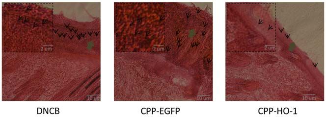
2                                  tang et al: CELL‑PENETRATING HO‑1 IN ATOPIC DERMATITIS

Materials and methods                                              Table I. Evaluation criteria of atopic dermatitis (8).

Chemicals. The H&E staining kit, SDS‑PAGE 12% gel                  Items                       Response intensity                     Score
preparation kit and acetone were obtained from Sangon
Biotech Co., Ltd. 2,4‑Dinitrochlorobenzene (DNCB) was              Erythema                         None                                0
purchased from Sigma‑Aldrich (Shanghai) Trading Co., Ltd.                                           Mild                                1
Acetone and olive oil were mixed at a ratio of 4:1. DNCB was                                        Moderate                            2
dissolved in the acetone/olive oil mixture at a concentration of                                    Severe                              3
1%. PBS, xylene and PBST (10% Tween‑20) were prepared by           Erosion                          None                                0
the authors' laboratory.                                                                            Mild                                1
                                                                                                    Moderate                            2
Animals. A total of 18 ICR mice (male, aged 4‑5 weeks; weight,
                                                                                                    Severe                              3
21±2 g) were purchased from Shanghai Slacker Laboratory
Animal Co., Ltd. [(approval no. SCXK(hu)2017‑005; Shanghai,        Scarring                         None                                0
China]. All animals were housed under specific pathogen‑free                                        Mild                                1
conditions at a controlled temperature of 20‑25˚C and 50‑60%                                        Moderate                            2
humidity with a 12‑h light/dark cycle. The animals were                                             Severe                              3
provided access to sterile food and water ad libitum.              Edema                            None                                0
    The present study was approved by the Ethics Committee                                          Mild                                1
of Huzhou University (Huzhou, China) and all animal care and                                        Moderate                            2
experiments were performed in strict accordance with the Guide
                                                                                                    Severe                              3
for the Care and Use of Laboratory Animals (published by the
National Institutes of Health and revised in 1996; no. 85‑23).

Induction of AD‑like lesions and drug treatment. After
2 weeks of acclimation, the mice were divided into three
groups (n= 6/group) as follows: AD, CPP‑enhanced green fluo‑
rescent protein (EGFP) (DNCB with 0.5 µg/µl CPP‑EGFP)
and CPP‑HO‑1 (DNCB with 0.5 µg/µl CPP‑HO‑1) groups.
    The method for inducing AD was modified from a previous
study (10). The dorsal skin hair was clipped and an area
~1x2 cm 2 was depilated with a hair removal cream. A total         Figure 1. Protocol for induction of AD in specific‑pathogen‑free mice.
of 50 µl 1% DNCB was added to the dorsal skin three times          DNCB, 4‑dinitrochlorobenzene; AD, atopic dermatitis; CPP, cell‑penetrating
per week for a total of 2 weeks. Treatment with 50 µl PBS,         peptide; HO‑1, heme oxygenase‑1; EGFP, enhanced green fluorescent protein.
50 µl CPP‑EGFP (0.5 µg/µl) or 50 µl CPP‑HO‑1 (0.5 µg/µl)
was applied 3 times for the first week 1 h following DNCB and
7 times for the second week. In order to increase cell pene‑       2.5 ml eluent was added to the pre‑balanced G25 desalination
trating efficiency, CPP‑HO‑1, CPP‑EGFP or PBS solution was         column (GE Healthcare) for desalination and was finally stored
kept on the skin for at least 3 h. The animals were sacrificed     in 10% glycerol (Sangon Biotech Co., Ltd.) at ‑80˚C.
on day 15, and dorsal dermal tissues were collected for further
analysis. The experimental schedule is summarized in Fig. 1.       Evaluation of AD severity. AD was observed in all mice and
                                                                   the score was recorded on the last day of the experiment,
Preparation of CPP‑HO‑1. The preparation method of the             according to the criteria described in Table I. The severity of
CPP‑HO‑1 was as follows: The CPP‑HO‑1 and CPP‑EGFP                 AD was determined based on four symptoms: i) Erythema;
genes were synthesized by Sangon Biotech Co., Ltd. and             ii) erosion; iii) scar; and iv) edema. The score of each clinical
inserted into the pET28b vector by the NdeI and EcoRI restric‑     symptom ranged from 0 to 3 (none, 0; mild, 1; moderate, 2; and
tion enzymes and T4 DNA ligase (Sangon Biotech Co., Ltd.),         severe, 3). The total AD score (maximum score, 12) was the
and subsequently transferred into the Novagen's Rosetta™ 2         sum of individual scores.
(pLysS) host strains from Hangzhou Biogroup Technology Co.,
Ltd. CPP‑HO‑1 was induced at 0.7 mm IPTG for 18 h at 37˚C,         Measurement of scratching behavior. All mice were accli‑
and was subsequently centrifuged at 13.8 x g for 15 min at 4˚C.    mated in acrylic cages for 15 min. Following acclimation, the
The resultant pellet was stored at ‑80˚C for at least 24 h. For    mice were videotaped for 30 min and the number of scratches
protein extraction, the E. coli were released with PBS buffer      was counted by the same observer.
(pH 7.4) containing 20 mM imidazole and sonicated with an
ultrasonic homogenizer (Ningbo Xinzhi Biological Technology        Histological analysis. The mice were anesthetized with an i.p.
Co., Ltd.) at 60% amplitude for 5 min on ice, for 15 cycles of     injection of a mixed solution containing xylazine (20 mg/kg;
5 sec on and 15 sec off. The supernatant was subsequently          Bayer) and ketamine (150 mg/kg; Bayer). All mice were decapi‑
centrifuged at 14.95 x g for 20 min at 4˚C and bound to a HiTrap   tated and the dorsal dermal skin tissues were collected and fixed
His column (1 ml, GE Healthcare) at a rate of 0.5 ml/min.          in optimal cutting temperature (OCT) compound (Sakura Finetek
Elution was performed using 500 mM imidazole. Subsequently,        USA, Inc.) for 24 h at ‑80˚C. Tissue samples were cut into 50‑µm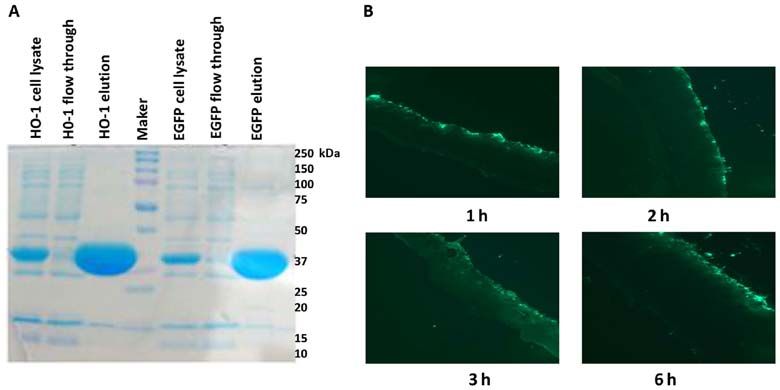
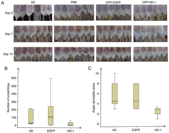
EXPERIMENTAL AND THERAPEUTIC MEDICINE 22: 941, 2021                                                          3

Figure 2. CPP‑HO‑1 purification. (A) Purified proteins were analyzed via 12% SDS‑PAGE (stained with Coomassie blue). (B) Fluorescence microscopy
examination at 1, 2, 3 and 6 h after application of EGFP. Scale bar, 100 µm. CPP, cell‑penetrating peptide; HO‑1, heme oxygenase‑1; EGFP enhanced green
fluorescent protein.

Figure 3. AD severity and scratching behavior. (A) Images of the AD, EGFP and HO‑1 groups were capturedon days 0, 7 and 14. (B) Scratching behavior
was assessed after 30 min. (C) AD scores of the AD, EGFP and HO‑1 groups. *P
4                                     tang et al: CELL‑PENETRATING HO‑1 IN ATOPIC DERMATITIS

Figure 4. Hematoxylin and eosin staining of skin lesion tissue from the AD, EGFP and HO‑1 groups (magnification, x1,000; scale bar, 10 µm). DNCB,
4‑dinitrochlorobenzene; AD, atopic dermatitis; HO‑1, heme oxygenase‑1; CPP, cell‑penetrating peptide; EGFP, enhanced green fluorescent protein.

Dermatitis severity and scratching behavior. The AD model                 keratinocytes, by suppressing NF‑κ B activation and inducing
was induced by DNCB, and 50 µl CPP‑EGFP (0.5 µg/µl) or                    HO‑1 expression in keratinocytes. Hung et al (10) demon‑
50 µl CPP‑HO‑1 (0.5 µg/µl) was added to study the therapeutic             strated that WA‑25 can protect against AD by increasing the
effect (Fig. 1). The AD group exhibited severe dermatitis                 expression levels of HO‑1 and Nrf2. Gene therapy of AD via
with erythema, scarring, edema and erosion on day 14                      targeting the HO‑1 gene has been reported in several studies;
(Fig. 3). The skin condition was significantly improved in                however, AD therapy using the HO‑1 protein is limited by its
CPP‑HO‑1‑treated mice compared with those in the AD group                 inability to enter cells. In previous studies, CPPs have been
(Fig. 3A). All mice were acclimated in acrylic cages for 15 min           conjugated with HO‑1 to form the CPP‑HO‑1 fusion protein,
and subsequently videotaped for 30 min on day 15, and the                 which can transfer the HO‑1 protein into cells and decrease
number of scratches was counted by the same observer. The                 the extent of I/R injury (14,15); however, to the best of our
results demonstrated that the number of scratching events was             knowledge, this method has not been reported in AD to date.
lower in the CPP‑HO‑1 group compared with the AD group                        In the present study, the CPP was conjugated to HO‑1 or
(Fig. 3B; P0.05). The score                    to form the fusion proteins CPP‑HO‑1 and CPP‑EGFP, and
was recorded on the last day of the experiment, according to the          CPP‑HO‑1 was studied in the therapy of AD in mice. The
criteria described in Table I (8). The results demonstrated that          CPP‑EGFP could effetely enter the skin time‑dependently, as
CPP‑HO‑1 decreased the dermatitis score of DNCB‑induced                   shown in Fig. 2A, similar to CPP‑EGFP penetration reported
skin lesions on day 15 compared with the AD group (Fig. 3C;               in heart tissues (20,21). In the present study, in order to increase
P0.05).                                           solution were kept on the skin for at least 3 h. The AD model
                                                                          was induced using DNCB, the immune phenotype of AD, such
H&E staining. Examination of H&E stained sections of the                  as upregulated iNOS protein expression, was not included in
skin lesions revealed decreased epidermal thickness and inflam‑           the present study, as it was described in a previous study (10).
matory cells in the CPP‑HO‑1 group. However, the epidermal                The DNCB‑treated skin area exhibited itchy, red, swollen and
thickness and inflammatory cell infiltration exhibited no signifi‑        cracked skin, which indicated that the AD model had been
cant differences between the CPP‑EGFP and AD groups (Fig. 4).             successfully established. In order to determine the optimal time
Taken together, these results indicated that topical application of       and concentration of CPP‑HO‑1 treatment, the benefits in mice
CPP‑HO‑1 can improve the histological signs of AD.                        were compared between different concentrations of CPP‑HO‑1
                                                                          (0.1 and 0.5 µg/µl) in a pre‑study (data not shown), and the result
Discussion                                                                revealed that 0.5 µg/µl was the most effective. However, the hair
                                                                          in the depilated area grew rapidly, and the frequent use of hair
It was previously indicated that induction of HO‑1 plays a                remover would also further damage the skin; therefore, in order
protective role in several inflammation‑related diseases (7),             to reduce the use of hair remover, the CPP‑HO‑1, CPP‑EGFP
including AD, and HO‑1 has been reported to exhibit thera‑                or PBS treatment was applied 1 h after DNCB induction.
peutic efficiency in AD (8‑11). Chen and Zhong (16) revealed              Furthermore, the treatment times were also increased in the
that HO‑1 combined with microRNAs may affect certain skin                 second week to increase the therapeutic efficacy, according to
diseases, such as ischemia, hypoxia, rheumatoid arthritis and             a previously published study (10). Compared with the PBS and
AD, by regulating the functions of T cells, dendritic cells and           CPP‑EGFP groups, CPP‑HO‑1 effectively alleviated scratching,
mast cells, and the release of chemokines and cytokines. Another          lowered skin score, and decreased skin swelling and inflam‑
study reported that the HO‑1 inducer, cobaltic protoporphyrin,            matory cell infiltration, as shown in Fig. 3. Similar conclusions
inhibited T‑cell‑dependent skin inflammation by suppressing               were also reported by Kirino et al (12) and Hung et al (10) via
antigen‑presenting cells (17). In 2016, Kim et al (18) reported           enhancing HO‑1 expression. H&E staining of the skin lesions
that 2,3‑dimethoxy‑2'‑hydroxychalcone, a derivative of                    also revealed that CPP‑HO1 decreased epidermal thickness
2'‑hydroxychalcone in the flavonoid family, can alleviate                 and inflammatory cell infiltration compared with the PBS and
skin inflammation by inhibiting TNF‑α‑induced intercellular               CPP‑EGFP groups (Fig. 4). Taken together, these results suggest
adhesion molecule‑1 expression and adhesion of monocytes to               that CPP‑HO‑1 may have a therapeutic effect on AD.
EXPERIMENTAL AND THERAPEUTIC MEDICINE 22: 941, 2021 5
However, there were certain limitations to the present study. 2. Kowalska‑Olędzka E, Czarnecka M and Baran A: Epidemiology
of atopic dermatitis in Europe. J Drug Assess 8: 126‑128, 2019.
A normal control group, which was not set in our research, 3. Silverberg JI: Atopic Dermatitis in Adults. Med Clin North
is required to better demonstrate the effect of HO‑1 on AD. Am 104: 157‑176, 2020.
The present study used 50‑µm OCT sections for HE staining, 4. Elias PM and Steinhoff M: ‘Outside‑to‑inside’ (and now back to
‘outside’) pathogenic mechanisms in atopic dermatitis. J Invest
although 5‑µm sections may exhibit a higher resolution and Dermatol 128: 1067‑1070, 2008.
thus will be used in future studies. To strengthen the evidence 5. Puar N, Chovatiya R and Paller AS: New treatments in atopic
on the benefits of HO‑1 for AD, immunohistochemistry and dermatitis. Ann Allergy Asthma Immunol 126: 21‑31, 2021.
6. Johnson BB, Franco AI, Beck LA and Prezzano JC:
ELISA must also be performed to investigate additional Treatment‑resistant atopic dermatitis: Challenges and solutions.
parameters of AD in mice, such as the serum levels of IL‑4, Clin Cosmet Investig Dermatol 12: 181‑192, 2019.
IL‑13 and inducible nitric oxide synthase. Furthermore, the 7. Pae HO, Lee YC and Chung HT: Heme oxygenase‑1 and carbon
monoxide: Emerging therapeutic targets in inflammation and
detailed mechanism underlying the therapeutic effect of HO‑1 allergy. Recent Pat Inflamm Allergy Drug Discov 2: 159‑165,
on AD must be further elucidated. 2008.
In conclusion, the results of the present study suggested 8. Wu W, Peng G, Yang F, Zhang Y, Mu Z and Han X: Sulforaphane
has a therapeutic effect in an atopic dermatitis murine model and
that CPP‑HO‑1 may have a therapeutic effect on AD, and thus activates the Nrf2/HO‑1 axis. Mol Med Rep 20: 1761‑1771, 2019.
may hold promise as a therapeutic strategy. 9. Choi JH, Jin SW, Han EH, Park BH, Kim HG, Khanal T,
Hwang YP, Do MT, Lee HS, Chung YC, et al: Platycodon gran‑
diflorum root‑derived saponins attenuate atopic dermatitis‑like
Acknowledgements skin lesions via suppression of NF‑κ B and STAT1 and activation
of Nrf2/ARE‑mediated heme oxygenase‑1. Phytomedicine 21:
Not applicable. 1053‑1061, 2014.
10. Hung HC, Feng CW, Lin YY, Chen CH, Tsui KH, Chen WF,
Pan CY, Sheu JH, Sung CS and Wen ZH: Nucleophosmin
Funding modulates the alleviation of atopic dermatitis caused by the
marine‑derived compound dihydroaustrasulfone alcohol. Exp
Mol Med 50: e446, 2018.
The present study was funded by the National Key Research and 11. Lee JH, Jo EH, Lee B, Noh HM, Park S, Lee YM, Kim DK
Development Program of China (grant no. SQ2020YFF0401041) and Park MC: Soshiho‑Tang, a Traditional Herbal Medicine,
and the Zhejiang Provincial Natural Science Foundation of Alleviates Atopic Dermatitis Symptoms via Regulation of
Inflammatory Mediators. Front Pharmacol 10: 742, 2019.
China (grant nos. LGF18C050001 and 2021C03036). 12. Kirino M, Kirino Y, Takeno M, Nagashima Y, Takahashi K,
Kobayashi M, Murakami S, Hirasawa T, Ueda A, Aihara M, et al:
Availability of data and materials Heme oxygenase 1 attenuates the development of atopic
dermatitis‑like lesions in mice: Implications for human disease.
J Allergy Clin Immunol 122: 290‑297, 297.e1‑297.e8, 2008.
The datasets used and/or analyzed during the current study are 13. Morris MC, Deshayes S, Heitz F and Divita G: Cell‑penetrating
available from the corresponding author on reasonable request. peptides: From molecular mechanisms to therapeutics. Biol
Cell 100: 201‑217, 2008.
14. He XH, Tang JJ, Wang YL, Zhang ZZ and Yan XT: Transduced
Authors' contributions heme oxygenase‑1 fusion protein reduces renal ischemia/reper‑
fusion injury through its antioxidant and antiapoptotic roles in
rats. Transplant Proc 47: 1627‑1632, 2015.
HL designed the study and edited manuscript; FT drafted the 15. He XH, Li QW, Wang YL, Zhang ZZ, Ke JJ, Yan XT and Chen K:
initial manuscript. WJ, TQ and SQ isolated the proteins; FT, Transduced PEP‑1‑heme oxygenase‑1 fusion protein reduces
JS, XM and MR performed the animal experiments. WJ and remote organ injury induced by intestinal ischemia/reperfusion.
Med Sci Monit 21: 1057‑1065, 2015.
TQ confirm the authenticity of all the raw data. All authors 16. Chen L and Zhong JL: MicroRNA and heme oxygenase‑1 in
have read and approved the final manuscript. allergic disease. Int Immunopharmacol 80: 106132, 2020.
17. Listopad J, Asadullah K, Sievers C, Ritter T, Meisel C, Sabat R
and Döcke WD: Heme oxygenase‑1 inhibits T cell‑dependent
Ethics approval and consent to participate skin inf lammation and differentiation and function of
antigen‑presenting cells. Exp Dermatol 16: 661‑670, 2007.
The present study was approved by the Ethics Committee of 18. Kim H, Youn GS, An SY, Kwon HY, Choi SY and Park J:
2,3‑Dimethoxy‑2'‑hydroxychalcone ameliorates TNF‑α‑induced
Huzhou University (Huzhou, China) and all animal care and ICAM‑1 expression and subsequent monocyte adhesiveness via
experiments were performed in strict accordance with the Guide NF‑kappaB inhibition and HO‑1 induction in HaCaT cells. BMB
for the Care and Use of Laboratory Animals (published by the Rep 49: 57‑62, 2016.
19. Brunet AA, Fuller-Carter PI, Miller AL, Voigt V, Vasiliou S,
National Institutes of Health and revised in 1996; no. 85‑23). Rashwan R, Hunt DM, Carvalho LS: Validating fluorescent
Chrnb4.EGFP mouse models for the study of cone photoreceptor
Patient consent for publication degeneration. Transl Vis Sci Technol 9: 28, 2020.
20. Ma J, Lau CK, Obed A, Dada A, Doenecke A, Fan ST, Schlitt HJ
and Tsui TY: A cell penetrating heme oxygenase protein protects
Not applicable. heart graft against ischemia/reperfusion injury. Gene Ther 16:
320‑328, 2009.
21. Li H, Zheng X, Koren V, Vashist YK and Tsui TY: Highly efficient
Competing interests delivery of siRNA to a heart transplant model by a novel cell
penetrating peptide‑dsRNA binding domain. Int J Pharm 469:
The authors declare that they have no competing interests. 206‑213, 2014.
This work is licensed under a Creative Commons
References Attribution-NonCommercial-NoDerivatives 4.0
International (CC BY-NC-ND 4.0) License.
1. Kapur S, Watson W and Carr S: Atopic dermatitis. Allergy
Asthma Clin Immunol 14 (Suppl 2): 52, 2018.You can also read