Cerebral cysticercosis in a wild Bengal tiger (Panthera tigris tigris) in Bhutan: a first report in non-domestic felids - UZH
←
→
Page content transcription
If your browser does not render page correctly, please read the page content below
Zurich Open Repository and
                                                                            Archive
                                                                            University of Zurich
                                                                            Main Library
                                                                            Strickhofstrasse 39
                                                                            CH-8057 Zurich
                                                                            www.zora.uzh.ch

 Year: 2021

    Cerebral cysticercosis in a wild Bengal tiger (Panthera tigris tigris) in
                Bhutan: a first report in non-domestic felids

Phuentshok, Yoenten ; Choden, Kinley ; Alvarez Rojas, Cristian A ; Deplazes, Peter ; Wangdi, Sonam ;
 Gyeltshen, Kuenzang ; Rinzin, Karma ; Thapa, Nirmal Kumar ; Tenzinla, Tenzinla ; Dorjee, Dechen ;
  Valitutto, Marc ; Gilbert, Martin ; Siriaroonrat, Boripat ; Jairak, Waleemas ; Piewbang, Chutchai ;
                  Sharma, Puspa Maya ; Dema, Tshewang ; Gurung, Ratna Bahadur

Abstract: The endangered Bengal tiger (Panthera tigris tigris) is a keystone species playing an essential
role in ecology as well as in the social and spiritual lives of the Himalayan people. The latest estimate of the
Bengal tiger population in Bhutan accounts for 103 individuals. Infectious organisms, including zoonotic
parasites causing high burden in human health, have received little attention as a cause of mortality
in tigers. Taeniosis/cysticercosis, caused by the cestode Taenia solium, is considered one of the major
neglected tropical diseases in Southeast Asia. We present here a case of neurocysticercosis in a Bengal
tiger showing advanced neurological disease outside Thimphu, the capital city of Bhutan. After palliative
care, the animal died, and necropsy revealed multiple small cysts in the brain. Here we show the presence
of two genetic variants of T. solium in the parasite material collected based on PCR and sequencing of the
complete cox1 and cytB genes. The sequences form a discrete branch within the Asia plus Madagascar
cluster of the parasite. On other hand, tests for feline morbillivirus, feline calicivirus, canine distemper
virus, Nipah, rabies, Japanese encephalitis, feline leukaemia and feline immunodeficiency virus were
negative. In contrast, PCR for feline herpesvirus was positive and a latex agglutination test revealed an
elevated antibody titer against Toxoplasma gondii (titer 1:256). The molecular examination of taeniid
eggs isolated from the tiger faeces produced sequences for which the highest homology in GenBank is
between 92% and 94% with T. regis and T. hydatigena. This fatal case of T. solium neurocysticercosis, a
disease previously unrecorded in tigers or other non-domestic felids, demonstrates an anthropogenically
driven transmission of a deadly pathogen which could become a serious threat to the tiger population.

DOI: https://doi.org/10.1016/j.ijppaw.2021.02.003

Posted at the Zurich Open Repository and Archive, University of Zurich
ZORA URL: https://doi.org/10.5167/uzh-203946
Journal Article
Published Version

The following work is licensed under a Creative Commons: Attribution 4.0 International (CC BY 4.0)
License.

Originally published at: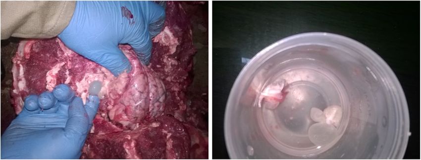
Phuentshok, Yoenten; Choden, Kinley; Alvarez Rojas, Cristian A; Deplazes, Peter; Wangdi, Sonam;
Gyeltshen, Kuenzang; Rinzin, Karma; Thapa, Nirmal Kumar; Tenzinla, Tenzinla; Dorjee, Dechen; Val-
itutto, Marc; Gilbert, Martin; Siriaroonrat, Boripat; Jairak, Waleemas; Piewbang, Chutchai; Sharma,
Puspa Maya; Dema, Tshewang; Gurung, Ratna Bahadur (2021). Cerebral cysticercosis in a wild Bengal
tiger (Panthera tigris tigris) in Bhutan: a first report in non-domestic felids. International Journal for
Parasitology: Parasites and Wildlife, 14:150-156.
DOI: https://doi.org/10.1016/j.ijppaw.2021.02.003

                                                    2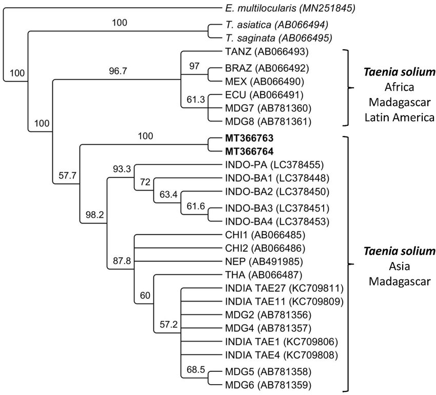
International Journal for Parasitology: Parasites and Wildlife 14 (2021) 150–156

                                                                     Contents lists available at ScienceDirect

                                       International Journal for Parasitology: Parasites and Wildlife
                                                           journal homepage: www.elsevier.com/locate/ijppaw

Cerebral cysticercosis in a wild Bengal tiger (Panthera tigris tigris) in Bhutan:
A first report in non-domestic felids☆
Yoenten Phuentshok a, **, 1, Kinley Choden b, 1, Cristian A. Alvarez Rojas c, Peter Deplazes c, *,
Sonam Wangdi b, Kuenzang Gyeltshen b, Karma Rinzin d, Nirmal Kumar Thapa e,
Tenzinla Tenzinla e, Dechen Dorjee f, Marc Valitutto g, Martin Gilbert h, Boripat Siriaroonrat i,
Waleemas Jairak i, Chutchai Piewbang j, Puspa Maya Sharma e, Tshewang Dema e,
Ratna Bahadur Gurung e
a
  Food and Agriculture Organization of the United Nations, Bangkok, 10200, Thailand
b
  Nature Conservation Division, Department of Forests and Park Services, Ministry of Agriculture and Forests, Taba, Bhutan
c
  Institute of Parasitology, Vetsuisse Faculty, University of Zurich, Zurich, Switzerland
d
  Animal Health Division, Department of Livestock, Ministry of Agriculture and Forests, Thimphu, Bhutan
e
  National Centre for Animal Health, Department of Livestock, Ministry of Agriculture and Forests, Serbithang, Bhutan
f
  World Wildlife Fund, Washington DC, 20037, USA
g
  EcoHeatlh Alliance, New York, NY, 10018, USA
h
  Cornell Wildlife Health Center, College of Veterinary Medicine, Cornell University, Ithaca, NY, 14853, USA
i
  Bureau of Conservation and Research, Zoological Park Organization, Bangkok, 10800, Thailand
j
  Faculty of Veterinary Science, Chulalongkorn University, Bangkok, 10330, Thailand

A R T I C L E I N F O                                       A B S T R A C T

Keywords:                                                   The endangered Bengal tiger (Panthera tigris tigris) is a keystone species playing an essential role in ecology as
Taenia solium                                               well as in the social and spiritual lives of the Himalayan people. The latest estimate of the Bengal tiger population
Bengal tiger                                                in Bhutan accounts for 103 individuals. Infectious organisms, including zoonotic parasites causing high burden in
Panthera tigris tigris
                                                            human health, have received little attention as a cause of mortality in tigers. Taeniosis/cysticercosis, caused by
Bhutan
Conservation medicine
                                                            the cestode Taenia solium, is considered one of the major neglected tropical diseases in Southeast Asia. We
Neurocysticercosis                                          present here a case of neurocysticercosis in a Bengal tiger showing advanced neurological disease outside
One health                                                  Thimphu, the capital city of Bhutan. After palliative care, the animal died, and necropsy revealed multiple small
First report                                                cysts in the brain. Here we show the presence of two genetic variants of T. solium in the parasite material
                                                            collected based on PCR and sequencing of the complete cox1 and cytB genes. The sequences form a discrete
                                                            branch within the Asia plus Madagascar cluster of the parasite. On other hand, tests for feline morbillivirus,
                                                            feline calicivirus, canine distemper virus, Nipah, rabies, Japanese encephalitis, feline leukaemia and feline im-
                                                            munodeficiency virus were negative. In contrast, PCR for feline herpesvirus was positive and a latex aggluti-
                                                            nation test revealed an elevated antibody titer against Toxoplasma gondii (titer 1:256). The molecular
                                                            examination of taeniid eggs isolated from the tiger faeces produced sequences for which the highest homology in
                                                            GenBank is between 92% and 94% with T. regis and T. hydatigena. This fatal case of T. solium neurocysticercosis, a
                                                            disease previously unrecorded in tigers or other non-domestic felids, demonstrates an anthropogenically driven
                                                            transmission of a deadly pathogen which could become a serious threat to the tiger population.

    The views expressed in this publication are those of the author(s) and do not necessarily reflect the views or policies of the Food and Agriculture Organization of
    ☆

the United Nations
  * Corresponding author.
 ** Corresponding author.
    E-mail addresses: vetyoen@gmail.com (Y. Phuentshok), deplazesp@access.uzh.ch (P. Deplazes).
  1
    Joint first authors.

https://doi.org/10.1016/j.ijppaw.2021.02.003
Received 4 January 2021; Received in revised form 3 February 2021; Accepted 3 February 2021
Available online 10 February 2021
2213-2244/© 2021 The Authors. Published by Elsevier Ltd on behalf of Australian Society for Parasitology. This is an open access article under the CC BY license
(http://creativecommons.org/licenses/by/4.0/).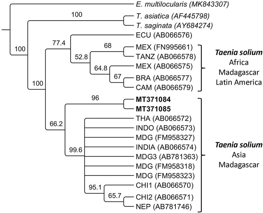
Y. Phuentshok et al. International Journal for Parasitology: Parasites and Wildlife 14 (2021) 150–156
1. Introduction
The International Union for Conservation of Nature (IUCN) has listed
tigers as endangered since 1969 with the global tiger numbers and range
continue to decline. By 2010, fewer than 3500 animals occupied
approximately 7% of their historical global range (Sanderson et al.,
2010; Seidensticker, 2010; Walston et al., 2010). With more than 70% of
its land covered by forest, Bhutan is a hotspot for wild felids (Tempa
et al., 2013). The Bengal tiger (Panthera tigris tigris, Linnaeus 1758) is a
keystone species playing an important role in ecology as well as the
social and spiritual lives of the Bhutanese people. An estimated popu-
lation of 103 adult tigers roam undisturbed in Bhutan (Ministry of
Agriculture and Forests, 2015) with their habitat ranging from the
southern foothills bordering India at 150 m above sea level (masl) to
alpine forests and the high Himalayas adjoining the Tibetan Autono-
mous Region to the north at 4300 masl (Jigme and Tharchen, 2012). The
tiger population in Bhutan is thriving compared to many other regions,
but poaching is still considered the main threat to their conservation. By
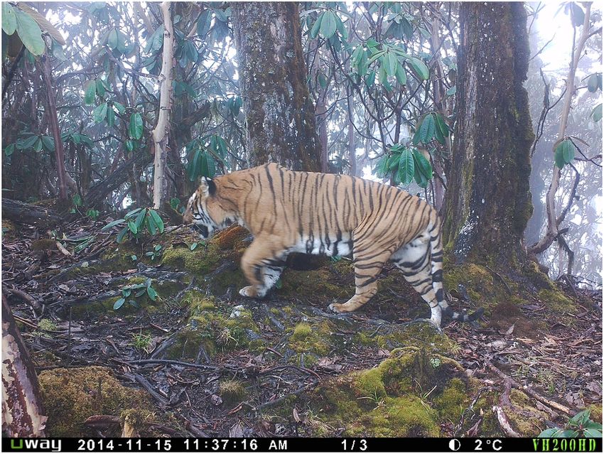
contrast, little attention has been given to the impact that zoonotic Fig. 1. Camera trap image of the tiger infected with Taenia solium reported in
pathogens and disease might represent to tiger conservation in Bhutan. this article captured on the 15th of November 2014. The image was taken
This situation warrants re-evaluation following the case of neuro- during a survey of the Bengal tiger population at national level in Bhutan.
cysticercosis reported in this article. Copyright: Nature Conservation Division, DoFPS, MoAF, Bhutan.
Cysticercosis due to infection with the cestode Taenia solium is a
widely distributed zoonosis with the highest transmission in South Residents of Chamjekha, a residential complex in Kabesa suburb located
America, India, Africa and Southeast Asia but is probably under- about 6 km north of Thimphu city shared photos and videos of the tiger
recognized in many other endemic countries (Flisser et al., 2011; Ito on social media, which subjectively showed the animal being indifferent
et al., 2019). Bhutan is considered part of an endemic area of infection to its surroundings. The Nature Conservation Division (NCD), Depart-
with northern India and Nepal (Ito et al., 2019). In Bhutan, there has ment of Forests and Park Services (DoFPS) of the Ministry of Agriculture
recently been an increasing number of epilepsy cases among human and Forests (MoAF) intervened and captured the tiger from Taba
patients attributed to neurocysticercosis (Brizzi et al., 2016; Bruno et al., (89◦ 38′ 45′′ E; 27◦ 30′ 49′′ N), another suburb adjacent to Kabesa in the
2017; Health, 2016). The life cycle of the tapeworm T. solium takes place early hours of March 22, 2018, using 450 mg of tiletamine and zolaze-
in two hosts, with humans acting as the definitive host and domestic pigs pam (Zoletil®100, Virbac, Pty. Australia); the dose (4 mg/kg body
as the intermediate host. Besides humans, intestinal infection has been weight) was based on an estimated body weight of 115 kg. The tiger
established experimentally in other primates including lar gibbons exhibited clinical signs consistent with a compromised nervous system
(Hylobates lar) and chacma baboons (Papio ursinus), as well as in including uncoordinated gait, stumbling, walking in clockwise circles
immunosuppressed laboratory animals (Flisser et al., 2011). However, with head held down, head pressing, lack of aggression and indifference
the potential for a sylvatic cycle of T. solium involving wild primates and to surrounding objects and people. A small bruised wound on the fore-
intermediate hosts such as wild boars (Sus scrofa) has yet to be docu- head skin was evident of constant pressing of the head on fixed objects.
mented. Humans acquire intestinal infections of T. solium through The tiger was cared for in the NCD Wildlife Rescue and Rehabilitation
ingestion of cysticerci in undercooked infected pork. The cysticerci Centre for fifteen days. Initially, he was treated orally with antibiotics
develop within 9–10 weeks into adult gravid tapeworms which can (Cefotaxime) and vitamin B. The tiger was active and eating (~10 kg
persist for several months as subclinical patent infections (Flisser et al., beef) and drinking (~4 L water) daily but the health conditions kept
2011). During the patency, eggs are excreted in proglottids or free in the deteriorating daily. On the 6th day, the animal became inactive,
stool of the infected humans. Pigs ingest embryonated eggs from developed sunken eyes and hyporexia, consuming only small volumes of
contaminated soil, water, food or through coprophagia. The eggs hatch water but no meat. Despite supportive care, on the 12th day, the tiger’s
in the pig’s intestine and the activated oncospheres penetrate the mu- neurological condition significantly advanced, as exhibited by an
cosa, migrate into the bloodstream and settle in tissues where they form inability to stand (but in sternal recumbency with the head elevated),
the larval stages containing an invaginated scolex. In humans exposed to staring, with no response to visual or auditory stimuli and an inability to
T. solium eggs, the oncospheres invade the mucosa following anti- hold food in its mouth. By the 13th day, the tiger’s condition progressed
peristalsis (patients with intestinal infections), by hand-mouth trans- to lateral recumbency with no response to touching or painful stimuli.
mission after contact with egg contaminated surfaces or by ingestion of The animal died on the 6th of April 2018; necropsy was performed
contaminated food, water or soil. The migrating oncospheres encyst in within 5 hours of death.
various organs leading to clinical signs associated with their location. The necropsy was performed following established protocols, and
Cysticerci developing in the central nervous system lead to neurological samples of different organs were collected and fixed in 10% formalin
symptoms (neurocysticercosis), causing epileptic seizures and increased and/or directly frozen. Oral, rectal and conjunctival swabs and faeces
intracranial pressure, which can be fatal (Garcia and Del Brutto, 2005; were also collected. The cysts recovered on necropsy were triturated and
García et al., 2010). frozen at −20 ◦ C and the material was sent to the Institute of Parasi-
tology, University of Zurich together with taeniid eggs isolated from
2. Materials and methods faeces.
2.1. Clinical presentation and necropsy
2.2. Histopathology
On March 21, 2018, an adult male tiger, subsequently identified
using old remote camera trap images as a known individual of 9 years of Formalin-fixed tissues from liver, lung, spleen, kidney, diaphragm,
age (Fig. 1) emerged from the forest close to the capital city of Thimphu testicle, pancreas, trachea, heart and hind pad were shipped to the
(at 2300 masl and home to approximately 115,000 inhabitants). College of Veterinary Medicine, Cornell University for histological
151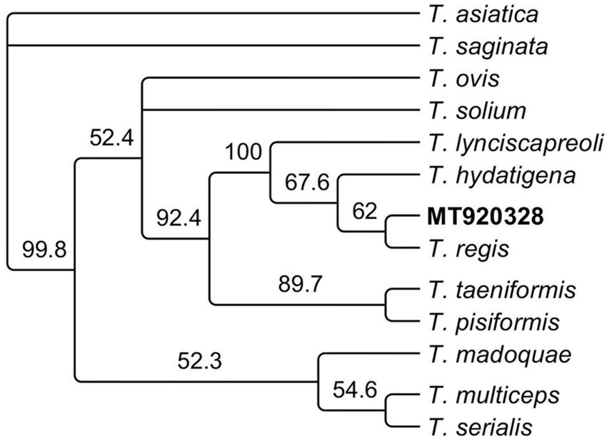
Y. Phuentshok et al.                                                                               International Journal for Parasitology: Parasites and Wildlife 14 (2021) 150–156

examination.                                                                              2.5. Microscopic and molecular analysis of faeces

2.3. Diagnosis of viral and parasitic infections in serum and tissues                         At the National Centre for Animal Health, Bhutan, faecal samples
                                                                                          from the tiger were processed following the floatation and sieving
    Ancillary diagnostics were performed in Thailand at the National                      method as described by Mathis et al. (1996). The sediment of the 21 μm
Institute of Animal Health, Mahidol University, Chulalongkorn Univer-                     filter was examined microscopically and fixed in ethanol 70% and sent
sity, and in the United States at Cornell University to determine the                     to the Institute of Parasitology (Zurich, Switzerland) for subsequent
presence or exposure to a variety of viral and apicomplexan pathogens.                    molecular analysis. The sediment was subjected to alkaline lysis to
Details of the methods used are provided in Table 1.                                      isolate DNA as previously described (Štefanić et al., 2004). The DNA was
                                                                                          used as a template for a multiplex PCR (Trachsel et al., 2007) to get an
2.4. Molecular analysis of the brain lesion                                               initial identification of the taeniid eggs. Additional PCR was performed
                                                                                          amplifying a section of the nadh gene (NADH dehydrogenase subunit 1)
    An aliquot of the sample was washed three times in PBS(physiolog-                     (Bowles and McManus, 1993). PCR products were sequenced and
ical phosphate buffered saline), and total DNA was extracted using the                    compared with the NCBI database. Cladograms were inferred with the
QIAGEN DNeasy Blood & Tissue Kit. DNA was used as a template for a                        neighbour-joining method using the same gene section from the refer-
multiplex PCR as previously described (Trachsel et al., 2007). PCR                        ence mitochondrial genome of other Taenia species.
products were visualised in agarose gel 2%, purified with the MinElute
QIAGEN kit and sequenced at Microsynth (Switzerland) using the PCR                        3. Results
primers. Additional PCRs were performed to amplify the full length of
the cox1 (cytochrome c oxidase I) and cytb (cytochrome b) genes of the                    3.1. Necropsy
parasite for phylogenetic analysis according to Nakao et al. (2002). PCR
products were cloned using the TOPO TA Cloning Kit (Thermo Fisher                             Healthy and intact coat was observed except a lacerated wound in
Scientific), five clones were sequenced per gene using vector primers.                    the middle of the forehead; the injury could have occurred from pressing
Sequences of the cox1 and cytB genes and all similar sequences of the                     its head against the cage. A mark of its own (lower) canine teeth at either
same length from T. solium were collected from GenBank and aligned                        side of the upper lips were observed; the right lower canine was missing
using the software Geneious R10 (https://www.geneious.com). Phylo-                        and the left incisor was broken at the root. The liver was congestive,
genetic trees were inferred for each gene with the neighbour-joining                      haemorrhagic and friable while extensive haemorrhages were noted in
method using Echinococcus multilocularis as an outgroup.                                  the internal mucosa of the small intestine. Both lobes of the lungs were
                                                                                          collapsed and ecchymotic. The mesenteric lymph nodes were slightly
                                                                                          enlarged and haemorrhagic. Both kidneys appeared slightly enlarged,
Table 1
                                                                                          and haemorrhages were noted in the cortex and medulla. The brain
Summary of virology and Toxoplasma results from analyses performed at
Mahidol University, Chulalongkorn University, Cornell University, and the Na-             contained two clear fluid-filled cysts approximately 1–2 cm diameter
tional Institute of Animal Health (NIAH). Tests performed included reverse                buried deep inside the brain tissue. The larger cyst was located on the
transcription-polymerase chain reaction (RT-PCR), polymerase chain reaction               left lobe of the cerebrum while the slightly smaller cyst was lodged in the
(PCR), enzyme-linked immunosorbent assay (ELISA), hemagglutinin inhibition                right brain (Fig. 2).
(HI), latex agglutination test (LAT), virus neutralisation test (VNT) and Western
blot (WB).                                                                                3.2. Molecular analysis of the cyst
  Pathogen                Method   Specimen          Lab              Result
                                   tested                                                     The PCR, according to Trachsel et al. (2007) produced a single band
  Molecular analysis:                                                                     (267bp) which sequence showed 99.5% homology with the corre-
  Canine distemper        RT-      Rectal swab,      Mahidol          Negative            sponding section of the T. solium full mitochondrial genome
    virus                 PCR      whole blood                                            (AB086256). The PCR products of the cox1 (1,620bp) and the cytB (1,
  Canine distemper        RT-      Brain             Cornell          Negative
                                                                                          068bp) were cloned and two different variants were identified per gene
    virus                 PCR
  Nipah virus             RT-      Nasal and oral    Mahidol          Negative            differing only in two nucleotides between them. Sequences were
                          PCR      swab                                                   deposited in GenBank Accession numbers: MT366763- MT366764 for
  Rabies virus            RT-      Oral swab         Mahidol          Negative            cox1 and MT371084-MT371085 for cytB. The cladograms showing the
                          PCR                                                             relationship between the sequences for cox1 and cytB from different
  Japanese                RT-      Blood             Mahidol          Negative
                                                                                          isolates of T. solium deposited in GenBank is shown in Figs. 3 and 4
    encephalitis virus    PCR
  Feline morbillivirus    RT-      Rectal swab       Chulalongkorn    Negative            respectively. The sequences of both genes cluster together with the Asia
                          PCR      and blood                                              + Madagascar group of T. solium previously described by Nakao et al.
  Panparamyxovirus        RT-      Nasal and         Chulalongkorn    Negative            (2002).
                          PCR      rectal swab
  Feline calicivirus      RT-      Nasal and         Chulalongkorn    Negative
                          PCR      conjunctival                                           3.3. Histopathology
                                   swab
  Feline herpesvirus      PCR      Oral and rectal   Chulalongkorn    Positive                The most striking finding was congestion of the liver (severe) and
                                   swab                                                   lung (mild). The liver congestion appeared diffuse (rather than targeted
  Feline leukaemia        ELISA    Serum             Cornell          Negative
                                                                                          to a specific region), with erythrocytes expanding the sinusoids leading
    virus
  Serological analyses:                                                                   to disarray and fragmentation of hepatic chords without signs of cellular
  Japanese                HI       Serum             Mahidol          Negative            necrosis. No significant lesions were identified in the other tissues.
    encephalitis virus
  Toxoplasma gondii       LAT      Serum             NIAH             Positive
                                                                                          3.4. Diagnosis of viral and parasitic infections in serum and tissues
                                                                      (1:256)
  Canine distemper        VNT      Serum             Chulalongkorn    Negative
    virus                                                                                    The results of molecular and serological analyses are summarized in
  Feline                  WB       Serum             Cornell          Negative            Table 1. All tests were negative except for a PCR for feline herpesvirus
    immunodeficiency                                                                      (FHV); and the latex agglutination test for antibodies against Toxoplasma
    virus
                                                                                          gondii (titer 1:256).

                                                                                    152
Y. Phuentshok et al.                                                                           International Journal for Parasitology: Parasites and Wildlife 14 (2021) 150–156

                              Fig. 2. Cysts identified at necropsy of the Bengal tiger in Bhutan (left) and isolated in a jar (right).

                                                                                                                                 Fig. 3. Cladogram showing the rela-
                                                                                                                                 tion between the sequences of the cox1
                                                                                                                                 gene of Taenia solium from the present
                                                                                                                                 study highlighted in bold (MT366763-
                                                                                                                                 MT366764) and unique published se-
                                                                                                                                 quences of the cox1 gene available in
                                                                                                                                 GenBank from Tanzania (TANZ), Brazil
                                                                                                                                 (BRA), Mexico (MEX), Ecuador (ECU),
                                                                                                                                 Madagascar (MDG), Indonesia (INDO-
                                                                                                                                 PA, INDO-BA), China (CHI), Nepal
                                                                                                                                 (NEP), Thailand (THA) and India
                                                                                                                                 (TAE). Sequences for the cox1 gene
                                                                                                                                 from Taenia saginata (AB066495) and
                                                                                                                                 T. asiatica (AB066494) were also
                                                                                                                                 included. The cox1 sequence of Echi-
                                                                                                                                 nococcus multilocularis (MN251845)
                                                                                                                                 was used as an outgroup.

3.5. Molecular analysis of taeniid eggs in faeces                                     sequence with T. regis is evident in the cladogram in Fig. 5 which in-
                                                                                      cludes other Taenia species.
    Faecal examination revealed the presence of taeniid eggs. A band of
267bp was amplified using a multiplex PCR to discriminate between                     4. Discussion
Echinococcus and other cestodes including Taenia. The closest homology
in GenBank for this sequence was around 94% with the corresponding                        Neurological disease in wild tigers has recently gained prominence
section of the complete genome of T. regis (AB905198) and T. hydatigena               following the recognition that Canine Distemper Virus (CDV) represents
(GQ228819). Subsequently, a section of the nadh gene (491bp) was                      a threat to the conservation of Amur tigers (P. tigris altaica) in the
amplified and its sequence (accession number: MT920328) showed a                      Russian Far East (Gilbert et al., 2015; Seimon et al., 2013). The lack of
homology around 92% with the corresponding section of the genome of                   any serological or molecular evidence for active or resolved CDV
T. hydatigena (FJ518620) and T. regis (AB905198). The proximity of this               infection in the tiger suggested an alternative aetiology for this case. The

                                                                                153
Y. Phuentshok et al.                                                                           International Journal for Parasitology: Parasites and Wildlife 14 (2021) 150–156

                                                                                                              Fig. 4. Cladogram showing the relation between the
                                                                                                              sequences of the cytB gene of Taenia solium from the
                                                                                                              present study highlighted in bold (MT371084-
                                                                                                              MT371085) and unique published sequences of the cytB
                                                                                                              gene available in GenBank from Ecuador (ECU),
                                                                                                              Tanzania (TANZ), Mexico (MEX), Brazil (BRA), Camer-
                                                                                                              oun (CAM), Thailand (THA), Indonesia (INDO),
                                                                                                              Madagascar (MDG), INDIA, China (CHI) and Nepal
                                                                                                              (NEP). The sequences for the cytB gene from Taenia
                                                                                                              saginata (AB066581) and Taenia asiatica (AB066580) are
                                                                                                              also included. The cytB sequence from Echinococcus
                                                                                                              multilocularis (MK843307) was used as an outgroup.

                                                                                      Madagascar group of T. solium, which is distinct from the group of iso-
                                                                                      lates from Africa, Latin America and Madagascar. However, they form a
                                                                                      separate branch within the Asian and Madagascar group. So far, this
                                                                                      group comprises samples from China, India, Indonesia, Taiwan and
                                                                                      Thailand and Madagascar. In the case of Madagascar, it is well known
                                                                                      that T. solium of African-American and Asian genotypes are present due
                                                                                      to animal trade from both regions towards the island (Michelet et al.,
                                                                                      2010). In the case of Bhutan, its geopolitical isolation has likely
                                                                                      contributed to the establishment of distinct variants of T. solium which
                                                                                      have not been previously described. Further examination of T. solium
                                                                                      isolates from humans, pigs or wild boar are needed to fully understand
                                                                                      the molecular epidemiology of T. solium in Bhutan and neighbouring
                                                                                      countries.
                                                                                          To our knowledge, this is the first case of neurocysticercosis recorded
                                                                                      in a wild or captive tiger. Cerebral cysticercosis, also known as neuro-
                                                                                      cysticercosis, caused by the larval stage (metacestode) cysticercus cel-
                                                                                      lulosae of T. solium (Cyclophyllida: Taeniidae), has been identified as a
                                                                                      major public health problem in low and middle-income countries
Fig. 5. Cladogram showing the relation between the sequence of the nad1 gene          (Donadeu et al., 2016). Besides its normal intermediate host, the do-
(491bp) amplified from the sediment of the 21 μm filter of the sieving method         mestic pig, the metacestode of T. solium has been reported from a wide
for isolation of taeniid eggs from faeces of the tiger analysed in this study         range of terrestrial (Abuladze and Mifʻal tirgume ha-madaʻ, 1970), as
highlighted in bold (MT920328); together with the sequence of the same gene
                                                                                      well as from a few marine mammals (De Graaf et al., 1980). However,
of different Taenia spp.
                                                                                      although these occasional infections are of clinical importance to the
                                                                                      infected individuals, they are thought to represent “dead-end hosts” and
identification of T. solium in two cerebral cysts and clinical signs                  are epidemiologically unimportant at a population level. Apart from
consistent with intracranial lesion suggested that neurocysticercosis was             human, cases of cerebral T. solium cysticercosis have been reported in
the only supported diagnosis in this case. There have been very few                   domestic dogs (Rogers et al., 1989; Rommel et al., 2000) and to a lesser
published reports of infectious disease in free-ranging tigers (Gilbert               extent in domestic cats (Carillo Melgar and Quiroz Romero, 1974;
et al., 2015) and our finding highlight the importance of considering                 Mazzotti et al., 1965). Interestingly, a case of T. solium neuro-
alternative aetiologies to CDV when wild tigers present with neurolog-                cysticercosis was found in a cat in South Africa (Schwan et al., 2002).
ical signs.                                                                           Clinical symptoms were observed around 6 h after deworming with
    The initial molecular analyses performed here confirmed that                      praziquantel, a drug which can increase the pathogenicity of the infec-
T. solium was responsible for the cerebral cysts present in the tiger.                tion by enhancing local inflammation around the dead parasite (Schwan
Additionally, two variants of each gene (cox1 and cytB) were identified               et al., 2002). Another rare case of a cerebral Taenia crassiceps cysticer-
in this study, since there were two cysticerci in the brain of the tiger              cosis in a domestic cat was described in the USA (Wünschmann et al.,
(which were pooled before molecular analysis). Each of them was likely                2003), a Taenia sp. probably also endemic for Bhutan. Rare cases of
caused by different variants of the parasite. Figs. 3 and 4 show that se-             coenurosis in cats caused by T. serialis have been reported (Hayes and
quences identified in the present study are part of the Asia and

                                                                                154
Y. Phuentshok et al. International Journal for Parasitology: Parasites and Wildlife 14 (2021) 150–156
Creighton, 1978; Huss et al., 1994; Orioles et al., 2014; Slocombe et al., Funding
1989; Smith et al., 1988). In Bhutan, T. multiceps is an important species
causing coenurosis with high mortalities in young yaks (Dorji, 2002). Cornell Wildlife Health Centre and Institute of Parasitology, Vet-
However, there have been no cases of T. multiceps in both domestic and suisse Faculty, University of Zurich for diagnostic investigations per-
wild felids in Bhutan. formed. WWF country office, Bhutan for support with camera in the
Domestic and wild cats are the definitive hosts of the apicomplexan animal enclosure for observation and the Ministry of Agriculture and
parasite Toxoplasma gondii, but clinical disease is rare in most feline Forests (MoAF), Royal Government of Bhutan for sample referral.
species (Elmore et al., 2010). Antibodies to T. gondii were detected in
62% (95% CI: 45.7–76.0%, n = 42) of wild Amur tigers sampled in the
Russian Far East (Goodrich et al., 2012) and so their detection in the Declaration of competing interest
Bhutan tiger was not unexpected. Although clinical toxoplasmosis has
been reported in a captive Amur tiger with profuse diarrhoea and The authors declare that they have no known competing interests
wasting (Dorny and Fransen, 1989), in the case presented, Toxoplasma that could have appeared to influence the work reported in this paper.
seropositivity alone is not indicative for the clinical presentation of the All authors approved the final manuscript and its submission.
animal. FHV causes an upper respiratory and ocular infection in cats
including tigers (Sun et al., 2014). Extended infections due to latency Acknowledgements
increase the likelihood of detection, and the absence of neurological
disease associated with FHV infections suggest the detection of the virus Authors thank Navapon Techakriengkrai, Kannika Phongroop,
was also incidental. Immunosuppression has contributed to other cys- Somporn Techangamsuwan (Chulalongkorn University); Benjaporn
ticercoses in atypical dead-end hosts as described for Versteria sp. or Bhusri, Phirom Prompiram, Kridsada Chaichoun (Mahidol University)
T. crassiceps cysticercoses in primates including humans (Deplazes et al., and Jessica Bodgener for their assistance in sample testing; Minister and
2019). However, in this case, other pathogens associated with immu- Secretary, Ministry of Agriculture and Forests, Bhutan for supporting the
nosuppression including CDV, feline immunodeficiency virus and feline study.
leukaemia were not detected.
Interestingly, the molecular analysis of taeniid eggs isolated from
References
faeces reveal sequences which share between 92 and 94% similarity
with T. regis and T. hydatigena. The electropherograms of these se- Abuladze, K.I., Mifʻal tirgume ha-madaʻ, h.-Y.r., 1970. Taeniata of Animals and Man and
quences showed clear single peaks and good quality which allow us to be Diseases Caused by Them. Israel Program for Scientific Translations, Jerusalem.
confident that these PCR products are not artefacts or the consequence Bowles, J., McManus, D.P., 1993. NADH dehydrogenase 1 gene sequences compared for
species and strains of the genus Echinococcus. Int. J. Parasitol. 23, 969–972.
of a mixed infection with different Taenia species. Phylogenetic analysis Brizzi, K., Pelden, S., Tshokey, T., Nirola, D.K., Diamond, M.B., Klein, J.P., Tshering, L.,
showed that it is likely that the Taenia eggs found in the tiger faeces Deki, S., Nidup, D., Bruno, V., Dorny, P., Garcia, H.H., Mateen, F.J., 2016.
could belong to a non previously described species (Fig. 5). However, Neurocysticercosis in Bhutan: a cross-sectional study in people with epilepsy. Trans.
R. Soc. Trop. Med. Hyg. 110, 517–526.
this could only be confirmed if a morphological and molecular analysis Bruno, V., Klein, J.P., Nidup, D., Nirola, D.K., Tshering, L., Deki, S., Clark, S.J., Linn, K.
of the adult worm could be carried out. Unfortunately, it was not A., Shinohara, R.T., Dorji, C., Pokhrel, D.R., Dema, U., Mateen, F.J., 2017. Yield of
possible to have access to such material. The sequence of the nad1 gene brain MRI in clinically diagnosed epilepsy in the Kingdom of Bhutan: a prospective
study. Ann. Glob. Health 83, 415–422.
from this study is also different from sequences from T. regis and T. Carillo Melgar, H., Quiroz Romero, S., 1974. Un caso de cisticercosis cerebelar de un
hydatigena isolated from carnivores and felids in Kenya (Zhang et al., gato. In: Revista de la Facultad de Medicina Veterinaria y Zootecnia de la
2007). Little is known about Taenia species infecting tigers in Asia. In the Universidad Autónoma de México, vol. 5, pp. 10–11.
De Graaf, A.S., Shaughnessy, P.D., McCully, R.M., Verster, A., 1980. Occurrence of Taenia
case of Africa, T. simbae, a rare species in lions, was characterized in the
solium in a Cape Fur seal (Arctocephalus pusillus). Onderstepoort J. Vet. Res. 47,
past based on morphology and was described to be similar to T. regis 119–120.
(Dinnik and Sachs, 1972). To date, there is no molecular data for T. Deplazes, P., Eichenberger, R.M., Grimm, F., 2019. Wildlife-transmitted Taenia and
Versteria cysticercosis and coenurosis in humans and other primates. Int. J. Parasitol.
simbae and also there is no record of its presence in Asia. Future char-
Parasites Wildl. 9, 342–358.
acterisation of taeniids from tigers could clarify whether the Taenia Dinnik, J.A., Sachs, R., 1972. Taeniidae of lions in east Africa. Z. Tropenmed. Parasitol.
species infecting the tiger intestine is related to T. simbae or is a different 23, 197–210.
species of Taenia. Donadeu, M., Lightowlers, M.W., Fahrion, A.S., Kessels, J., Abela-Ridder, B., 2016.
Taenia Solium: WHO Endemicity Map Update. WHO, pp. 585–600.
The mechanism for T. solium exposure in this unusual case of neu- Dorji, T., 2002. Yak production systems in Bhutan. Proceedings of the 5th TAPAFON
rocysticercosis in a wild Bengal tiger is unknown. It is possible that the Meeting. Food and Agriculture Organization of the United Nations, Bajo, Bhutan.
tiger may have ingested infective T. solium eggs through direct con- Dorny, P., Fransen, J., 1989. Toxoplasmosis in a Siberian tiger (Panthera tigris altaica).
Vet. Rec. 125, 647.
sumption of human faeces or consumption of water or food contami- Elmore, S.A., Jones, J.L., Conrad, P.A., Patton, S., Lindsay, D.S., Dubey, J.P., 2010.
nated with human faeces. Camera trapping records of the Nature Toxoplasma gondii: epidemiology, feline clinical aspects, and prevention. Trends
Conservation Division shows that the home range of this tiger extended Parasitol. 26, 190–196.
Flisser, A., Craig, P.S., Ito, A., 2011. Oxford Textbook of ZoonosesBiology, Clinical
to the outskirts of Thimphu, which is the most populated city in Bhutan Practice, and Public Health Control, Cysticercosis and Taeniosis: Taenia Solium,
with a population of around 115,000 inhabitants. Bhutan is known to be Taenia Saginata and Taenia Asiatica. Oxford University Press.
an endemic area of T. solium neurocysticercosis with cases throughout Garcia, H.H., Del Brutto, O.H., 2005. Neurocysticercosis: updated concepts about an old
disease. Lancet Neurol. 4, 653–661.
the country including urban populations (Brizzi et al., 2016). Open
García, H.H., Gonzalez, A.E., Rodriguez, S., Tsang, V.C.W., Pretell, E.J., Gonzales, I.,
defecation in forests or bushes is a common practice. With an increasing Gilman, R.H., Cysticercosis Working Group in, P., 2010. Neurocysticercosis:
number of trekkers and commuters passing through the tiger’s habitat, unraveling the nature of the single cysticercal granuloma. Neurology 75, 654–658.
Gilbert, M., Soutyrina, S.V., Seryodkin, I.V., Sulkhan, N., Uphyrkina, O.V.,
this environmental contamination is a plausible mechanism for expo-
Goncharuk, M., Matthews, L., Cleaveland, S., Miquelle, D.G., 2015. Canine distemper
sure. Considering the escalating pressures on the tiger habitat and their virus as a threat to wild tigers in Russia and across their range. Integr. Zool. 10,
increasing proximity to people, it is unknown whether the Bhutanese 329–343.
case was an unusual and incidental finding or an indication of a broader Goodrich, J.M., Quigley, K.S., Lewis, J.C.M., Astafiev, A.A., Slabi, E.V., Miquelle, D.G.,
Smirnov, E.N., Kerley, L.L., Armstrong, D.L., Quigley, H.B., Hornocker, M.G., 2012.
problem. With global tiger populations now below 1000 breeding fe- Serosurvey of free-ranging Amur tigers in the Russian far east. J. Wildl. Dis. 48,
males (Sanderson et al., 2010; Seidensticker, 2010; Walston et al., 186–189.
2010), this case illustrates the need for better understanding of infec- Hayes, M.A., Creighton, S.R., 1978. A coenurus in the brain of a cat. Can. Vet. J. 19,
341–343.
tious causes of mortality and its impact on tiger conservation in land- Health, M., 2016. In: H.M.I.S.P.P. (Ed.), Annual Health Bulletin in: Division. Royal
scapes that are increasingly dominated by people. Government of Bhutan, Thimphu.
155Y. Phuentshok et al. International Journal for Parasitology: Parasites and Wildlife 14 (2021) 150–156
Huss, B.T., Miller, M.A., Corwin, R.M., Hoberg, E.P., O’Brien, D.P., 1994. Fatal cerebral Seidensticker, J., 2010. Saving wild tigers: a case study in biodiversity loss and
coenurosis in a cat. J. Am. Vet. Med. Assoc. 205, 69–71. challenges to be met for recovery beyond 2010. Integr. Zool. 5, 285–299.
Ito, A., Li, T., Wandra, T., Dekumyoy, P., Yanagida, T., Okamoto, M., Budke, C.M., 2019. Seimon, T.A., Miquelle, D.G., Chang, T.Y., Newton, A.L., Korotkova, I., Ivanchuk, G.,
Taeniasis and cysticercosis in Asia: a review with emphasis on molecular approaches Lyubchenko, E., Tupikov, A., Slabe, E., McAloose, D., 2013. Canine Distemper Virus:
and local lifestyles. Acta Trop. 198, 105075. an emerging disease in wild endangered Amur Tigers (Panthera tigris altaica). mBio 4.
Jigme, K., Tharchen, L., 2012. Camera-trap records of tigers at high altitudes in Bhutan. Slocombe, R., Arundel, J., Labuc, R., Doyle, M., 1989. Cerebral coenuriasis in a domestic
Cat. News 56, 14–15. cat. Aust. Vet. J. 66, 92–93.
Mathis, A., Deplazes, P., Eckert, J., 1996. An improved test system for PCR-based specific Smith, M.C., Bailey, C.S., Baker, N., Kock, N., 1988. Cerebral coenurosis in a cat. J. Am.
detection of Echinococcus multilocularis eggs. J. Helminthol. 70, 219–222. Vet. Med. Assoc. 192, 82–84.
Mazzotti, L., A, D., Martínez Marañón, R., 1965. Infecciones naturales y experimentales Štefanić, S., Shaikenov, B.S., Deplazes, P., Dinkel, A., Torgerson, P.R., Mathis, A., 2004.
por Cysticercus cellulosae en diferentes especies de mamiferos. Rev. Inst. Salubr. Polymerase chain reaction for detection of patent infections of Echinococcus
Enferm. Trop. 25, 151–162. granulosus (“sheep strain”) in naturally infected dogs. Parasitol. Res. 92, 347–351.
Michelet, L., Carod, J.-F., Rakontondrazaka, M., Ma, L., Gay, F., Dauga, C., 2010. The pig Sun, H., Li, Y., Jiao, W., Liu, C., Liu, X., Wang, H., Hua, F., Dong, J., Fan, S., Yu, Z.,
tapeworm Taenia solium, the cause of cysticercosis: biogeographic (temporal and Gao, Y., Xia, X., 2014. Isolation and identification of feline herpesvirus type 1 from a
spacial) origins in Madagascar. Mol. Phylogenet. Evol. 55, 744–750. South China tiger in China. Viruses 6, 1004–1014.
Nakao, M., Okamoto, M., Sako, Y., Yamasaki, H., Nakaya, K., Ito, A., 2002. Tempa, T., Hebblewhite, M., Mills, L.S., Wangchuk, T.R., Norbu, N., Wangchuk, T.,
A phylogenetic hypothesis for the distribution of two genotypes of the pig tapeworm Nidup, T., Dendup, P., Wangchuk, D., Wangdi, Y., Dorji, T., 2013. Royal Manas
Taenia solium worldwide. Parasitology 124, 657–662. National Park, Bhutan: a hot spot for wild felids. Oryx 47, 207–210.
Orioles, M., Beltran, E., Stewart, J., Boufana, B., Holloway, A., 2014. Cerebral coenurosis Trachsel, D., Deplazes, P., Mathis, A., 2007. Identification of taeniid eggs in the faeces
in a cat. Vet. Record Case Rep. 2, e000124. from carnivores based on multiplex PCR using targets in mitochondrial DNA.
Rogers, S.E., Pandey, V., Bleakley, J., 1989. Neurocysticercosis in a dog. Ann. Soc. Belg. Parasitology 134, 911–920.
Med. Trop. 69, 337–338. Walston, J., Karanth, K.U., Stokes, E., 2010. Avoiding the Unthinkable: what Will it Cost
Rommel, M., Eckert, J., Kutzer, E., W, K., Schnieder, T., 2000. Veterinärmedizinische to Prevent Tigers Becoming Extinct in the Wild. Wildlife Conservation Society, New
Parasitologie, fifth ed. York.
Sanderson, E., Forrest, J., Louckes, C.J., Ginsberg, J., Dinerstein, E., Seidensticker, J., Wünschmann, A., Garlie, V., Averbeck, G., Kurtz, H., Hoberg, E.P., 2003. Cerebral
Leimbruber, P., Songer, M., Heydlauff, A., O’Brien, T., Bryja, M., Klenzendorf, S., cysticercosis by Taenia crassiceps in a domestic cat. J. Vet. Diagn. Invest. 15,
Wikramanayake, E., Tilson, R., Nyhus, P.J., 2010. Setting Priorities for Conservation 484–488.
and Recovery of Wild Tigers: 2005-2015. Zhang, L., Hu, M., Jones, A., Allsopp, B.A., Beveridge, I., Schindler, A.R., Gasser, R.B.,
Schwan, V., de Scally, M., L van Rensburg, C., T Durand, D., 2002. Cerebral cysticercosis 2007. Characterization of Taenia madoquae and Taenia regis from carnivores in Kenya
in a cat. J. S. Afr. Vet. Assoc. 73, 219–221. using genetic markers in nuclear and mitochondrial DNA, and their relationships
with other selected taeniids. Mol. Cell. Probes 21, 379–385.
156You can also read