Chronic food restriction enhances dopamine-mediated intracranial self-stimulation
←
→
Page content transcription
If your browser does not render page correctly, please read the page content below

1128 Original article
Chronic food restriction enhances dopamine-mediated
intracranial self-stimulation
Federico G. Gnazzoa, Devry Mourraa,b, Christopher A. Guevaraa,d and
Jeff A. Beelera,b,c
Dopamine-mediated reinforcement and behavioral self-stimulation responding. These data demonstrate the
adaptation is essential to survival. Here, we test the effects utility of oICSS for examining changes in reinforcement
of food restriction on dopamine-mediated learning and learning concomitant to neuroadaptations induced in
reinforcement using optical intracranial self-stimulation dopamine signaling by experimental manipulations such
Downloaded from http://journals.lww.com/neuroreport by BhDMf5ePHKav1zEoum1tQfN4a+kJLhEZgbsIHo4XMi0hCywCX1AWnYQp/IlQrHD3i3D0OdRyi7TvSFl4Cf3VC1y0abggQZXdgGj2MwlZLeI= on 08/06/2021
(oICSS), an optogenetic version of conventional electrical as food restriction. NeuroReport 32: 1128–1133 Copyright
ICSS (also known as brain stimulation reward, BSR). © 2021 Wolters Kluwer Health, Inc. All rights reserved.
Using mouse genetic lines to express channelrhodopsin NeuroReport 2021, 32:1128–1133
selectively in midbrain dopamine neurons, we demonstrate
Keywords: dopamine, food restriction, intracranial self-stimulation,
that genetically expressed channelrhodopsin can mediate optogenetics, reinforcement learning, ventral tegmental area
optically evoked dopamine release and support self-
a
stimulation in a lever-pressing paradigm. Using this Department of Psychology, Queens College, bCUNY Neuroscience
Collaborative, The Graduate Center, City University New York, cBiology
midbrain dopamine oICSS preparation, we compare Program, The Graduate Center, City University New York and dDepartment of
acquisition and rate of pressing in ad libitum versus food Neuroscience, Icahn School of Medicine at Mount Sinai, New York, New York,
USA
restricted mice. Food restriction facilitated both more
rapid acquisition of self-stimulation behavior and higher Correspondence to Jeff A. Beeler, PhD, Department of Psychology, Queens
College, City University of New York, 65-30 Kissena Boulevard, Flushing, NY
rates of responding; reversing food status after acquisition 11367, USA
modulated response vigor in already established behavior. Tel: +718 997 3200; e-mail: jbeeler@qc.cuny.edu
These data suggest that food restriction enhances both Received 20 May 2021 Accepted 10 June 2021
the acquisition and expression of dopamine-reinforced
Introduction to circumvent this challenge is to test dopamine and
The ability to adapt to different environmental conditions reinforcement learning in the absence of food reward
determines survival. Among evolutionary challenges, through intracranial self-stimulation (ICSS) paradigms,
adapting to food scarcity is fundamental. In many organ- also known as brain stimulation reward (BSR) [14,15].
isms, dopamine plays a central role in mediating rein- Traditional ICSS studies using electrical stimulation have
forcement learning and modulating motivated behaviors demonstrated that food restriction lowers the stimula-
that facilitate adaptive behavior. Consistent with a role for tion threshold required for reward [15–17] and increases
dopamine in adapting to environmental conditions, such animals’ willingness to work harder for self-stimulation
as food scarcity, the midbrain dopamine system changes [18,19]. However, the electrical stimulation used in ICSS
under chronic food restriction [1–4]. Under food restric- excites all neurons in its vicinity indiscriminately, making
tion, animals exhibit lower tonic dopamine transmission it difficult to specifically assess dopamine reinforcement.
[5–7], increased dopamine D1-expressing medium spiny Here, we selectively stimulated dopamine neurons in the
neuron excitability [7,8], and increased levels of dopa- ventral tegmental area using optical intracranial self-stim-
mine transporter (DAT) [5,9], yielding a sensitized dopa- ulation (oICSS) by selectively expressing channelrho-
mine system. These food restriction induced alterations dopsin-2, a light-activated sodium channel, in dopamine
sensitize the brain to drugs of abuse [2,10,11]. For exam- cells [20,21].
ple, food restriction increases the rate of drug self-admin-
istration in rodents [10,12,13].
Methods
Investigating how food restriction alters dopamine and Animals
dopamine-mediated behavior can be challenging because Thirteen mice of 270–300 days old were kept on a 12-h
animal behavioral tasks typically use food restriction to light/dark cycle. Crossing mice homozygote for floxed
motivate the animal to participate in the task. As a conse- channelrhodopsin (ChR2+/+; Jackson 012569) with
quence, there is a confound between the increased value mice expressing cre-recombinase (Cre) under the con-
of the reinforcer (i.e. the food) to a hungry animal and trol of the DAT promoter (DATcre/wt; JAX 020080) pro-
changes to the dopamine system itself. One approach duced mice heterozygous for both ChR2 and DAT-Cre
0959-4965 Copyright © 2021 Wolters Kluwer Health, Inc. All rights reserved. DOI: 10.1097/WNR.0000000000001700
Copyright © 2021 Wolters Kluwer Health, Inc. Unauthorized reproduction of this article is prohibited.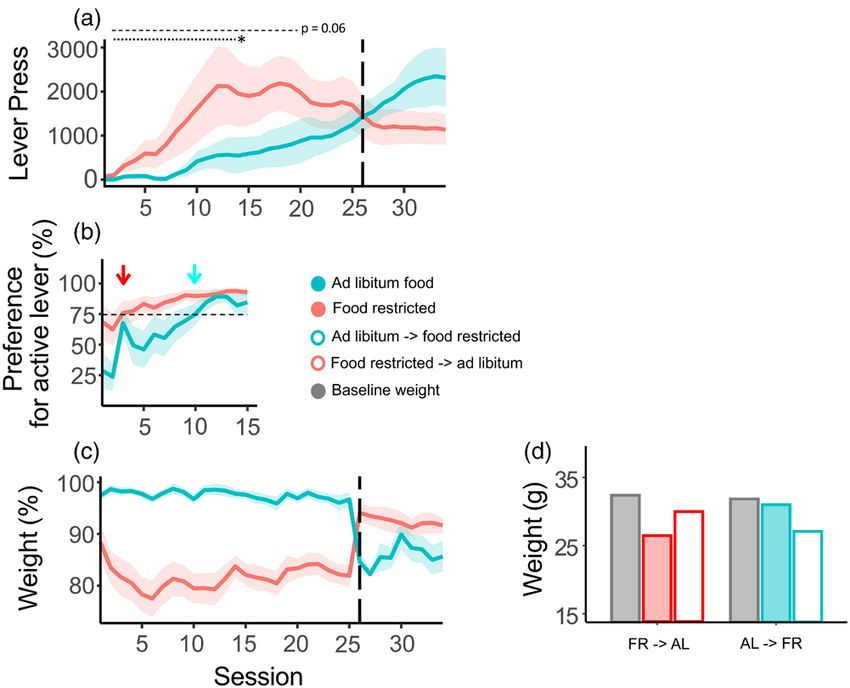
Food restriction and oICSS Gnazzo et al.   1129

(ChR2+/−;DATcre/wt), selectively expressing ChR2 in            40 Hz, with 2 min between each optical test stimulation.
dopamine cells. Animal protocols were approved by the          Oxidation and reduction peaks at 0.6 V and −0.2 V was
Institutional Animal Care and Use Committee of Queens          identified using a cyclic voltammogram. Microelectrodes
College, City University of New York.                          were calibrated following experiments using a micro flow
                                                               cell and a 1-μM dopamine solution [22].
Diet
Mice (seven males and six females) were assigned to            Behavioral testing
either ad libitum or food restricted groups counterbal-        Mice were singly housed during the course of the experi-
anced by initial weight and sex. Food restricted mice          ments. Operant chambers were made from black, opaque
were maintained at 85% of their baseline weight by pro-        plastic boxes (22 cm × 35 cm × 23 cm). Chambers included
viding 4 h of free access to chow starting 2 h after each      an active and inactive lever and a pellet dispenser (Med-
daily session during the light cycle. Food restriction         Associates, St. Albans, Vermont, USA). Above each cham-
began 4 days prior to the start of behavioral testing. When    ber was a PlexBright Compact LED module attached
the animals’ feeding conditions were reversed, they were       to a PlexBright LED Commutator which allowed the
maintained under the new conditions for 4 days prior to        fiber optic cable to spin freely as the animals moved in
resuming behavioral testing to allow stabilization in con-     the chambers (Plexon, Dallas, Texas, USA). Mice were
sumption and body weight. Sample size did not have             weighed daily immediately prior to testing. 60-min ses-
sufficient power to detect sex differences and therefore       sions began after the fiber optic was attached and the
sexes were collapsed in the analysis.                          animal placed in the chambers. Mice were allowed to
                                                               press both the active and inactive lever at will during the
Fiber optic surgery                                            session.
Mice were given an intraperitoneal injection of ket-
amine/xylazine (50 mg/kg and 5 mg/kg, respectively)            Optogenetics
for placement into stereotaxis and then maintained             A blue 465 nm λ LED light (Plexon) was used to excite
under isoflurane anesthesia (4% induction, 1.5% main-          ChR2. Each lever press yielded a 500 ms burst of 20 pulses
tenance). Coordinates targeting the ventral tegmental          lasting 10 ms each at 40 Hz delivered by a Plexon control-
area (VTA) were AP: −3.16 mm, lateral (LAT): +0.55 mm,         ler (OPTMN0002e; PlexBright 4 Channel Optogenetic
dorsal-ventral (DV): −4.6 mm relative to bregma. A fiber       Controller; Plexon Inc, Brentwood, New York, USA).
optic (FP200URT; Thorlabs, Newton, New Jersey, USA)            Light intensity at the tip of the ferrule was ~5 mW, tested
attached to a ferrule (CFLC230-10; Thorlabs) was slowly        daily prior to starting sessions.
lowered into the brain. The ferrule was fixed to the skull
with C&B Metabond Quick Adhesive Cement System                 Imaging
(Parkell Inc., Brentwood, New York, USA).                      Brains were perfused and fixed in 4% paraformaldehyde
                                                               and placed in a 25% sucrose solution for 48 h. Frozen
Fast-scan cyclic voltammetry                                   brains were cut using a cryostat (Leica Biosystems,
Mice were given an intraperitoneal injection of urethane       Buffalo Grove, Illinois, USA) in 50 μm sections.
(1.8 g/kg) and placed in a stereotaxic frame. A carbon fiber   Channelrhopdosin was visualized via coexpression of
microelectrode was lowered into the nucleus accumbens          enhanced yellow fluorescent protein (EYFP) and imaged
at the following coordinates, AP: +1.3 mm, LAT: +1.1 mm,       with an Olympus Fluoview FV10i confocal microscope.
DV: −4.4 mm relative to bregma and a chloride-coated sil-      The sections were also used to verify correct placement
ver wire reference electrode was implanted and secured         of the fiber in the VTA (Fig. 1).
contralateral to the microelectrode using a stainless-steel
screw and dental cement. A fiber optic attached to a           Results
ferrule was lowered directly above the VTA using the           Genetically expressed channelrhodopsin mediates
VTA coordinates noted above. A cycling potential was           optically stimulated dopamine release
applied to the carbon fiber microelectrode (–0.4 V to 1.3 V    Commonly, expression of channelrhodopsin in dopa-
and back) at a scan rate of 400 V/s while being held at        mine cells is achieved via viral expression, resulting in
– 0.4 V between scans. To stabilize background current,        high expression levels [20,23]. Here, we use genetically
the microelectrode was cycled at 60 Hz for 15 min then         expressed channelrhodopsin. Mice heterozygous for
10 Hz for 10 min prior to starting experimental stimula-       ChR2 and DAT-Cre (ChR2+/−; DATcre/wt) express chan-
tion protocols. Optical stimulation power was 20 mW.           nelrhodopsin in the midbrain as reflected by coexpressed
Evoked dopamine data were collected sampling at 10 Hz          EYFP (Fig. 2a). The DATcre line has previously been
for 15 s. In each recording, background was digitally sub-     demonstrated to be selective for dopamine cells [24,25].
tracted using 10 scans immediately preceding optical           We tested optical stimulation of dopamine cells using
stimulation. After establishing an optimal dopamine sig-       fast-scan cyclic voltammetry, optically stimulating in
nal, dopamine was evoked using 5, 10, 20 pulses at 5 and       the VTA at 5 and 40 Hz with 5, 10, and 20 pulses and

    Copyright © 2021 Wolters Kluwer Health, Inc. Unauthorized reproduction of this article is prohibited.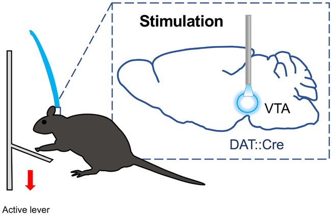
1130     NeuroReport   2021, Vol 32 No 13

Fig. 1

Verification of placement of optic fibers used in oICSS experiments. Blue and red dots indicate placement of fiber tip in ad libitum fed and
restricted mice, respectively; AP coordinates relative to Bregma. oICSS, optical intracranial self-stimulation.

Fig. 2

Genetically expressed channelrhodopson-2 supports optically stimulated evoked dopamine release. (a) Expression of enhanced yellow fluorescent
protein fused to channelrhodopsin with the VTA outlined in red. (b) Schematic of fast scan cyclic voltammetry recordings in the nucleus accum-
bens using optical stimulation of dopamine cells in the VTA. (c) Average evoked dopamine release after 5, 10, and 20 pulses at 5 and 40 Hz. (d)
Averaged voltammograms at 5 and 40 Hz showing oxidation (0.6 mV) and reduction (−0.2 mV) peaks indicative of dopamine. (e) Average of peak
evoked dopamine across frequencies and pulses. VTA, ventral tegmental area.

recording dopamine release in the nucleus accumbens                         increased with pulse numbers (Fig. 2c–e). In the oICSS
(Fig. 2b). At 5 Hz, dopamine release was minimal and did                    studies described here, we stimulated with 20 pulses at
not scale with pulse number, but at 40 Hz evoked release                    40 Hz.

            Copyright © 2021 Wolters Kluwer Health, Inc. Unauthorized reproduction of this article is prohibited.
Food restriction and oICSS Gnazzo et al. 1131
Fig. 3 Chronic food restriction facilitates rapid acquisition of
optical self-stimulation
Mice were maintained on either food restriction (3 h/day
access) or ad libitum access to food and tested in opti-
cal self-stimulation paradigm where every press on the
active lever activated an LED that selectively stimulated
midbrain dopamine cells (Fig. 3). In terms of day to day
increases in lever-pressing, acquisition of oICSS was
enhanced in food restriction mice compared to ad libi-
tum mice (Fig. 4a, first 14 days, main effect, F(1,9) = 4.95,
P = 0.05; group × day, F(1,165) = 18.83, P < 0.001). The food
restriction group reached asymptotic pressing by 14 days,
exhibiting a group average of 1950 presses per session
(Fig. 4a, days 12–14, 1950 ± 415). In contrast, in the ad libi-
tum group, the much slower session by session increases
never reached asymptote, achieving a maximal average
Schematic of oICSS behavioral paradigm. A press on the active of only 1012 per session (Fig. 4a, days 23–25, 1012 ± 200).
lever is followed by optical stimulation of VTA dopamine cells in
ChR2+/−;DATcre/wt mice. DAT, dopamine transporter; oICSS, optical
Another measure of learning is the proportion of pressing
intracranial self-stimulation; VTA, ventral tegmental area. on the active versus the inactive lever, where the percent-
age of presses on the active lever should increase substan-
tially with reinforcement via dopamine stimulation. The
Fig. 4
Optical self-stimulation of ventral tegmental area dopamine in food restricted and ad libitum fed mice. (a) Mean daily responding on the active lever
for each group across sessions. Dashed vertical line indicates reversal of feeding conditions. (b) Preference for active lever compared to inactive
lever. Arrowheads indicate when each group reached 75% preference, dashed horizontal line indicates 75% preference. (c) Average weight from
baseline by group across sessions, dashed vertical line indicates reversal of feeding conditions. (d) Average weights by group and condition.
N = 6, 7 for food restricted and ad libitum, respectively. *P = 0.05, Error bars, SEM.
Copyright © 2021 Wolters Kluwer Health, Inc. Unauthorized reproduction of this article is prohibited.1132 NeuroReport 2021, Vol 32 No 13
food restriction mice as a group reached 75% preference acutely change responding for self-stimulation, assessing
for active lever by day 3 compared to the ad libitum mice how drugs alter self-stimulation dose-response (electrical
that did not reach an equivalent preference until day 10 ‘dose’ to lever pressing) curves, quantified as a single read-
(Fig. 4b, t(1,11) = −2.05, P = 0.066). These data suggest that out of threshold. It is difficult, however, to assess rates of
chronic food restriction increases the efficacy of stimu- acquisition of learning in paradigms with constantly shift-
lated dopamine release in reinforcing an instrumental ing levels of stimulation. By providing a constant level of
action in the absence of external reward such as food. optical stimulation, our simple paradigm offers an intuitive
assessment of the efficacy of dopamine-mediated rein-
Current food restriction status modulates the vigor of forcement under different environmental and organismal
responding for optical self-stimulation conditions. Consistent with prior work by others, these
While the data for initial acquisition indicate a differ- data demonstrate that chronic food restriction alters the
ence in rate of learning, or reinforcement efficacy, to test dopamine system, highlighting that studying dopamine in
whether the vigor of the acquired response is modulated animals under food restriction does not necessarily reflect
by food availability, we reversed feeding conditions. The ‘normal’ reward function [1–4]. Use of oICSS paradigms
ad libitum group were placed on 3 h of access per day (ad can provide a route to examine dopamine-mediated
libitum → food restriction) while the food restriction group motivation and learning without the confounding effects
were provided ad libitum access to chow (food restriction of chronic food restriction and associated chronic stress.
→ ad libitum). After 4 days for weight adaptation and sta-
Although oICSS selectively stimulates dopamine neu-
bilization, testing continued as before (in plots, open sym-
rons, we cannot conclude with certainty the observed
bols reflect period after reversal of feeding conditions).
effects are mediated by dopamine per se. Specifically, glu-
The change in feeding conditions induced a significant
tamate can be released by dopamine cells and has been
weight adjustment for both groups (Fig. 4c and d, food
shown to mediate reinforcement independent of dopa-
restriction → ad libitum, F(1,60) = 672.29, P < 0.01; ad libi-
mine [27,28]. Thus, we cannot determine here whether
tum → food restriction, F(1,71) = 94.78, P < 0.01). The food
increased reinforcement efficacy under food restriction
restriction → ad libitum group decreased lever pressing
approximately 67% (Fig. 4a, days 23–25 vs. 32–34, 1690 arises from altered release of dopamine, glutamate, or
→ 1132), while the ad libitum → food restriction group both from dopamine cells in the midbrain.
increased pressing and stabilized at a group average of
2316 presses per session (Fig. 4a, days 23–25 vs. 34–35, Acknowledgements
1247 → 2316). After reversal, the now food restricted ad This work was supported by a grant from the National
libitum→ food restriction mice pressed 205% more than the Institute on Drug Abuse (J.B., DA046058) and a PSC-
now ad libitum food restriction → ad libitum mice, reflect- CUNY Award, jointly funded by The Professional Staff
ing a statistical trend (Fig. 4a, days 32–34, main effect, Congress and The City University of New York (J.B.).
F(1,9) = 3.60, P = 0.09).
Conflicts of interest
Discussion There are no conflicts of interest.
Chronic food restriction increased both learning rate and
vigor of responding for oICSS in the absence of food References
1 Fulton S. Appetite and reward. Front Neuroendocrinol 2010; 31:85–103.
reward. Food restriction mice were able to distinguish 2 Carr KD. Augmentation of drug reward by chronic food restriction:
between active and nonactive levers faster and more behavioral evidence and underlying mechanisms. Physiol Behav 2002;
rapidly reached peak levels of lever pressing than ad 12:353–364.
3 Stouffer MA, Woods CA, Patel JC, Lee CR, Witkovsky P, Bao L, et al.
libitum mice. In addition, food restriction increased the Insulin enhances striatal dopamine release by activating cholinergic
overall number of lever presses per session. The finding interneurons and thereby signals reward. Nat Commun 2015; 6:8543.
that food restriction increases vigor of responding is con- 4 Carr KD. Chronic food restriction: enhancing effects on drug reward and
striatal cell signaling. Physiol Behav 2007; 91:459–472.
sistent with previous studies showing that food restric- 5 Zhen J, Reith ME, Carr KD. Chronic food restriction and dopamine trans-
tion increases the amount of effort an animal is willing porter function in rat striatum. Brain Res 2006; 1082:98–101.
to expend for ICSS [18,19]. Altogether, these findings 6 Pothos EN, Creese I, Hoebel BG. Restricted eating with weight loss
selectively decreases extracellular dopamine in the nucleus accumbens and
reflect the body of evidence showing that food restric- alters dopamine response to amphetamine, morphine, and food intake. J
tion causes neuroadaptations that increase the efficacy of Neurosci 1995; 15:6640–6650.
dopamine reinforcement [3,10,26]. 7 Haberny SL, Carr KD. Comparison of basal and D-1 dopamine receptor
agonist-stimulated neuropeptide gene expression in caudate-putamen and
Our behavioral paradigm measures the rate of acquisition nucleus accumbens of ad libitum fed and food-restricted rats. Brain Res
Mol Brain Res 2005; 141:121–127.
of self-stimulation, which contrasts with typical ICSS stud- 8 Ouyang J, Carcea I, Schiavo JK, Jones KT, Rabinowitsch A, Kolaric R,
ies that measure stimulation threshold by varying either et al. Food restriction induces synaptic incorporation of calcium-per-
strength or frequency of electrical stimulation. By calcu- meable AMPA receptors in nucleus accumbens. Eur J Neurosci 2017;
45:826–836.
lating stimulation threshold, conventional ICSS is used 9 Lindblom J, Johansson A, Holmgren A, Grandin E, Nedergård C,
to determine how putative drugs of abuse administered Fredriksson R, Schiöth HB. Increased mRNA levels of tyrosine hydroxylase
Copyright © 2021 Wolters Kluwer Health, Inc. Unauthorized reproduction of this article is prohibited.Food restriction and oICSS Gnazzo et al. 1133
and dopamine transporter in the VTA of male rats after chronic food restric- of d-amphetamine reward by food restriction but no effect of a “sensi-
tion. Eur J Neurosci 2006; 23:180–186. tizing” regimen of d-amphetamine. Psychopharmacology 2004; 175:
10 D’Cunha TM, Daoud E, Rizzo D, Bishop AB, Russo M, Mourra G, et al. 106–113.
Augmentation of heroin seeking following chronic food restriction in the 20 Deisseroth K. Optogenetics. Nat Methods 2011; 8:26–29.
Rat: differential role for dopamine transmission in the nucleus accumbens 21 Stauffer WR, Lak A, Yang A, Borel M, Paulsen O, Boyden ES, Schultz W.
shell and core. Neuropsychopharmacology 2017; 42:1136–1145. Dopamine Neuron-Specific optogenetic stimulation in rhesus macaques.
11 Carr KD. Homeostatic regulation of reward via synaptic insertion of Cell 2016; 166:1564–1571.e6.
calcium-permeable AMPA receptors in nucleus accumbens. Physiol Behav 22 Sinkala E, McCutcheon JE, Schuck MJ, Schmidt E, Roitman MF, Eddington
2020; 219:112850. DT. Electrode calibration with a microfluidic flow cell for fast-scan cyclic
12 Carrol ME, Meisch RA. Increased drug-reinforced behavior due to food voltammetry. Lab Chip 2012; 12:2403–2408.
deprivation. Adv Behav Pharmacol 1984; 4:47–88. 23 Tye KM, Deisseroth K. Optogenetic investigation of neural circuits
13 Shalev U. Chronic food restriction augments the reinstatement of extin- underlying brain disease in animal models. Nat Rev Neurosci 2012;
guished heroin-seeking behavior in rats. Addict Biol 2012; 17:691–693. 13:251–266.
14 Olds J, Milner P. Positive reinforcement produced by electrical stimulation of 24 Liu Z, Brown A, Fisher D, Wu Y, Warren J, Cui X. Tissue specific expression
septal area and other regions of rat brain. J Comp Physiol Psychol 1954; of cre in rat tyrosine hydroxylase and dopamine active transporter-positive
47:419–427. neurons. PLoS One 2016; 11:e0149379.
15 Carlezon WA Jr, Chartoff EH. Intracranial self-stimulation (ICSS) in rodents 25 Zhuang X, Masson J, Gingrich JA, Rayport S, Hen R. Targeted gene expres-
to study the neurobiology of motivation. Nat Protoc 2007; 2:2987–2995. sion in dopamine and serotonin neurons of the mouse brain. J Neurosci
16 Cabeza de Vaca S, Carr KD. Food restriction enhances the central reward- Methods 2005; 143:27–32.
ing effect of abused drugs. J Neurosci 1998; 18:7502–7510. 26 Cadoni C, Solinas M, Valentini V, Di Chiara G. Selective psychostimulant
17 Abrahamsen GC, Berman Y, Carr KD. Curve-shift analysis of self-stim- sensitization by food restriction: differential changes in accumbens shell
ulation in food-restricted rats: relationship between daily meal, plasma and core dopamine. Eur J Neurosci 2003; 18:2326–2334.
corticosterone and reward sensitization. Brain Res 1995; 695:186–194. 27 Zell V, Steinkellner T, Hollon NG, Warlow SM, Souter E, Faget L, et al. VTA
18 Elder ST, Montgomery NP, Rye MM. Effects of food deprivation and Glutamate neuron activity drives positive reinforcement absent dopamine
methamphetamine on fixed-ratio schedules of intracranial self-stimulation. co-release. Neuron 2020; 107:864–873.e4.
Psychol Rep 1965; 16(Suppl 3):1225–1233. 28 Yoo JH, Zell V, Gutierrez-Reed N, Wu J, Ressler R, Shenasa MA, et al.
19 Cabeza de Vaca S, Krahne LL, Carr KD. A progressive ratio sched- Ventral tegmental area glutamate neurons co-release GABA and promote
ule of self-stimulation testing in rats reveals profound augmentation positive reinforcement. Nat Commun 2016; 7:13697.
Copyright © 2021 Wolters Kluwer Health, Inc. Unauthorized reproduction of this article is prohibited.You can also read