Click to edit Master title style - GLS & Echocardiogaphy
←
→
Page content transcription
If your browser does not render page correctly, please read the page content below

Click to edit Master title style

    Global Longitudinal Strain (GLS) &
            Echocardiography

                LVEF & Beyond

      Chi-Ming Chow MDCM                 FRCPC FCCS FASE ABIM
                      Director of Echo Lab,
          Division of Cardiology, St. Michael’s Hospital
                  Professor in Medicine, U of T
                    Co-Founder, USquareSoft

Objectives

• LV Function Assessment
• Basic Principles of Strain & GLS
• Technical Requirements to perform GLS
• Clinical Applications esp. related to Cardio-
  Oncology

Ventricular Function
•   Pump Performance

    •    SV, SV index, CO, CI

•   Myocardial Contractility

    •    Basic property of myocardium

    •    Load independent

•   Contractile Function

    •    EF

    •    Dependent on load, fibre length, inotropic state
LV EF Assessment
•   Eyeball Method

•   Modified / Simplified Quinones Method

•   Simpson’s Monoplane and Biplane

•   Newer Modalities
    •   Echo contrast (EBD)

    •   3-D (Real time vs. non-real time)

    •   AFI (Automatic Function Imaging)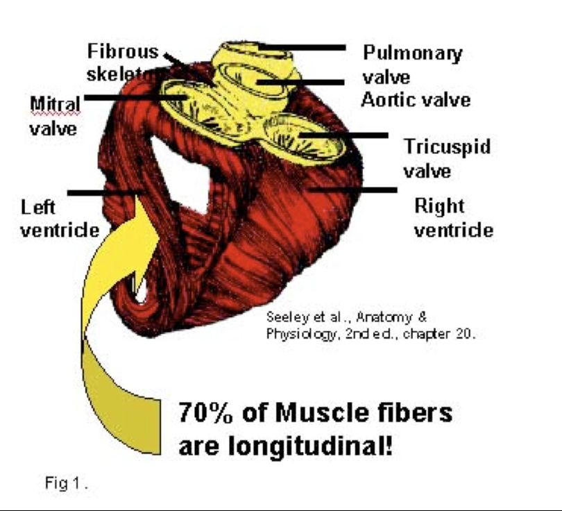
LV EF - Good IQ
Biplane Simpson’s Method

                            Thomas Simpson
                              1710 - 1761

      C/W MUGA r = 0.88, SEE 7.1%
Interobservers r = 0.77, mean % diff 22.9%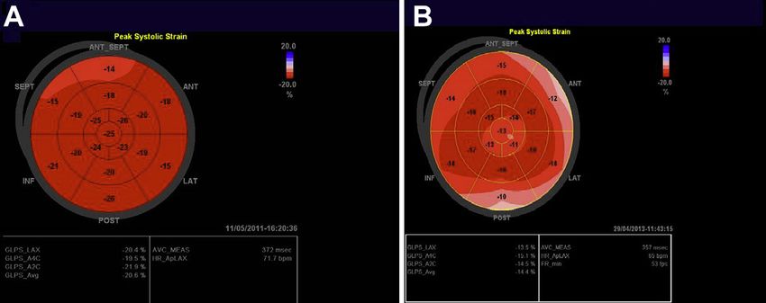
LV EF - Bad IQ
Coming to Rescue

To LVEF And Beyond
Echo Contrast
CMR vs. Echo Contrast
     LV End-Diastolic Volume

      LV End-Systolic Volume

       LV Ejection Fraction

Hundley EG et al. Administration of an intravenous perfluorocarbon contrast agent improves echocardiographic determination of left ventricular volumes and
ejection fraction: comparison with cine magnetic resonance imaging. JACC V32 No. 5, 1998
Tissue Velocity Imaging
Myocardial architecture
Myocardial fibers are arranged in spiral
fashion around LV cavity
Epicardial fibers twist clockwise, helical
orientation from base to apex
Endocardial fibers twist counterclockwise
Subendocardial and subepicardial fibers
run longitudinally
Midwall fibers run circumferentially
Yu CM et al. Tissue Doppler imaging: a new prognosticator for cardiovascular diseases. J Am Coll
Cardiol 2007; 49: 1903-14.Tissue Velocity Imaging
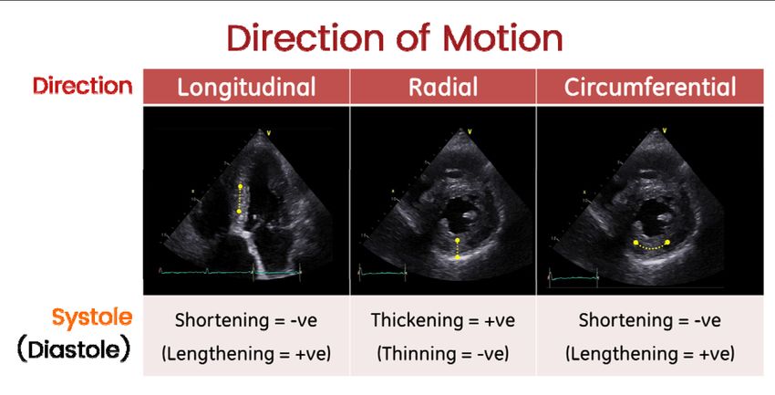
Myocardial architecture
Two components including longitudinal
shortening and rotation and radial
contraction
Myocardial velocities from apical views
reflects longitudinal shortening and
relaxation
Myocardial velocities from parasternal
views reflects radial shortening and
relaxation
Yu CM et al. Tissue Doppler imaging: a new prognosticator for cardiovascular diseases. J Am Coll
Cardiol 2007; 49: 1903-14.Stress & Strain
Strain
           Echocardiography: Basics

•1966- Mirsky and Parmley- introduced
 the concept to understand elastic stiffness

•Dimensionless quantity that represented
 the percent change in dimension from a
 resting state to one achieved following
 application of a force (stress).
Strain Imaging
Echocardiography: Basics
Strain and strain rate are measures
of myocardial compression or
deformation
Quantifies the regional function of the
myocardium
Movement of 1 tissue relative to
another in sample volume is strain as
opposed to TVI which reflects
movement of 1 site relative to
transducer
Marwick TH. Measurement of strain and strain rate by echocardiography: ready for prime time?
J Am Coll Cardiol. 2006; 47: 1313-27.Strain Imaging
Echocardiography: Basics
Measures deformation of myocardium between 2 points
2 ways to display strain :
• Strain: Percent of deformation (%)
• Strain rate: Speed of deformation (1/s)
Marwick TH. Measurement of strain and strain rate by echocardiography: ready for prime time?
J Am Coll Cardiol. 2006; 47: 1313-27.Strain Imaging
Echocardiography: Basics
• 2 points in the myocardium
• At end systole, points 4 mm apart
• At end diastole, points 2 mm apart
• What is the % change?
•50% strain
• How long did it take?
•2.0 s-1 strain rate
Marwick TH. Measurement of strain and strain rate by echocardiography: ready for prime time?
J Am Coll Cardiol. 2006; 47: 1313-27.Strain Measurements
Strain Imaging
                      Echocardiography: Basics
     • Strain
         • Local % of deformation
         • Correlates with EF
     • Strain rate
         • Rate at which myocardial deformation occurs
         • Correlates with dP/DT
     Similar to TVI, strain imaging can be acquired using
     either Doppler or speckle tracking
Marwick TH. Measurement of strain and strain rate by echocardiography: ready for prime time?
J Am Coll Cardiol. 2006; 47: 1313-27.
Left Ventricle
LV Global Systolic Function
ML is myocardial length at
end-systole (MLs) and end-
diastole (MLd). Peak GLS is
a negative number because
MLs is smaller than MLd.
Speckle-tracking echocardiographical images illustrating GLS obtained from the apical long-axis view (A), four-chamber-view (B), and two-chamber-view (C) an
strain curves and bullseye plot. Each segment has a numeric and color-coded strain value.
Expert consensus for multimodality imaging evaluation of adult patients during and after cancer therapy: a report from the American Society of
Echocardiography and the European Association of Cardiovascular Imaging. (J Am Soc Echocardiogr 2014;27:911-39.)Strain Imaging
Clinical Pearls and Technical Tips
• Strain is NEGATIVE and practically assessed in long axis
• Normal being -20%. The large the number the better systolic function
• High frame rates >= 80 fps
• HR should be the similar (+/-5 bpm) in all 3 views
• Could use 3D probe to get the 3 2D views
• Quiet respiration / breath holding
• Longitudinal motion in apical viewsLeft Ventricle
LV Global Systolic Function
2015 ASE/EACVI Chamber Quantification GuidelinesAutomated Function Imaging
AFIThe Goal is to go From Subjective Expert Evaluation
To Quick Quantitative Information
LAD
!

                               AFI (3-5 min)
How does it work
AFI is based on the 2D Strain technique
• In 2D strain the approach is to analyze motion by tracking “stable” features
from frame to frame, in a way similar to the analysis in tagged MRI. We can
look at this as “natural acoustic tagging”.
Time (sequential frames)How does it work
Natural Acoustic Tagging
• The algorithm selects natural acoustic tags within the myocardium. The green points in the figure below illustrate the
selection.
• The algorithm then looks for the position of each selected feature in the next frame using SAD (Sum of Absolute
Differences).
• Results are then spatially and temporally smoothed in order to reduce noise.
ZOOM ZOOM
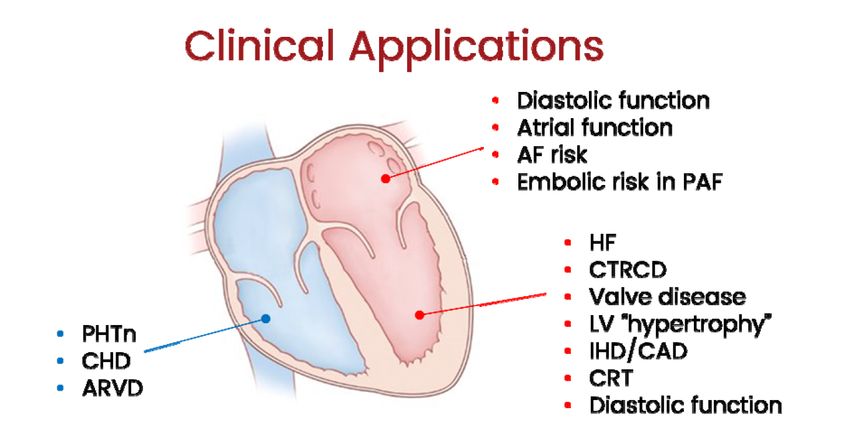
FeatureClinical Applications
Baseline: GLS -20.6%                                                 3 Months Chemo Tx: GLS -14.4%

Figure 9 Bull’s-eye plot showing GLS of the patient shown in Figure 8. (A) GLS and regional longitudinal strain at baseline. (B) GLS
and regional longitudinal strain 3 months during trastuzumab-based therapy after anthracyclines. GLS has decreased from
20.6% to 14.4% (30% decrease). The decrease in GLS is therefore considered of clinical significance (>15% vs baseline)
Restrictive Cardiomypathy
                      Cardiac Amloidosis

Apical sparing on deformation imaging

                                               ASE/EACVI LV Diastolic Function Guidelines, JASE 2016
Click to edit Master title style Thank You.
You can also read