Consensus of the 3rd Round Table Barcelona June 2013 - Ortho Solutions
←
→
Page content transcription
If your browser does not render page correctly, please read the page content below

Consensus of the 3rd Round Table
       Barcelona June 2013

              Mark Rogers FRCS (Tr & Orth)

               Derek Park FRCS (Tr & Orth)

                Dishan Singh FRCS (Orth)

    Aspects of Orthopaedic Foot & Ankle Surgery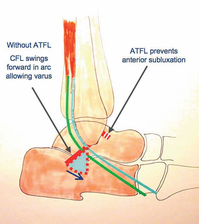
Preface

The 1st Round Table meeting was held in Padua in June 2011, followed by the 2nd Round Table
meeting in Paris in June 2012. The 3rd Round Table in Barcelona in June 2013 has once again followed
a format where all attendees review the literature and present their individual experience on a topic
with ample time for an informal discussion of the subject. There is no distinction between faculty and
delegates.

Mark Rogers and Derek Park were responsible for recording opinions and capturing the essence of
the debates, many of which resulted in consensus being reached on areas of foot and ankle practice.
This booklet collates the literature review and the views of all those who participated. The opinions
on consent, particularly, will hopefully guide practice and form the basis for a wider discussion at
BOFAS.

This booklet does not represent Level 1 evidence derived from prospective randomized controlled
trials but represents the compilation of anecdotal reports and small case studies based on the
combined experience of 34 British orthopaedic surgeons as well as Judith Baumhauer from the USA
and Harvinder Bedi from Australia.

I hope that you will find something of use and relevant to your own practice.

Dishan Singh, MBChB, FRCS, FRCS (Orth)

Consultant Orthopaedic Surgeon

Royal National Orthopaedic Hospital

Stanmore, UK

Consensus of the 3rd Round Table
                                                  Barcelona 2013

                                                            Mark Rogers

                                                             Derek Park

                                                           Dishan Singh

                                     Aspects of Orthopaedic Foot & Ankle Surgery

          1.    Consent in Foot & Ankle Surgery
          2.    Peroneal Tendon Disorders
          3.    Management of the Mangled Foot
          4.    Ankle Instability
          5.    Gastrocnemius Tightness

Convenors:

                     Mr Dishan Singh

                     Mr Paul Cooke

                     Mr Nick Geary

                     Mr Fred Robinson

Hosts:

                     Ortho Solutions

Distilled in this document are the thoughts and opinions with consensus where possible of 30 Orthopaedic Foot and Ankle Consultant
Surgeons who gathered from across the United Kingdom, USA and Australia. Though eminence rather than true evidenced based
medicine this represents the concepts of over 200 years of combined experience. A basis of invited lectures introduced open and frank
discussion from which consensus was sought. The statements herein only represent those of individuals and no claim is made that they
are irrefutable. All the percentage figures quoted represent the proportion of the surgeons present who voted on the subject in
discussion.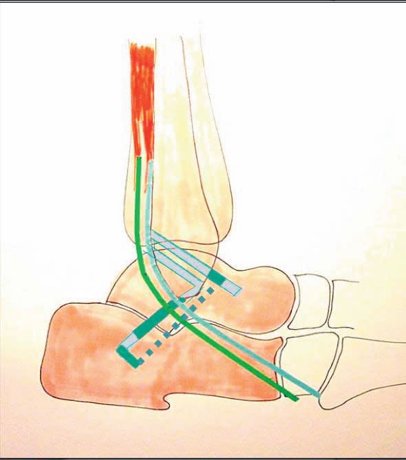
Consensus Session 1: Consent in Foot & Ankle Surgery
Chairman: Paul Cooke
The session explored several areas of the consent process including attitudes toward the consent
process, practical issues of how and when consent should be taken and evidence used to inform
patients of risks and benefits when taking consent.
Introduction: Paul Cooke
For consent to be valid the following essentials must be met:
o The patient must be competent to give consent
o They must have received sufficient information to make their decision
o They must not be acting under duress (including time duress, and hence the
rationale of the DOH recommendation for consent to be taken in advance of surgery
ie in clinic or pre-assessment clinic)
The surgeon should mention any significant risk that would affect the decision of a reasonable
patient.
Misconceptions in consent:
The 1% rule is law
Listing a complication on the consent form relieves you of the responsibility to avoid it
Listing a complication on the consent form is evidence that it has been discussed
Questions regarding current consent practice:
1. Do you ask the patient to sign the consent form in advance of the day of surgery?
Always 8 27%
Usually 14 47%
Sometimes 4 13%
Never 4 13%
2. Is the person taking consent capable of performing the procedure?
Always 12 40%
Usually 16 53%
Sometimes 2 7%
Never 0 0%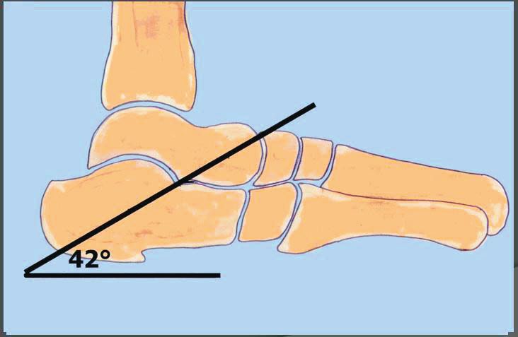
3.   Do you discuss the option of doing nothing?
                Always          23      77%
                Usually         7       23%
                Sometimes       0         0%
                Never           0         0%

    4. Do you provide written material to support the consent process?
              Always          4       14%
              Usually         10      36%
              Sometimes       11      40%
              Never           3       10%

There was broad agreement among delegates that patients undergoing inpatient surgery should be
consented in advance of their admission, in some form of pre-assessment or consent clinic. There
are inevitably local variations between Trusts as to how this is implemented, with some surgeons not
having routine access to a pre-assessment clinic: only 17/30 (57%) of the delegates have a regular
“pre-assessment” clinic, of which only 12 (40%) were “consultant led”.

A further interesting question was raised during the discussion: Should we take consent for low
complexity day case procedures, eg bunion surgery, at the outpatient consultation (ie when the
patient is listed for surgery)? These patients may have no medical need to attend a “pre-
assessment” clinic and we are being advised by the DoH guidelines to consent them in advance of
their attendance for surgery. One response might be that all patients should come to a consent clinic
but this may be needlessly burdensome and costly. Another solution would be to allocate more time
to outpatient slots to potentially consent day case patients when they are listed for surgery.
There was concern expressed that this may “pressurise” patients to sign a consent form on a first
clinic attendance and there would be a need to increase time allocations to clinic slots, reducing
throughput.

Question: How much extra time would you allocate to an outpatient attendance to consent a
patient for bunion surgery?

15 mins        0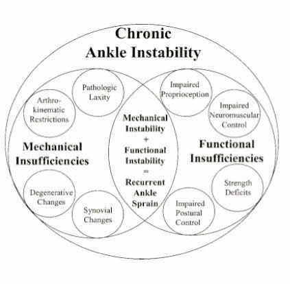
Consent for Bunion Surgery - Mark Rogers

Remarkably for a common procedure such as hallux valgus correction, there is no current agreement
on which complications that should be listed on the consent form for bunion surgery.

Consent practice for hallux valgus correction varies widely:
At the 2012 BOFAS meeting in Newport, 25 Foot and Ankle Consultants were asked to complete a
questionnaire which aimed to determine which complications of surgery were routinely written on
the consent form. The form offered 22 possible surgical complications and respondents were asked
to indicate which complications they would “always”, “sometimes”, “never” or “never but should”
include on the consent form for a hallux valgus correction.

The number of complications “always” listed for bunion surgery varied between 4 and 18, with a
mean of 12. Table 1 shows the possible response options and their relative frequency of inclusion on
the consent form by the 25 respondents. The only possible complication recorded by all respondents
was infection.

The speaker presented his personal view and practice of hallux valgus consent:
Recurrence, hallux varus and transfer metatarsalgia are all distinct complications that the surgeon
has a degree of control over and hence should be mentioned by name.

There is wide variability in the use of several terms that cover nerve injury: numbness, sensitivity,
hyperaesthesia, CRPS etc. It is not clear what is meant by “CRPS” and doubtful whether a patient or
even surgeon could satisfactorily explain it. The speaker uses the term “numbness including painful
numbness” which he feels covers both anaesthetic skin from the division of cutaneous nerves and
also the hypersensitivity and pain that may be part of a pain syndrome. His view was that patients
may understand this term better.

The use of the term “dissatisfaction” covers individual issues, discussed during the consent process
regarding the return to activity and daily living following surgery and whilst surgically a stable well
aligned foot will have been achieved, the patient may still require the use of an orthosis or be unable
to wear fashion shoes.

The speaker now discusses the following when consenting patients for hallux valgus correction:

Infection
DVT/PE
Recurrence
Hallux Varus
Transfer Metatarsalgia
Dissatisfaction
Stiffness
Numbness Including Painful Numbness
Further surgery
Ongoing Pain
Table 1. Results of Survey of Consent Practice of 25 Consultant Foot and Ankle Surgeons, surveyed
at BOFAS Newport Meeting, 2012.

          COMPLICATION        ALWAYS       SOMETIMES       NEVER       NEVER BUT
                                                                        SHOULD!

Infection                        25             0             0             0

Recurrence                       22             0             2             1

Numbness                         21             0             4             0

Stiffness                        17             2             6             0

Pain                             17             2             6             0

Failure                          15             3             7             0

DVT/PE                           15             3             6             1

Swelling                         13             4             8             0

Dissatisfaction                  13             3             8             1

Hyperaesthesia/sensitivity       13             6             5             1

Further surgery                  13             2            10             0

Non union/delayed union          12             6             7             0

Malunion                         10             4            10             1

Scar                              9             2            14             0

Bleeding                          8             3            12             2

Hallux varus                      6             5            12             2

Transfer metatarsalgia            6             8             9             2

Complex Regional Pain             5             6            12             2
Syndrome

Removal of Metalwork              5             10            9             1

Risk of anaesthesia               5             0            18             2

Risk of amputation                1             0            21             3

Death                             0             0            24             1
Discussion

There was broad agreement that the term CRPS was potentially unhelpful as the condition is poorly
understood. However there is a need to make reference to a post-operative condition of pain,
swelling and or numbness that could be interpreted as a pain syndrome.

The question was raised of whether the risk of amputation should be discussed with the patient.
Several delegates were of the opinion that if an elective uncomplicated bunion correction
subsequently resulted in amputation, there would likely be other flaws in the patient’s care that
would be of more importance than whether the risk of amputation was discussed or not.

Question: In the case of an uncomplicated bunion correction in a patient under the age of 70
years, would you consent the patient for the risk of amputation?
        Yes     1       ( 3%)
        No      29      (97%)

-----------------------------------------------------------------------------------------------------------------------------------

Consent for Ankle Arthroscopy - Paul Hodgson

Adverse outcome following ankle arthroscopy (and indeed any surgery) could be thought of under 3
headings:
       1. Side effects: pain, swelling , stiffness
       2. Failure to achieve the desired aim
       3. Complications.

A meta-analysis (Zengerink M, Van Dijk C. Complications in ankle arthroscopy. Knee Surg Sports
Traumatol Arthrosc 2012;0:1420-31) listed the complications of ankle arthroscopy. The most
common complication was nerve injury, average 3.7% (1-27%). Most are transient and resolve in 1
year. The risk will be dependent upon the portal used, but the intermediate branch of the superficial
peroneal nerve is that which is most commonly reported as injured via the anterolateral portal.

          Nerve injury                    3.7% (1-27%)

          Infection

                     Superficial          1.6% (0-8%)

                     Deep                 0.6% (0-3%)

          CRPS                            0.3% (0-2%)

          Instrument breakage             0.3% (0-2%)

          Sinus/delayed healing 0.9% (0-4%)

          DVT/PE
Consent for Ankle Fusion - Raman Dega

The message that consent is a process was re-iterated. This begins in the clinic and continues
through the pre-assessment clinic and onto the ward on the day of operation. An interesting analogy
was raised that consent is a “ritual” and as with many rituals there may be disagreement about its
conduct.

Consent may be thought of as having objectives/aims for both patient and surgeon: The patient
must be given adequate information about their condition such that they can understand it and
make an informed choice between treatment options. Risks and benefits of these choices must be
discussed and the patient be given realistic expectations of outcome. Specifically with ankle arthritis,
a discussion about the role and indication for ankle replacement as an alternative to fusion must be
discussed.

For this process to be effective, the surgeon must have adequate knowledge to inform the patient as
well as be able to discuss facts about outcome, including chances of success or failure.

The decision of when to operate and the merit in exhausting available non-operative measures is
important to consider and well as deciding on a reason to operate.

Risks were presented as general (infection, DVT/PE, CRPS, risk of anaesthesia) and specific (nerve
injury, non-union, malunion).

Several papers are useful as a basis for informing patients about the outcome of ankle fusion:

    1. Myerson et al. Ankle Arthrodesis vs TTC Arthrodesis. Patient outcomes, satisfaction and
       return to activity. Foot Ankle Int 2013;34:636-4
       Fusion achieves good functional outcomes and satisfaction. Certain activities become more
       difficult following fusion such as tennis and soccer but some activities are improved: driving,
       walking, golf.

    2. Van Dijk et al. Medium to long term outcome of ankle arthrodesis. Foot Ankle Int
       2011;32:940-47.
       At a mean follow up 9 years, a satisfaction rate of 90% was reported. Union was achieved in
       91% of patients. There was a 1% infection rate.

    3. Trichard T et al. Long term behaviour of ankle fusion: assessment of the same series at 7 and
       23 year follow up. Rev Chir Orthop Reparatrice Appar Mot 2006;92:701-07
       Half of 52 patients were lost to follow up at 23 years however there was little clinical
       expression of hindfoot arthritis in the triple complex of remaining patients although there is
       radiographic deterioration of these joints.

    4. Soohoo et al. Comparision of reoperation rates following ankle arthrodesis and total ankle
       arthroplasty. Journal Bone Joint Surg (Am) 2007;89:2143-49
       TAR showed 9% revision surgery rates at 1 year and 23% at 5 years, compared to ankle
       fusion group where revision rates were 5% at 1 year and 11% at 5 years.
Consent for Ankle Fracture Fixation - Al Best There are numerous small studies on outcome of ankle fractures that may inform the consent process, but two notable large data base studies are by Soohoo1 and Koval2. Between them they represent the outcome of approximately 90,000 patients. The series of Soohoo is broadly representative of UK practice as a whole, whilst that of Koval, with a mean patient age of 76 years, represents outcome of ankle fracture in the elderly. The 90 day complication rates of all ankle fractures are as follows: Infection (deep) 1.44% (1.4-6.8%) PE (DVT) 0.34% (0.12-2.7%) Re-fixation 0.82% Amputation 0.16% Mortality 0.37-1.37% Nerve injury 3.8-20% (note a 10% incidence even in nonoperative group) Non union
This confers a wound infection rate approximately 5 times higher than in non-smokers.

It is reasonable to conclude that a closed ankle fracture in a young healthy patient will have
low rates of complication, but that there are specific patient groups highlighted above in
which ankle fracture is not a benign injury.

References

1. SooHoo NF, Krenek L, Eagan MJ, Gurbani B, Ko CY, Zingmond DS. Complication rates
   following open reduction and internal fixation of ankle fractures. J Bone joint Surg Am.
   2009;91:1042-49
2. Koval KJ, Zhou W, Sparks MJ, Cantu RV, Hecht P, Lurie J. Complications after ankle
   fracture in elderly patients. Foot Ankle Int 2007;12:1249-55
Consensus Session 2: Peroneal Tendon Disorders

Chairman: Fred Robinson

Peroneal Tendon Tears - Alex Wee

The clinical history to suggest a diagnosis of peroneal tendon disorder may be relatively short: a
twisting injury in an athlete or an alteration in training regime or running style may precipitate a
peroneal tendon injury.

The presentation of a tendinopathic pathology may be more insidious. The patient may complain of
swelling, pain, crepitus or a clicking/flicking sensation behind the lateral malleolus. There may be an
associated history of ankle instability.

Asking the patient to localise the pain with one finger may be beneficial, as the specific localisation
of pain may be indicative of the pathology and help to clinically rule out differential diagnoses such
as pain from the subtalar joint or ankle.

Examination findings specific to the peroneal tendon disorder are swelling, elicited pain on direct
palpation or passive stretch in the presence of synovitis, and the replication of tendon subluxation
by ankle dorsiflexion and eversion.

Other features of the foot and ankle examination are also important - such as hindfoot alignment,
noting the presence of a cavus foot, assessment of ankle stability etc as these may point to other
pathologies or the aetiology of the peroneal pathology.

Several differing radiological modalities may be employed to confirm the diagnosis:

Ultrasound by a skilled musculoskeletal radiologist1 has been shown to be highly sensitive (100%),
specific (85%) and accurate (90%). It has the advantages of a dynamic investigation, allows
demonstration of subluxation and guides simultaneous injection if indicated.

MRI is useful if the pain around the ankle is more global or if multiple pathologies are suspected
(ankle instability, chondral injury etc) and as such may confirm or refute differential diagnoses such
as intra-articular pathology of the ankle and subtalar joint. The sensitivity and specificity of MRI for
peroneal tendon pathology is lower than that of US2 with reported sensitivity of 83% and specificity
of 75%.

Peroneal tendon tears have been classified by Brandes3 into anatomical zones, by Sobel4 according
to the size and extent of the tear and by Brodsky5 into those tears involving either less than or more
than 50% of the tendon substance.

Management

A trial of non-operative management should be employed and encompass activity modification,
analgesia, immobilisation with a boot in the acute period, an orthotic incorporating a lateral
hindfoot wedge and an explanation to the patient that the majority of cases will respond to non-
operative management with an expected recovery of several months.

Surgical intervention may be considered in those who fail non-operative treatment. Importantly the
complications of open peroneal tendon surgery are not rare. Although publications are typically
small retrospective case series, it is notable that Redfern and Myerson6 demonstrated 9/28 patients
with complications of infection, neuritis, CRPS symptoms, adhesions and wound healing problems.

However the small series in the literature do report reasonable results with Myerson’s paper6
demonstrating 91% of patients regaining moderate to full peroneal tendon strength and post op
AOFAS scores of 82 being representative.

Open surgery involves tendon debridement, tear excision or repair, tubularisation and repair of the
retinaculum. A figure of 50 % or more of dysfunctional tendon (based on Brodsky’s classification) is
taken as indicating the need for tenodesis. In the context of two dysfunctional tendons, there may
be a role for FDL/FHL single stage transfer7.

References

     1. Grant TH, Kelikian AS, Jereb SE, McCarthy RJ. Ultrasound diagnosis of peroneal tendon tears.
        A surgical correlation. J Bone Joint Surg Am 2005;87:1788-94
     2. Lamm BM, Myers DT, Dombek M, Mendicino RW, Catanzariti AR, Saltrick K. Magnetic
        resonance imaging and surgical correlation of peroneus brevis tears. J Foot Ankle Surg
        2004;43:30-6
     3. Brandes CB, Smith RW. Characterization of patients with primary peroneus longus
        tendonopathy: a review of twenty two cases. Foot Ankle Int 2000;21:462-68
     4. Sobel M, Geppert MJ, Olson EJ, Bohne WH, Arnoczky SP. The dynamics of peroneus brevis
        tendon splits: a proposed mechanism, technique of diagnosis and classification of injury.
        Foot Ankle 1992;13:413-22
     5. Krause JO, Brodsky JW. Peroneus brevis tendon tears: pathophysiology, surgical
        reconstruction and clinical results. Foot Ankle In. 1998;19:271-9
     6. Redfurn D, Myerson M. The management of concomitant tears of the peroneus longus and
        brevis tendons. Foot Ankle Int 2004;25:695-707
     7. Jockel JR, Brodsky JW. Single stage flexor tendon transfer for the treatment of severe
        concomitant Peroneus Longus and Brevis tendon tears. Foot Ankle Int 2013;34:666-72

------------------------------------------------------------------------------------------------------------------------------------

Peroneal Tendon Subluxation and Groove Deepening - Maneesh Bhatia

The peroneal tendons lie in a triangular shaped fibro-osseous tunnel formed by the fibula anteriorly,
the superior peroneal retinaculum and fibrocartilagenous lip laterally and the talo-fibular and
calcaneo-fibular ligaments medially.

The posterior surface of the fibula is typically concave but there is anatomical variation. Edwards1
demonstrated a flat or even convex posterior surface to the fibula in 18% of cases.
Peroneal tendon subluxation is uncommon. Acute traumatic dislocation may occur in forced
dorsiflexion injuries of the ankle with violent peroneal contraction. Whilst it may occur in isolation, it
can be associated with ankle instability or hindfoot varus.
Patients typically report hearing a “pop” or “snap” acutely subsequently followed by pain and
swelling. They may report a subsequent feeling of “flicking” with certain activities or positions of the
ankle. The diagnosis may be confirmed with USS and classified according to Oden2 as below.
Raikin3 further described an “intra-sheath” type of subluxation subdivided into type A (P. Longus and
Brevis flicking over each other within the sheath) and type B (P longus herniating through a tear in
brevis).
Oden’s Classification of peroneal tendon tears. Peroneus Brevis (1), Peroneus Longus (2).
Non operative management with casting has been described but carries failure rates of up to 75%.
Several procedures have been described to treat peroneal tendon subluxation including groove
deepening procedures4, bone block advancement procedures5 and rerouting the tendons behind the
calcaneo-fibular ligament6.Deepening of the peroneal groove is most popular and may be achieved through direct or indirect
methods.
Direct groove deepening involves osteotomising the distal fibula and elevating an intact cortical
hinge of bone to allow curettage of the underlying cancellous bone to deepen the groove.
Indirect deepening is achieved by sequential passes of increasing drill diameters in the longitudinal
axis of the distal fibula before “tamping down” the posterior cortex.
The literature records several small case series of outcome following a variety of procedures4-6 all of
which have been reported as successful.
References
1. Edwards M. The relation of the peroneal tendons to the fibula, calcaneus and cuboideum.
Am J Anat 1927;42:213-252
2. Oden RR. Tendon injuries about the ankle resulting from skiing. Clin Orthop Rel Res
1987;(216):63-9
3. Raikin SM, Elias I, Nazarian LN. Intrasheath subluxation of the peroneal tendons. Journal
Bone Joint Surg Am 2008;90:992-9
4. Mendicino RW, Orsini RC, Whitman SE, Catanzariti AR. Fibula groove deepening for
recurrent peroneal subluxation. J Foot Ankle Surg 2001;40:252-63
5. Micheli LJ, Waters PM, Sanders DP. Sliding fibula graft repair for chronic dislocation of the
peroneal tendons. Am J Sports Med 1989;17:68-71
6. Boykin RE, Ogunseinde B, McFeely ED, Nasreddine A, Kocher MS. Preliminary results of
calcaneo-fibular ligament transfer for recurrent peroneal subluxation in children and
adolescents. J Pediatr Orthop 2010;30:899-903
-----------------------------------------------------------------------------------------------------------------------------
Peroneus Quartus – Dishan Singh
Peroneus quartus is an accessory muscle of the lateral compartment of the leg. It has been shown to
be present in between 6%1 and 22% of individuals2.
It may originate from the peroneus brevis or longus muscles or the surface of the fibula and insert
variably into the retrotrochlear eminence, base of 5th metatarsal or cuboid.
Associated pathology includes longitudinal tears of the peroneus brevis, peroneal tendon
subluxation or a prominent retrotrochlear eminence.
It must not be misinterpreted on imaging of the ankle as a split in the adjacent peroneal tendons.
If seen at operation, it should be excised to reduce the volume of the contents of the peroneal
compartment.References
1. Zammit J, Singh D. The peroneus quartus muscle. Anatomy and clinical relevance. J Bone
Joint Surg Br 2003;85:1134-7
2. Sobel M, Levy ME, Bohne WH. Congenital variations of the peroneus quartus muscle: an
anatomic study. Foot Ankle 1990;11:81-9
Discussion
The role of injection for peroneal tendon disorder provoked extensive debate. There was discussion
of the detrimental effect of both steroid and local anaesthetic to the collagen structure of tendon.
There was broad agreement that if steroid injections were to be administered within the peroneal
sheath, then they should be of low dose steroid, using a high volume of local anaesthetic and
ensuring the injection was in the sheath and not intra-substance to the tendon as demonstrated by
the lack of resistance to injection.
13 delegates reported having seen a rupture of the peroneal tendons after an injection.
Concerning current practice:
1. 3 (10%) would inject “blindly” in a clinic setting with no radiological guidance
2. 17 (59%) would inject under ultrasound guidance
3. 9 (31%) would never inject around the peroneal tendons (for reasons of concern regarding
potential rupture)
However, no member thought it negligent to inject around the peroneal tendons.
Concerning the indication for injection:
1. 20 (62%) would offer injection for an intact tendon with tenosynovitis
2. 12 (38%) would inject in cases of partial tear of a peroneal tendon
3. No member would inject in cases of a tendon tear and subluxation
8 members would immobilise a patient following peroneal tendon sheath injection with a walking
boot.
With regard to surgical intervention:
1. Only 2 members routinely repair any tear at surgery, irrespective of its size.
2. 23 members use the “50% rule” based on Brodsky’s classification and tenodese the Brevis
and Longus if more than 50% of a tendon was felt to be degenerate.
3. There was 100% agreement that a prominent peroneal tubercle should be debulked.
--------------------------------------------------------------------------------------------------------------------------------
Painful Os Peroneum - Senthil Kumar
Present in up to 30% of individuals1, the Os Peroneum represents a sesamoid bone of the Peroneus
longus. It may be unipartite or bipartite, articulates with the cuboid and calcaneus and may have
multiple fibrous attachments to calcaneus, base of 5th metatarsal and the adjacent peroneus brevis.As such, it is thought to be comparatively “tethered” compared to other sesamoid bones and
therefore be subjected to greater stress, perhaps explaining the aetiology of injury.
The symptomatically painful Os Peroneum is rare: the literature containing only case reports
describing its presentation and treatment.
Sobel2, in 1994, coined the phrase “Painful Os Peroneum Syndrome” (POPS) but this term is also
used to include disorders of the peroneus longus.
Injury to the os peroneum may be acute ie a fracture through a unipartite os or a diastasis of a
bipartite os. In the acute setting, it is important to note that the function of peroneus longus is lost.
Chronic repetitive sprain type injuries do occur but here the function of the peroneus longus is
preserved, perhaps because the brevis has compensated for the loss of function.
Presentation may be with pain and swelling over the lateral border of the foot and in the chronic
setting, there may be a history of recurrent “giving way”. A high index of suspicion is required.
Investigations may include plain radiography, USS or MRI, all of which may be diagnostic.
In the acute setting management may be conservative with a below knee cast or walking boot, as
the displacement between fragments may not be excessive.
If presentation is greater than a month from injury, then operative intervention (fixation or
excision of the fragment and tendon repair) is probably indicated.
References
1. Muehleman C, Williams J, Bareither ML. A radiologic and histologic study of the os
peroneum: prevalence, morphology and relationship to degenerative joint disease of the
foot and ankle in a cadaveric sample. Clin Anat 2009;22:747-54
2. Sobel M, Pavlov H, Geppert MJ, Thompson FM, DiCarlo EF, Davis WH. Painful os peroneum
syndrome: a spectrum of conditions responsible for plantar lateral foot pain. Foot Ankle Int
1994;15:112-24
--------------------------------------------------------------------------------------------------------------------------------------
Cuboid Syndrome - Ioan Jones
This syndrome describes a rather non-specific pain localised to the lateral border of the hindfoot,
over the calcaneo-cuboid joint or bases of the 4th and 5th metatarsals. It is referred to in the
literature under a variety of names, including a “dropped cuboid”, “locked cuboid”, “peroneal cuboid
syndrome” and “subluxed cuboid”.
It is felt to be due to a subtle instability of the calcaneo-cuboid joint with a traumatic aetiology but
this is debated. There is also thought to be an association with instability of the first ray which may
cause excessive mechanical loading of the lateral column in midstance to toe off of the gait cycle.
It is typically a diagnosis of exclusion and radiological investigations including MRI are normal.The diagnosis may be confirmed by a positive diagnostic response to local anaesthetic/steroid
injection to the calcaneo-cuboid joint.
Management is non-operative with orthotics.
Question and Discussion
How many delegates believe in the entity of cuboid syndrome?
YES: 18/30 (60%)
NO: 12/30 (40%)
Although there was no consensus that cuboid tunnel syndrome exists with a discrete aetiology of
instability at the calcaneo-cuboid joint, there was broad agreement that there does exist a syndrome
of “lateral foot pain” in patients in whom radiological investigations are normal.
The aetiology of the pain was debated as being either neurogenic, synovitic, mechanical/instability,
or from adhesive capsulitis. There was a proposal that the lateral foot pain was secondary to
instability of the medial column and overload of the lateral rays.
--------------------------------------------------------------------------------------------------------------------------------------
The Cavo-Varus Foot and Peroneal Tendons - Fred Robinson
A cavo-varus foot position may cause overloading of the peroneal tendons during activity causing
tendonosis and tears, particularly of the peroneal longus tendon. Therefore, when assessing the
patient with peroneal tendon pathology, care should be taken to assess the foot shape and in
particular hindfoot alignment.
There is certainly an association between hindfoot varus and lateral ligament injury of the ankle and
Strauss1 demonstrated that 24% of failed lateral ankle ligament reconstructions were in varus
aligned heels that had not been corrected. Similarly, there is an association between lateral ligament
instability and varus ankle arthritis and althought there is a long latency time of 34 years2, correcting
the hindfoot alignment halts the progression toward arthritis.
Little attention is given in the literature regarding the management of peroneal tendon pathology in
the context of a varus hindfoot, other than descriptions of bony procedure to correct the foot shape.
Discussion centred around simultaneous peroneal tendon exploration and repair at the time of cavo-
varus foot correction. The argument that correcting the shape of the foot defunctions the peroneal
tendons and hence negates the need to reconstruct them was presented. No consensus was
reached.
References
1. Strauss JE, Frosberg JA, Lippert FG3rd. chronic lateral ankle instability and associated
conditions: a rationale for treatment. Foot Ankle Int 2007;28:1041-44
2. Valderrabano V, Hintermann B, Horisberger M, Fung TS. Ligamentous posttraumatic ankle
osteoarthritis. Am J Sports Med 2006;34:612-20Consensus Session 3: Management of the Mangled Foot Chairman: Fred Robinson The mangled foot - Rick Brown and Simon Clint The mangled foot as defined by a foot sustaining a substantial mechanism of injury, with multiple fractures, and potential soft tissue compromise is thankfully rare in the UK. The experience of a busy district general hospital was presented. Thirteen injuries were treated in the preceding year equating to an incidence of 1.8/100,000 population per year. The mechanism of injury is typically industrial, agricultural, road traffic accidents or falls from height. Predicting the outcome of such injuries is difficult and in the most severe cases the difficult decision of salvage and reconstruction versus acute amputation presents itself. In 1990, the Mangled Extremity Severity Score1 (MESS) was published. Based on the four parameters of the energy involved in the injury, the limb ischaemia time, the degree of shock and the age of the patient, it attempts to predict the outcome of such injury and to guide decision making on the likely success of salvage of such injuries. The authors demonstrated a high MESS score was indicative of the need for amputation. However, the experience of the US Army over the last 10-15 years from the conflicts in the Gulf and Afghanistan has shown the MESS score to be unreliable for the prediction of limb salvage or amputation2. The US Army experience would also suggest that the outcome of a delayed amputation is likely to be as good as a primary “acute” amputation but the key to their success probably lies in the benefit conferred by intensive rehabilitation programmes2. The “civilian” experience of the mangled foot and ankle was published earlier this year3. The LEAP (Lower Extremity Assessment Project) study included 174 feet with severe open hindfoot or ankle injuries, treated by salvage or by immediate amputation. The principal measure of outcome was the Sickness Impact Profile. When compared to patients treated with below knee amputation, salvage patients who had required free flaps and/or ankle arthrodesis had significantly worse 2 year outcomes. The UK experience of open foot fractures has been published by Court-Brown and McQueen4. 348 open foot fractures were treated at their institution in the preceding 23 years. They identified 3 severity levels if injury: Level 1: isolated open fractures of the forefoot Level 2: multiple open forefoot fractures Level 3: open fractures of the midfoot and hindfoot. Open fractures of the calcaneus, talus and midfoot conferred amputation rates of 16.6%, 18.2% and 30% respectively.
!3.3% of patients with open multiple metatarsal fractures underwent forefoot amputation.

Management of the soft tissues in severe foot injury

The principals of management of open fractures of the foot and ankle are summarised in the joint
publication from the British Orthopaedic Association (BOA) and British Association of Plastic,
Reconstructive and Aesthetic Surgeons, available to download on from the BOA website5.

These guidelines emphasise the challenging nature of these injuries, the need for definitive internal
fixation with soft tissue coverage if possible and the consideration of primary amputation in severe
injuries.

    1. Helfet D, Howey T, Sanders R, Johansen K. Limb salvage versus amputation. Preliminary
       results of the Mangled Extremity Severity Score.Clin Orthop Rel Res 1990;56:80-6
    2. Shawen SB, Keeling JJ, Branstetter J, Kirk KL, Ficke JR. The mangled foot and leg: salvage
       versus amputation. Foot Ankle Clin 2010;15:63-75
    3. Ellington JK, Bosse MJ, Castillo RC, MacKenzie EJ. The mangled foot and ankle: results from a
       2-year prospective study. J Orthop Trauma 2013;27:43-8
    4. Court-Brown C, Honeyman C, Bugler K, McQueen M. The spectrum of open fractures of the
       foot in adults. Foot Ankle Int 2013;34:323-28.
    5. Standards for the management of open fractures of the lower limb. Downloadable at
       http://www.boa.ac.uk/Publications/Documents/Lower%20Limb%20Guide.pdf

Compartment Syndrome of the Foot

Compartment syndrome is estimated to occur in approximately 6% of cases of foot and ankle
trauma compared to 1.6% of tibial fractures.

The literature regarding the diagnosis, treatment and outcome is scarce but historically there has
been a drive toward prompt decompression with open fasciotomies through a variety of surgical
approaches. 1,2

The volume of literature on the outcome of foot fasciotomy is minimal and based on low numbers of
patients. Anecdotally, the outcome following fasciotomy is poor and Myerson2 has shown
appreciable limitation of function in patients who have had fasciotomies.

The vast majority of the evidence for fasciotomy has been extrapolated from data regarding tibial
fractures.

There is genuine concern regarding converting a closed foot injury to an open one with fasciotomies
and infection rates in the foot following fasciotomy may be high.

References

    1. Myerson M. Acute compartment syndromes of the foot. Bull hosp Jt Dis Orthop Inst
       1987;47:251-6.
2. Myerson M. Management of compartment syndromes of the foot. Clin Orthop Rel Res
1991;271:239-48.
Consensus discussion and voting
22/30 delegates regularly undertook foot and ankle trauma surgery.
8/30 delegates work in a major trauma centre.
3/30 (10%) had performed a fasciotomy of the foot in the preceding year.
12/30 (40%) had performed below knee amputation for trauma in the preceding year.
3/30 (10%) of delegates would base the decision for fasciotomy on a pressure reading alone.
11/30 (33%) would base their decision for fasciotomy on clinical grounds (pain, swelling).
20/30 (67%) delegates have seen patients made worse by foot compartment decompression.
Discussion focused on why the outcome of foot fasciotomy was so much worse than that in the
lower limb for tibial fractures. There was agreement that the fracture patterns, nature of injury and
soft tissue injury to the foot was not comparable to that of a tibial fracture.
The consequences of NOT performing lower limb fasciotomy for tibial fracture would result in the
significant sequalae of neurovascular compromise and muscle contracture which carries significant
morbidity.
In the foot, the sensory nerves are outside the fascial envelope so sensory loss is less and the
limitation to function posed by clawed toes is much less than that a Volkmann’s contracture of the
calf.
There was agreement that the risk of NOT doing something in the foot is not as great as the risk of
NOT doing something in the leg whilst the dangers are higher.
Questions:
In a patient with clinical signs and symptoms of a foot compartment syndrome or one in whom
elevated compartment pressures are measured, who thinks it is mandatory to perform fasciotomy?
Yes: 3 (10%)No: 27 (90%)
Who thinks measuring the compartment pressure in a foot with potential compartment syndrome is
mandatory?
Yes: 0 (0%)
No: 30 (100%)
The Establishment Of The Trauma Network And Its Impact On Foot And Ankle Trauma.
By the turn of this century it was apparent that trauma care in the UK was lagging behind that in
North America and other parts of Europe and a series of reports from the BOA, Royal College of
Surgeons, CEPOD and the National Audit Office all pointed to the quality of trauma care in the UK
being variable and too often poorly performed.
In April 2009, Keith Willet was appointed as “Trauma Tsar” with the subsequent establishment of
trauma networks throughout the UK, going “live” in April 2012. The network is based around
regional centres, the Major Trauma Centres, supported by local Trauma units. The function of the
network is to ensure that appropriate patients go to the MTC but that just as quickly patients may
return to their local trauma units for ongoing care and rehabilitation.
Which patients should go to the MTC? The Clinical Advisory Group concluded those with an Injury
Severity Score(ISS) >16 are appropriate for transfer, but the ISS can only be calculated
retrospectively.
The paramedics now use a triage tool based on suspected injury and physiological parameters,
designed to slightly over triage patients. Therefore, ideally all patients identified as “major trauma”
patients should go to the MTC as long as they can be transferred safely within 45 minutes have an
airway and not be at risk of exsanguination.
Criteria now used to triage patients to the MTC include:
Open pneumothorax
Crushed, de-gloved or mangled limb, proximal to the ankle or wrist
Suspected major pelvic fracture
Neck or back injury with paralysis
How does this affect foot and ankle trauma provision?
The average ISS for patients in McQueen’s paper was 5. The vast majority of foot and ankle trauma
is seen in isolation and not part of the polytrauma patient.
Has the establishment of the trauma network changed the pattern of foot and ankle trauma seen in
the local trauma units?The Cheltenham and Gloucester experience was presented. A review of trauma patients with foot
and ankle injuries for a year pre and post introduction of the Trauma Network was presented.
No significant difference was reported in the percentage of open ankle fractures treated, the
percentage of patients requiring external fixation of the foot and ankle, or the number of severe
open ankle injuries that required subsequent input from plastic surgery in the year following the
introduction of the Trauma Network.
Similarly, a review of all patients who were transferred to the regional MTC (Frenchay Hospital) from
all the local trauma units who had a co-existent foot and ankle injury as part of their injury profile,
demonstrated that the vast majority of transferred patients had multiple injuries and/or open
fractures that required plastic surgical support.
Therefore in conclusion, the majority of severe foot and ankle trauma was managed by the local
trauma units, the Trauma Network has had no significant impact on the numbers of patients with
foot and ankle trauma presenting to the local units and hence the local Trauma units need to be
staffed with Foot and Ankle surgeons to provide foot and ankle trauma care locally.
Questions
The questions explored what to do when significant foot and ankle trauma is admitted to your unit
and a non foot and ankle specialist is on call:
1. What advice would you give to a non foot and ankle surgeon who has admitted a 44 year old
man with a closed but significantly displaced Lisfranc injury, but without skin “compromise”?
Rest, elevate, splint and ice (await availability of foot and ankle surgeon) 25 (84%)
Perform reduction and percutaneous fixation with K wires 4 (13%)
Apply an external fixator 1 (3%)
Perform a definitive fixation 0
2. What advice would you give in the same scenario if the skin was compromised?
Rest, elevate, splint and ice (await availability of foot and ankle surgeon) 0
Perform a closed reduction and fixation with burried K-wires or screw fixation 10 (33%)
Apply an external fixator 3 (10%)
Undecided on method of fixation (dependent upon individual experience)
but in agreement of need to intervene surgically 17 (57%)Medial and Lateral Column Trauma to the Foot -Chris Blundell and Mark Davies
The bones (of the foot) are aligned in two functional columns. The lateral column includes the
calcaneus, cuboid, and fourth and fifth metatarsals. The medial column includes the talus, navicula,
cuneiforms, and the first, second, and third metatarsals.1 Dillwyn Evans made reference to the
columns of the foot in his paper in 1961, but there is no description of this in Sarrafian’s Foot &
Ankle anatomy textbook.2
Until fairly recently most reports in the literature regarding midfoot trauma focused primarily on
talar fractures, with authors advocating non-operative management with closed reduction and
plaster cast treatment, and discussing triple fusion as a salvage technique.3,4 Main and Jowett in
1975 classified midfoot trauma according to the direction of deforming force. They described
medial, longitudinal, lateral, plantar, or crush injuries of the foot but made no direct reference to
columns of the foot.5
Kelikian later described the Lisfranc joint and three-column theory, dividing it into:
Medial: medial cuneiform and 1st metatarsal
Middle: middle and lateral cuneiforms, and 2nd & 3rd metatarsals
Lateral: cuboid and 4th & 5th metatarsals
The emphasis moved to stabilisation of the medial and lateral columns, with either open reduction
and internal fixation, open reduction and external fixation, or primary arthrodesis. There was
certainly a move away from closed reduction and plaster cast imoobilisation.6
The importance of the column theory of the foot grew, with Hansen emphasising the following key
principles.7
Avoidance of lateral column shortening,
Restoration of the medial column,
Reduction of the naviculocuneiform joint.
The cuboid and navicula are crucial in maintaining the integrity of the lateral and medial columns of
the foot respectively. The cuneiforms also play an important role as the cornerstone of the
transverse arch of the foot.8 Based on this review by Pinney and Sangeorzan, the following summary
points were made:
Key points in managing trauma of the foot:
1. Obtain the correct diagnosis - use of CT scan
2. Maintain appropriate medial and lateral column length
3. Maintain appropriate relationship between forefoot and hindfoot
4. Preserve Talonavicular joint
5. Preserve Cuboid-4th metatarsal and Cuboid-5th metatarsal joints
6. Stable internal fixation for reduction, consider primary arthrodesis if necessary
7. Allow adequate time frame for soft tissue and bony healingTo achieve this:
1. Plate from talus to 1st metatarsal
2. Plate from calcaneum to 4th & 5th metatarsals
3. Remove metalwork, start physiotherapy
4. No role for external fixation
Klaue et al in a case series of various midfoot and hindfoot fractures alludes to column length and
advocates surgical approaches that allow visualisation of the talonavicular and calcaneocuboid
joints. In cuboid injuries, consider an AO distractor to restore column length. If the injury is too
severe primary arthrodesis should be considered. In navicular fractures, some shortening may be
accepted. Avoid bone block reconstruction that may result in inadvertent medial column
lengthening. Ultimately the salvage procedure is a triple arthrodesis.9
Sword et al wrote about the importance of the navicula and cuboid in column length and the
transverse arch. The talus is considered to be in the medial column. Bridging the talonavicular joint,
and stabilising from the talus to the 1st metatarsal may be necessary to maintain medial column
length.10
In terms of evidence for treatment of Lisfranc injuries, there are conflicting reports in the literature.
Coetzee and Ly found that primary arthrodesis of ligamentous Lisfranc injuries appears to be better
treated with primary fusion rather than ORIF.11 Rammelt found that primary ORIF with anatomic
reconstruction is better than salvage arthrodesis.12 Henning et al found no real difference in
outcomes between primary arthrodesis and ORIF.13 Coetzee et al, in a systematic review concluded
that both primary arthrodesis & ORIF have satisfactory and equivalent results.14
In assessing medial column injuries, it is important to have a high index of suspicion as these injuries
are easily missed. Associated injuries should be ruled out. A proper neurovascular assessment is
important and soft tissues should be treated with respect, especially in crush injuries. True medial
column only injuries are often stable, and tend to be crush injuries rather than high speed injuries. In
these cases where the lateral column is intact, no external fixation is required and definitive surgery
can be performed once the soft tissues settle.
The Sheffield philosophy:
Initial focus on soft tissues, allow time to settle with temporary back slab POP, elevation, cold
flowtron boots, and VTE thromboprophylaxis. They have moved away from compartment
fasciotomies for compartment syndrome of the foot.
The timing of surgery:
SCAN – (SPAN) – PLAN: If soft tissues not severely compromised
SPAN – SCAN – PLAN: If soft tissues at risk
The Sheffield approach:
A la carte approach to foot trauma
Direct (Topliss approach)
Open exposure and reduction
Simple external fixators to restore column length Locking plates to span joints if necessary
Percutaneous reduction and sometimes percutaneous fixation, away from exposure
= Focusing on restoration of columnar length, with particular attention to the navicular
Consensus statements for Management of the Mangled Foot
Discussion revolved around case presentations of complex midfoot and forefoot fractures and
general management strategies. As each case was different, it was difficult to take away any general
consensus statements.
There was a split of opinion that non-Foot & Ankle surgeons should apply an early external fixator in
the absence of a Foot & Ankle surgeon.
References
1. Dumontier TA, Falicov A, Mosca V, Sangeorzan B. Calcaneal lengthening: investigation of
deformity correction in a cadaver flatfoot model. Foot Ankle Int 2005;26:166-70.
2. Evans D. Relapsed club foot. J Bone Joint Surg Br 1961;43:722-733.
3. Kenwright J, Taylor RG. Major injuries of the talus. J Bone Joint Surg Br 1970;52:36-48.
4. Eichenholtz SN, Levine DB. Fractures of the tarsal navicular bone. Clin Orthop Relat Res
1964;34:142-57.
5. Main BJ, Jowett RL. Injuries of the midtarsal joint. J Bone Joint Surg Br. 1975;57:89-97.
6. Kelikian A. Operative Treatment of the Foot and Ankle. Appleton and Lange. 1999.
7. Hansen ST. Functional Reconstruction of the Foot and Ankle. Lippincott Williams & Wilkins.
2000.
8. Pinney SJ, Sangeorzan BJ. Fractures of the tarsal bones. Orthop Clin North Am. 2001;32:21-
33.
9. Klaue K. Chopart fractures. Injury 2004;35 Suppl 2:SB64-70.
10. Swords MP, Schramski M, Switzer K, Nemec S. Chopart fractures and dislocations. Foot Ankle
Clin 2008;13:679-93.
11. Ly TV, Coetzee JC. Treatment of primarily ligamentous Lisfranc joint injuries: primary
arthrodesis compared with open reduction and internal fixation. A prospective, randomized
study. J Bone Joint Surg Am 2006;88:514-20.
12. Rammelt S, Schneiders W, Schikore H, Holch M, Heineck J, Zwipp H. Primary open reduction
and fixation compared with delayed corrective arthrodesis in the treatment of
tarsometatarsal (Lisfranc) fracture dislocation. J Bone Joint Surg Br 2008;90:1499-506.
13. Henning JA, Jones CB, Sietsema DL, Bohay DR, Anderson JG. Open reduction internal fixation
versus primary arthrodesis for lisfranc injuries: a prospective randomized study. Foot Ankle
Int 2009;30:913-22.
14. Sheibani-Rad S, Coetzee JC, Giveans MR, DiGiovanni C. Arthrodesis versus ORIF for Lisfranc
fractures. Orthopedics 2012;35:e868-73. doi: 10.3928/01477447-20120525-26.Consensus session 4: Ankle Instability Chairman: Nick Geary (Note: where the speaker expresses a personal opinion or preference this is highlighted in italics to distinguish from consensus statements.) Ankle ligamentous injuries are amongst the most common types of injury sustained during sporting activities. The majority of these injuries involve the lateral ligamentous complex, which consists of the anterior talofibular ligament (ATFL), the calcaneofibular ligament (CFL), and the posterior talofibular ligament (PTFL). The ATFL is the most frequently injured ligament. Conservative Management of Ankle Instability - Mark Herron Chronic ankle instability is the persistence of mechanical and functional instability. Mechanical instability presents as recurrent episodes of instability with documented pathological laxity after an ankle ligament injury. Functional instability presents as recurrent episodes of ankle instability and sensation of insecurity or apprehension in the ankle joint due to proprioceptive and neuromuscular deficits, sometimes in the absence of demonstrable ankle laxity or mechanical instability. Hertel recognised that chronic ankle instability can arise from an interplay between mechanical and functional insufficiencies (Figure 1).1 Figure 1. The interplay between mechanical and functional insufficiencies in chronic ankle instability. The mainstay of conservative management of ankle instability centres on addressing functional insufficiencies arising from proprioceptive and neuromuscular problems. Proprioceptive problems include damage to the mechanoreceptors in the joint capsule, damage to peroneal muscle spindle cells, and damage to cutaneous sensation of the foot from soft tissue injury. Neuromuscular problems include peroneal muscle weakness or damage, weakness to ankle extensor muscles and problems arising from the central nervous system.
The majority of patients treated with neuromuscular proprioceptive rehabilitation programmes will
improve and approximately 20% will continue to have ongoing symptoms.2,3,4 It is unclear how many
patients with chronic ankle instability go on to develop ankle osteoarthritis, with conflicting reports
in the literature.5,6

Proprioceptive training (e.g. wobble boards), and neuromuscular exercise (e.g. strengthening
peroneal muscle complex), are the mainstay of physiotherapy treatment.7,8,9,10,11 Lack of a universally
accepted definition for chronic ankle instability, significant variation in treatment protocols and the
use of a variety of different outcome measures make review of the literature difficult. A systematic
review by McKeon found that completing at least 6 weeks of balance training after an acute ankle
sprain substantially reduced the risk of recurrent ankle sprains.12 A Cochrane review in 2011 found
only 4 suitable randomised controlled trials using neuromuscular rehabilitation programmes of 4 to
6 weeks duration that showed a small short-term improvement in recurrent instability.13

Ankle braces are also used ranging from rigid, semi-rigid and soft braces with one study suggesting a
semi-rigid brace to be most effective in controlling ankle inversion.14 There are conflicting reports in
the literature regarding the effectiveness of bracing, and the decision to use an ankle brace is largely
patient dependent.

Ankle taping probably contributes to mechanical limitation of movement, joint stabilisation, and
improved proprioception. A literature review by Verhagen et al found that ankle taping, bracing and
neuromuscular training were all effective in reducing the incidence of ankle sprain recurrences in a
proportion of patients.15

-------------------------------------------------------------------------------------------------------------------------------------

Surgical Role for Acute Ligamentous Injury - Rhys Thomas

The majority of acute ligamentous ankle injuries affect the lateral ligamentous complex. In the
general population, patients with acute ankle sprains do well with functional rehabilitation, with a
small proportion experiencing ongoing symptoms. In the high demand athlete, accelerated
rehabilitation, an objectively stable ankle, and an early return to sporting activity are often the
priorities. This must be balanced by the need to avoid chronic problems of pain, swelling and
recurrent instability. Surgery may be an advantage if it decreases recurrent ankle sprains, avoids
chronic problems, and confers subjective and objective ankle stability.

A Cochrane review in 2007 found no difference in outcome in surgical versus conservative treatment
for acute injuries of the lateral ligament complex of the ankle in adults.4 A randomized controlled
trial of surgical versus functional treatment for acute grade III injuries found no significant difference
in ankle scores and no difference in stress radiographs between the two groups. The non-surgical
group had a higher re-injury rate with a risk difference of 32%. There was a higher rate of grade II
osteoarthritis (27% risk difference) in the ankles treated surgically.16 A cohort study by Takao et al
found no difference in ankle scores and stress radiographs when comparing groups of patients
managed with nonoperative functional treatment alone versus functional treatment after primary
surgical repair.17 There was no significant difference between the two groups in terms of ankle
scores or stress radiographs. However there was a significant decrease in instability in the surgical group, and a significant decrease in time to return to full athletic activity in the surgical group. A systematic review by Peterson et al concluded that the majority of lateral ankle ligament injuries could be managed non-operatively. The main advantages to acute surgical ligament repair were that objective instability and recurrence rates were less common when compared with non-operative treatment.18 An ESSKA- AFAS consensus meeting in 2011 supported the view that future clinical studies should compare surgical results in experienced hands with the results of functional rehabilitation. Rehabilitation following surgical repair is divided into 5 stages: Stage 1 involves surgical recovery and early motion. Stage 2 involves establishing ankle joint control, improving strength and neuromuscular training. Stage 3 involves proprioceptive exercises. Stage 4 involves a step-wise progression of increasing function. Stage 5 involves sports-specific activity with at least 2 weeks of contact training before return to sport. The speaker’s preference is to rely almost solely on clinical assessment of instability supplemented by an examination under anaesthetic before surgery. A direct repair is performed, with suture anchor augmentation if there is an element of bony avulsion. An ankle arthroscopy is usually performed prior to surgical repair to assess for chondral injuries. -------------------------------------------------------------------------------------------------------------------------------------- Ankle Instability: Failed Surgery; What next? - Nick Geary Ankle instability can arise from a number of different conditions. They include lateral ankle ligament failure, medial ankle ligament failure, lateral subtalar ligament failure, osteochondral defects of the talus, peroneal tendon dislocation, and anteroinferior tibiofibular ligament failure. The subtalar joint is an S shaped joint that pivots around an axis with an average angle of inclination of 42 relative to the horizontal plane (Figure1). The head of the talus is roughly 16 internally rotated with respect to the calcaneum. Subtalar joint motion therefore occurs in inversion and eversion, and also demonstrates linear motion (anteroposterior movement), like a spiral screw moving forwards in inversion and backwards in eversion. The variable axis of the subtalar joint is mirrored by the axis of the calcaneofibular ligament. The ATFL prevents anterior subluxation and stops the CFL from swinging too far anteriorly in an arc at heelstrike (Figure 2). The ATFL is tight in plantarflexion and the CFL is tight in dorsiflexion.
You can also read