Developing the Next Generation Rapid Diagnostic Microfluidic Devices for Measuring the Protein Levels in Multiple Myeloma Patients for Early ...
←
→
Page content transcription
If your browser does not render page correctly, please read the page content below
Developing the Next Generation
     Rapid Diagnostic Microfluidic Devices
      for Measuring the Protein Levels in
     Multiple Myeloma Patients for Early
     Detection of Disease Re-Appearance
                  The Harvard community has made this
               article openly available. Please share how
               this access benefits you. Your story matters

Citation              Labovitis , Nicolas. 2021. Developing the Next Generation Rapid
                      Diagnostic Microfluidic Devices for Measuring the Protein Levels
                      in Multiple Myeloma Patients for Early Detection of Disease
                      Re-Appearance. Master's thesis, Harvard University Division of
                      Continuing Education.

Citable link          https://nrs.harvard.edu/URN-3:HUL.INSTREPOS:37367602

Terms of Use          This article was downloaded from Harvard University’s DASH
                      repository, and is made available under the terms and conditions
                      applicable to Other Posted Material, as set forth at http://
                      nrs.harvard.edu/urn-3:HUL.InstRepos:dash.current.terms-of-
                      use#LAA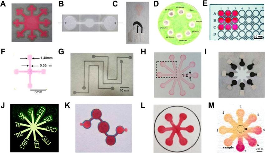
Developing the Next Generation Rapid Diagnostic Microfluidic Devices

for Measuring the Protein Levels in Multiple Myeloma Patients for Early Detection of

                              Disease Re-Appearance

                                 Nicolas Labovitis

            A Thesis in the Field of Bioengineering and Nanotechnology

            for the Degree of Master of Liberal Arts in Extension Studies

                                Harvard University

                                    March 2021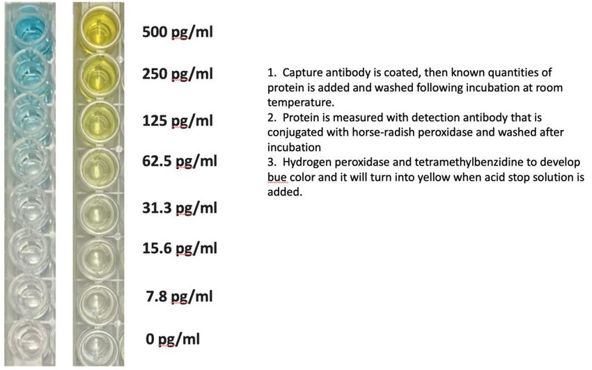
Copyright 2021 Nicolas Labovitis
Abstract

       The objective of this research is to determine whether rapid diagnostics paper

microfluidic devices can be easily fabricated in the lab and be viable for in house testing,

for multiple myeloma patients. Numerous paper microfluidics diagnostics tools exist for

pregnancy test, diabetes, various infectious diseases but we lack self-administered FDA

approved paper diagnostics test kits for multiple myeloma patients. Our construct is

designed to detect IgG levels, after chemotherapy treatment most patients relapse and

followed by a disease resurgence. Current healthcare model required patients to visit

hospital settings to perform weekly checkup tests. Our aim is to use develop in colorimetric

paper micro-fluidics chips which readouts can be easily visualized to quantitate human

immunoglobulin levels in serum or in urine without visiting a clinical laboratory. Our

device offers great detectability and sensitivity and capable of reaching the potential of

becoming non-invasive, user-friendly, low-cost and reliable in-home self-administered

diagnostics test kits. Further development and advances in paper-based diagnostics devices

are going have a subtle effect in early disease detection and addressing global health issues.

The technology will have a profound impact on applications that might go beyond

healthcare such as global environmental, water and even food testing and disrupting the

current diagnostics landscape.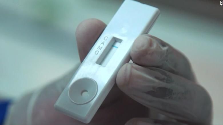
Table of Contents

List of Tables ..................................................................................................................... vi

List of Figures ................................................................................................................... vii

I. Introduction ..................................................................................................................... 1

                  Multiple Myeloma and The Importance of Rapid Diagnostics Testing ...........1

                  Existing Diagnostics Technology .....................................................................2

                  Exploring the Development of New Devices ...................................................3

                  Accesibility of paper based microfluidic devices .............................................4

                  New methodologies and techniques to fabricate μPADs .................................5

                  The complexity of paper devices ......................................................................6

                  Lab on a paper .................................................................................................7

                  Paper diagnostics testing ..................................................................................8

                  Multipurpose devices .......................................................................................9

                                                                     iv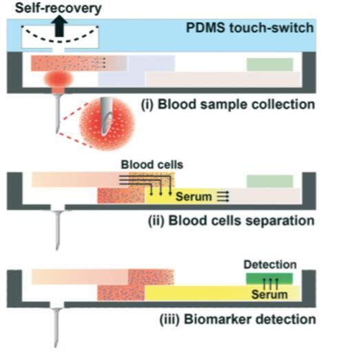
Comparing traditional devices with μPADs ...................................................11

                 Overcoming challenges of paper diagnostics devices ....................................13

                 Engineering better rapid diagnostics testing devices ......................................14

II. Materials and Methods............................................................................................... 16

                 Configuring the optimum fabrication techniques ...........................................17

                 Constructing various microfluidic paper-based devices .................................20

                 Controlling complex fluid flows.....................................................................23

                 Sample processing and analysis .....................................................................26

                 Fabrication of paper-based analytical device .................................................28

III. Results ....................................................................................................................... 30

                 Visualy quantifying human immonoglubulin levels ......................................30

                 Sufficiently detecting IgG levels ....................................................................32

                 Design prototype model example ...................................................................33

IV. Discussion .................................................................................................................. 34

V. References ..................................................................................................................47

                                                                  v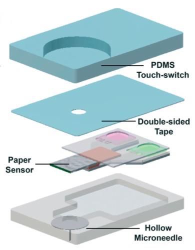
List of Tables

Table 1. Limitations and Advantages of Various Fabrication Techniques….…….…….24

Table 2. The Visualized Quantitation of Human Immunoglobulin….………………….30

                                       vi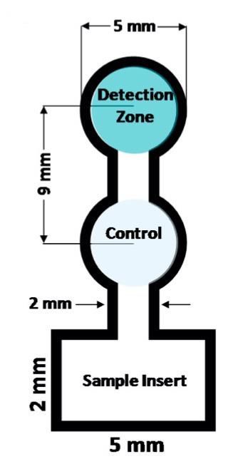
List of Figures

Figure 1. Fabrication techniques .......................................................................................17

Figure 2. Various two-dimensional μPAD’s.....................................................................20

Figure 3. The microneedle integration ..............................................................................26

Figure 4. Layout concept for the μPAD colorimetric assays ............................................28

Figure 5. Colorimetric detection of IgG ...........................................................................32

Figure 6. Prototype device construct ................................................................................33

                                                           vii
Chapter I.
Introduction
Mature plasma cells in the bone and bone marrow can become cancerous and lead
to the development of multiple myeloma. It is the most common hematological malignancy
in patients over 69 years of age, with an incidence rate of six per one hundred thousand
patients per year in Europe and the U.S. The median survival rate has improved to six years
in the past decades due to improvements in supportive care and early diagnosis (Röllig,
2015).
The multiple myeloma disease is usually characteristic on countries with high
income individuals and it begins as a non symptomatic condition that gradually progresses
and patients develop multiple deformities on their genetic code that include anomalies in
their heavy chain immonoglubulin. There are numerous drugs and treatments on the
market for this disease that its true origin is still unknown and not fully understood from
the scientific community. Patients that suffer from this condition usually experience
various health conditions such as renal failure, bone lesions, anaemia and hypercalcaemia.
Ongoing research in this field gives hopes to patients for better and more advanced
treatments in the near future, and current developments in the diagnostics field help
physicians to early diagnose this disease which directly leads to more lives saved and
extended livespan.
Patients that are suffering from multiple myeloma have an increased mortality as
well as morbidity rates, due to the fact that this disease leads to multiple complications on
1patients that include high rates of infections, gastrointestinal upsets and bone diseases. All
of these challenges increase the likelihood for patients to not be able to recover from this
sickness and most often they relapse after they have received various medical treatments
and there is a high rate of disease resurfacing within a short period of time upon
chemotherapy. In the early days of human civilization patients that had this disease died
within sixty days, and during the last few decades with the invention of novel therapies this
have reduced significantly mortality rates and have increased patient survival and even in
some instances a full recovery. The disease affects the human body in various ways starting
with cardiovascular, renal, bone disease, infections, and peripheral neuropathy. Since
multiple myeloma can lead to various and complex health complications, scientists and
physicians are developing systemic treatments that leads to better symptoms management,
lowering toxicity risks from administering multiple medicines simountaneously to disrupt
the progression of this disease.
The inability of having non reliable and non rapid diagnostics devices for detecting
myltiple myeloma, especially in some parts of the world where healthcare is not very
advanced, realible and easily accessible can lead to disease progression that significantly
lowers survival rates. Collaboration among various research institutions and various
governments for effectively sharing data and statistics among each other most often leads
to a collaborative multidisciplinary effort to combat and develop better diagnostics and
treatments for this devastating health condition which claims numerous lives on a yearly
basis worldwide. Multiple myeloma patients when they relapse, health officials conduct
charts and models to understand statistically the underlying causes of this issue. They
assess the situation by looking for patients’ characteristics, previous treatment charts,
2tumor characteristics, as well as social economical status and accessibility to quality health
care facitities. The ability of scientists to sequence the whole patient genome relatively
easy and rapidly is leading to personalized healthcare and most patients do received
personalized medicine to combat this disease (Van de Donk, 2021).
Multiple myeloma patient needs to have access to reliable diagnostics testing,
especially rapid, efficient and realiable testing, for monitoring their disease progress during
treatment as well as upon successfully completing their drug regimen. Microfluidic devices
increasingly demonstrate their ability to be effective in addressing the unment demand for
having quick methods for patients to test their daily disease status at home without the need
to go to the hospital frequnetly and perform extensive tests. Detecting biomarkers in blood
and urine via rapid diagnostics paper microfluidic devices can show patients if their
treatment works or if they need to immediately see a doctor for further extensive laboratory
testing. The bioengineering science is making impactful progress in developing
microfluidic devices that can detect biomarkers, validate and quantify their concentrations
and instantly produce quality results.
Multiple myeloma often progresses rapibly into the body and as it spreads and
affects various organs, physiological signals are good indicators that show the health status
of patients. Physicians by training can rapidly detect malfunctions in our physiological
appearances and order laboratory tests for further evaluations and technicians usually run
various assays to detect biomarkers in the blood or urine while testing for various diseases
and abnormalitites in the human body. There are two categories of biomarkers, the most
common one is testing for high fever, blood pressure, heart rate, breathing conditions and
various physiological traits that are easily detected upon the initial patient evaluation
3procedures. Genetic testing, molecular level disease research, and advances in technology
are expending the reach of diagnostics and biomarkers now can analyze our cells proteins,
nucleic acids, metabolites and numerous other indicators. These innovations lead to
improving disease detection, screening, prognosis while providing the scientific
community with multiple options for treating their patients and delivering personalized
healthcare.
Rapid diagnostics paper microfluidic testing is a novel and innovative approach
that is able to streamline the meticulous requirements for using biomarkers to detect,
authenticate, and discover reliable assays for detecting and monitoring diseases. Patients
upon expressing medical signs and changes in their physiological status often times they
seek immediate medical attention in which the medical professional orders an array of tests
for diagnostics purposes. There are three main categories of biomarkers and they are
classified as such: protein biomarkers which usually are evaluated under mass spectrometry
electrophoresis. Then, there is nucleic acid biomarkers that require genetic testing
technology for evaluation such as multiplex PCR/RT-PCR and DNA/RNA array essays.
As well as small molecular biomarkers that require metabolomic technology such as Mass
spectrometry NMR (Hung, 2014). Our approach is using biomarkes found in blood and
urine for disease diagnosis using paper based microfluidic devices.
Numerous advances in multiple myeloma diagnostics are helping patients to better
detect the disease early. Most of these advances do require expensive instrumentation,
lengthly times for getting the test results and various resources. There is a high demand for
developing reliable testing that patients can self administer at home and be able to read the
data effectively independently without the need to see their doctor on a daily basis during
4treatment or after relapse. Some tests for multiple myeloma patients are invasive such as
the bone marrow testing, or the new HevyLite testing which is a new serologic testing that
required drawing certain quantities of blood. Diagnostic imaging is also helping doctors to
see more accurately and visualize the disease and advances in the field provide high quality
imaging that permits accurate disease monitoring and rapid intervation when conditions
worsen rapidly (Barley, 2016).
In certain geographical regions where multiple myeloma patients do not have
access to adequate diagnostics testing, it is crucial delivering point of care devices that can
rapidly and cheaply diagnose diseases via the use of microfluidics technology. Paper is an
excellent construct due to its low cost, and ability to dispose easily and friendly to work
with and engineer around it. Paper-based microfluidic devices can be shipped around the
globe very effortesly and their portability makes them ideal candidates for analyzing small
volume samples instantly in the field. Healthcare workers require minimal training on
learning how to effectively administer simple testing solutions to their patients and with
clear instructions the patients can even self administer the test at home. Urine can be
applied directly to paper diagnostics devices and patients can have an instant readout via
colorimetric detection. Blood is a little bit more challenging due to its viscocity, low travel
speeds on the paper construct and the need to administer microneedles to get small droplets
of blood on the device (Raju, 2018).
Scientists are always exploring various methodologies and techniques on how to
successfully and reliably fabricate paper based microfluidic devices in ways that are
efficient, user friendly and via designing scalable mechanisms for successfully transferring
the technology across the globe. Some of the most common and well studied methods
5include inkjet-printing for the creation of hydrophilic channels as well as composing areas
that will host the dry reagents for colorimetric detection. Other techniqies include
microwave irradiation, heating and plasma treatment for developing μPADs. Shelf life is
an important aspect that requires special attention since these devices might be produced
at one continent and then have to be shipped to another region of the world that is difficult
to deliver new supplies upon short notice. A simple example of a paper microfluidic device
is one that you combine in a sandwich method a filter paper and a hydrophobized paper.
Inkjet printing can help create hydrophilic channels and zones that can be used for the
sample area, test zone and negative control. Then via administering urea on these channels
that can distribute the sample evently the scientists can test and experiment via changing
various variables until they design a fully functional prototype that generates reliable data
(Malekghasemi, 2016).
New emerging technologies such as self-contained system build with microfluidic
channels embedded into them are able to facilitate complete and fully sunctional biological
assays. Simple test devices are easier to build then more complex structures that require a
more refined engineering approach for multilevel testing purposes on more then one
variable simultaneously. Flow variables, shear stress, environmental temperatures,
variation in chemicals concentration, and microscale geometries require precise
engineering for paper based microfluidic devices to function at their optimum capabilities,
be robust and realible at all times. Some good examples of self-contained testing devices
are the self-administering pregnancy tests. They use capillary forces to move urine through
the channels, and the sample binds to the antibodies that are placed on the device for
colorimetric rapid reading and instant results.
6Multiple myeloma requires a more complex testing mechanism since working with
blood is more challenging than urine due to its convoluted properties. Blood often times
requires lengthier sample preparation procedures such as sample mixing and filtration, and
testing success is often times attributed to simulntaneously processing and using various
reagents. Microfluidic technologies and its constituents are crucial in designing a fully
operational self-containing testing device that can function as a single point diagnostics
platform. 3D printing is enabling the incorporation of miniature constructs on these devices
which scientists can add easily on their design, such as various mixers, valves and even
pumps for experimenting and testing numerous variables (Boyd-Moss, 2016).
Lab-on-a-paper is a new concept that aims to mimic an actual laboratory on a paper
construct, meaning that users can perform on a simple paper diagnostics device as a similar
level of experimentation as they will to use a fully equipped laboratory. Research and
development in the last decade show that technology is enabling us to replace traditional
laboratory procedures with new low-cost alternatives that can deliver data of a satisfactory
levels. Paper-based microfluidics are instrumentations that can be used beyond just rapid
diagnostics devices but as drug delivery vehicles as well or they can work simuntaneously
and in harmony with the diagnostics and drug delivery modality. Scientists are constantly
creating new and innovative point-of-care diagnostics using various paper models and
showing applications of these devices in the field of infenctions disease diagnosis,
environmental testing, veterinary applications, as well as food testing for the presence of
pathogens.
Micro- and nanotechnology is helping the paper diagnostics as well as the targeted
drug delivery field that can be initiated from the same diagnostics device into the patient
7in a more complex mechanism. Paper is advantageous over other material due to its
excellent ability to be used simultaneously for transporting fuids effectively, while filtering
various and specific sample components. Paper can also store various dry reagents at
derirable concentrations and scientists can easily integrate various microfluidic systems
and build a high-quality paper diagnostics point-of-care device for testing and generating
rapidly reliable data. The fact paper is inenxpensive and easily to fabricate allows scientists
to experiment multiple models and build various prototypes and having the abundance to
choose the most reliable and efficient model for further experimentation before
commercialization (Mao, 2020).
Paper diagnostics testing is being used in real world and there are numerous case
studies that we can use as a reference to discuss the evolution of this engineering field as
we analyze the data from past events. In Uganda rapid diagnostics testing was introduced
to test for malaria, thus addressing the lack of laboratory equipment and skilled personnel
is certain geographical areas. The goal is to perform the test in low-resource settings from
workers or even individuals that do not have advanced skills or access to instrumentations
and be able to generate realiable data. World Health Organization is continuously
promoting the invention of new diagnostics tools that are low cost, have prolonged shelf
life, they are temperature robust, and easily transportable across the globe. Nonprofit
organizations as well as the private sector is conducting several pilots with various
developing countries like Cambodia and is training community health workers to
effectively administer the tests and easily dispose them without the need of biohazard waste
collection due to the fact that a simple paper test can be effectively disposed via even
burning it.
8Pilots are excellent educational vehicles because promote a new behavior in regard
to mass testing and allows the communities to understand the importance of early disease
detection. Multiple developing countries have a free healthcare system, this is
advantageous because once cooperations with governments is established rapid diagnostics
testings can be quickly established in all healthcare centers across the nation. To pilot in
developing countries, it is also important to successfully get approval from the nation’s
ethical committees and gain all the necessary permits thus complying with local and
international laws. Equal distribution and mass accessibility for rapid diagnostics testing
can help significantly developing regions that are fighting certain topical diseases to work
collectively on eradication of certain illneses (Chandler, 2011).
Paper based microfluidic diagnostics devices are being used to diagnose a wide
array of viruses, for example ebola which traditional testing requires high technical skills
and is of a high cost. Laboratories are not easily accessible in Ebola struck regions and
nucleic acid amplification tests can only reach a small fraction of the overall population
due to their complexity. In Africa Ebola caused up to eleven thousand deaths and
endangered the life of thousands more, thus our paper microfluidic rapid diagnostics
approach is a life saver since it can help these countries early detect new cases and save
nations from humanitarian crisis. Scientists are conducting research by using sdvanced
molecular biology tools while combining bioengineering and nanotechnology expertise to
increase the sensitivity of these tests and bring them as close to the accuracy of nucleic acid
amplification tests.
During pandemic outbreaks, the most common methodology of detecting RNA
viruses, is performed by extracting RNA and performing real-time Polymerase Chain
9Reaction and reverse transcription. This method helps to replicate the genetic material and
search if the virus is present in-patient samples. The current technologies require skilled
personnel, expensive instrumentation and it takes a while for the results to become
available to the patients and health
officials. Nucleic Acid Amplification Tests are expensive, there are logistical issues
transporting and setting up instrumentations in countries with developing infrastructure,
and consumables for running these experiments are not readily available. Sample
preparation can also be an issue and is an add on burden due to the RT/PCR tests are more
complex in nature then easy to use point of care paper based microfluidic diagnostics
devices with colerimetric readouts. Immunoassays is where paper microfluidics technology
has been recently successfully applied, especially in real time events that the diseases were
highly contagious, and samples were difficult to withstand high temperatures when
collected from the field and transported for analysis to the laboratory.
Paper microfluidic multiplex devices are of a high demand and successful
commercialization of more complex and reliable designs will open the door for these point
of care instruments to be more widely adopted from the scientific and healthcare
communities. The ability to simultaneously test the presence of the pathogen as well as the
stage progression of the infection or disease can mean that not only these devices can tell
weather the patient is a carrier or not but also the degree to at what exact stage their
condition status is at the time of the test. Multiplex devices require elaborate and complex
fluidic connections that properly integrate with all device channels and are able to
successfully transport the sample at various end points and generate data instantly on the
10spot. Reagents are freeze dried and stored on the device so upon sample introduction they
react and are capable via colorimetric changes to spot the presence or lack of antigens.
Paper microfluidic devices when compared to traditional RT/PCR tests must be
capable of producing reproductable data with a statistical significance for the scientific
community to be able to trust and start using these new novel technologies for their routine
testing protocols. Multilayer devices as well as more simplistic paper strip tests that have
controls embedded in them are momentarily used as preliminary data sets and when signals
are detected patients proceed with second laboratory tests to precisely validate the initial
test results (Magro, 2017). We aim to construct a multiple myeloma paper microfluidic
device that will be able to tell us weather the disease is present or not, as well as provide
more analytical data to the status level of the antibody’s levels in patients blood or urine
samples. Proof of concept will occur once our construct is able to reliably determine
weather patient IgG antibodies are statistically present to indicate the appearance or
reappearance of the disease and then bioengineer the device to indicate the severity of the
illness. Urgent cases will be instantly reported, and further testing will determine and
validate the results of the initial rapid diagnostics testing through our point of care devices.
More testing being performed on instant point of care paper microfluidic devices
and a lot of data that is statistically significant collected, analyzed and compared with more
traditional instrumentation testing, will be able to determine the efficacy and reliability of
our rapid diagnostics paper microfluidic devices. Consistent performance results will show
the scientific community that our model is reliable and constantly delivers results capable
of early detecting disease reappearance on multiple myeloma patients. At home user
friendly devices provide patients with low-cost tests that can be performed at any time,
11thus indirectly helping the patient with their peace of mind as well and reducing anxiety
when they get reassurance that their disease is under control and have access to testing
instantly from the comfort of their home.
The fact that nucleic acid testing techniques are the most crucial modalities for the
medical diagnosis field, positions paper microfluidics testing in a competitive edge capable
of successfully overcoming the current models’ limitations such as the multistep levels
required for generating test results data in environments where speed is crucial in patient’s
accessibility to treatment. Recent years innovation allows paper microfluidics devices to
implement in a single point of care instrument stepps such as nucleic acid extraction, as
well as amplification followed by instant real time disease detection. Traditional elaborate
and time-consuming techniques such as extraction can be replaced with successfully
implementing on the paper construct a sample pad to perform this step. PCR machine can
be replaced with conjugated gold nanoparticles and electrophoresis with colerimetric
measurement channels that lead to a secondary absorption pad with the entire task being
completed in under a minute. Traditional instrumentation of the step’s extraction,
amplification and detection require an average of five hours without taking into the account
sample collection and reagent preparation for running the tests. In addition to colorimetric
detection, for increased accuracy patients can use personal smartphone sensors for data
redout and validation of their test results in real time (Choi, 2015).
Lateral-flow and dipstick microfluidic devices have dominated the rapid
diagnostics market in the past few decades. Current limitations in quantitative
measurements and specificity are pressuring the current demand for available assay kits in
the consumer market. Paper-based microfluidic devices are the new upcoming diagnostics
12platform that outperforms existing assays in surface influenced settings. Paper
microfluidics is the new lightweight, low-cost, disposable, and reliable technology, which
are easy to fabricate while engineering them for rapid diagnostics tests (Yetisen, 2013).
Existing technologies include lateral flow and dipsticks first introduced to measure
glucose levels in urine in the 1950s-60s (Free, 1957). Nowadays, urinary dipsticks are used
in an array of applications ranging from urinary infections, carbohydrate metabolism
disorder, as well as kidney and liver disorders. The serological lateral flow tests were born
from the invention of the now-famous pregnancy tests from the discovery of the hCG beta-
subunit radioimmunoassays (Vaitukaitis, 1972). Current lateral flow immunoassays are
complex and require the connection of multiple compartments to become fully functional
devices. They need a conjugate pad, sample pad, absorbent pad, and reaction membrane.
They are time-consuming, with a lack of specific quantitative measurements of the
substrate of interest and the need for skilled personnel to operate the instrument (Tisone,
2009).
Lateral flow tests have advantageous characteristics such as low sample
preparation, portable, they are light and relatively inexpensive, but there is an increased
demand for improved sensitivity, be more quantitative and multiplex (Wong, 2005).
Creating paper microfluidic devices that outperform current lateral flow assays requires the
processing of biological samples in relatively small volumes, to the ability to produce
instant results, increased specificity, and quantitative capabilities. The nonspecific
background on the paper microfluidics should be minimal in comparison to the sharp
capture lines that will quantitatively detect the antibody levels of myeloma patients (Lee,
2010). We aim to engineer a superior paper-based microfluidic device that only requires
13the user to add the sample and quickly interpret the results. Our assay will require minimal
effort from the user, introduce increasing levels of simplicity, and be inexpensive. The
constructing fluidic channels will be designed to separate the sample into multiple
channels, thus creating a multiplex paper-based microfluidic device for precisely
measuring the multiple myeloma antibody levels (Noh, 2010).
There are limitations of using paper microfluidic devices for rapid tests due to the
issue of red blood cells being filtered out, and the plasma separated (Yang, 2011). To
address this problem, we aim at using chromatography paper equipped with blood
separation membranes and sealing them using the wax dipping technique (Songjaroen,
2012). Traditional lateral flow tests use glass fiber to separate blood, and it comes with
engineering restraints, such as the inability to adjust the pH, filter out the undesired
analytes, and effectively separate red blood cells (RBC) from the plasma. Paper
microfluidics address all of these confinements and adequately separate RBC's from the
plasma without the need of using centrifugation (Alter, 1996).
We aim to use conjugate and absorbent pads to capture colloidal particles that will
display color in controlled conduct. These particles will be hydrophobic particles that are
negatively charged and covered with antibodies for enabling target analyte uptake. Our
research will be focusing on experimenting with various pore sizes, protein binding
affinity, thickness, and flow characteristics to improve upon the existing rapid test devices
currently on the market to enhance their stability on storage and create more defined test
lines. Our revolutionary approach will use fast chemical activation techniques on the paper
surface area for quick reaction rates on antibody mobilization. We aim to experiment with
relocating the readout lines closer to the origin for improving kinetics and decrease the
14sample analyte needed for each test (Yetisen, 2013). Hydrophobic barriers can also help to
avert the mix of reagents and promote multiplex assay development. We are going to
experiment with all three available techniques and determine which methods of wax
dipping or screen printing are going to help us achieve the ideal analytical microfluidic
paper-based device (Dungchai, 2010). Overall paper-based microfluidic instruments for
rapid diagnosis offer unique advantages over conventional lateral-flow and dipstick
technology. We aim to improve reaction rates, accuracy, and reliability and improve the
current state of rapid diagnostics paper-based microfluidic devices.
15Chapter II.
Materials and Methods
Current lateral flow immunoassays and one-dimensional paper-based analytical
models require external data analysis while simultaneously being involved and not user-
friendly for patients to individually utilize the technology. We aim to solve the problem by
creating the next wave of rapid testing paper-based diagnostic assays, equipped with the
complexity required to perform compelling analysis as well as providing to the end user
reliable results which are easy to interpret (Cate, 2015). The advantages of using paper are
numerous, including the high surface area to volume ratio for enhancing colorimetric
detection capabilities, the capacity of storing active reagents, and flawless fluid transport
via capillary action (Martinez, 2007).
Our research is focusing on experimenting and choosing the right paper material
for allowing our analyte to flow uninterruptedly and perform optimally on our quality
control and assurance test (Yetisen, 2013). We will be studying capillary flow models to
analyze the flow behavior in paper-based constructs. Some of the factors that influence
flow patterns are surface roughness, pore size variation, liquid infiltration is tortuous, and
noncircular capillary models. As paper-based microfluidic devices become more complex,
flow predictability is a vital research compartment for successfully developing the next
generation rapid test diagnostics instruments (Shou, 2014).
While the paper is a phenomenal substrate for having control over the fluid
direction without the need of external mechanisms, its physical characteristics make it
challenging to manage the direction and flow rate. We aim to strategically alter channel
16construct and engineer pathways that lead to transitioning from a wide to narrower
geometry for increasing the flow rate (Toley, 2013). Engineering porous channels by
cutting the μPADs either perpendicular or parallel to the flow direction enable us to control
the flow direction as well as rate by constructing a variety of cutting slits with other
orientation, length, size, and quantity (Giokas, 2014).
Figure 1. Fabrication techniques for creating a microfluidic paper-based analytical device
(μPAD). From "Recent Developments in Paper-Based Microfluidic Devices" by Cate,
2015, Analytical Chemistry, 87(1), 19-41.
Our vision is the creation of the ultimate multistep processing μPAD assay that is
fully functional and possesses capabilities of generating comprehensive data during one
sample analysis. The testing parameters are wicking rate, substrate-analyte chemistry,
17material grit, and fabrication techniques such as drawing, wax stamping, polymer ink
drawing/stamping; laser cutter, craft cutter; wax printing, inkjet etching, inkjet printing;
wax screen printing, photolithography, or wax dipping (Fig.1) (Cate, 2015). Thus, we aim
to investigate the integration of multiple reagents at various detection regions with
numerous constant diameter regions of filter paper for every reagent processing step. The
ideal paper-based microfluidic device will be compatible, user-friendly, and more
advantageous than current market μPAD assays. We will be incorporating dry state
reagents within the paper pads to decrease the complexity of transporting solutions with
our test kits, thus enduring as well as variations in environmental conditions (Fridley,
2014).
Surface chemistry is crucial for improving reagent chemical stability and fluid
transport, and this is a limitation that we aim at addressing by creating on paper pads with
nanoporous membranes. The ion concentration polarization technique will help us
construct nanoporous layers on the μPADs that will preserve continuous flow transport
even on the most saturated channels. This technique aims to address the current limitations
with folding valves, which can post a problem for multiplex paper-based microfluidic
devices not designed for sequential assay analysis (Yang, 2015).
We aim to incorporate smartphones as an add on feature that will remotely transfer
readout data to processing facilities for further evaluation upon determining that the
multiple myeloma patient antibody levels are increasing significantly. Since we will be
using colorimetric readouts, the commercially available smartphones are all equipped with
flashlight capabilities aiming to quantify assay data by a sole source of light illumination,
thus solving the ambient light challenges that patients might encounter while administering
18the self-test kit. The reagents will transform from low intensity too high-intensity
fluorescence, and consistent light illumination enables the end-user to quantify assay data
(Thom, 2013).
Creating custom handheld μPADs is cost-effective and user-friendly in contrast to
using prominent benchtop instruments for point-of-care settings. Incorporating numerous
colorimetric assays synchronously to measure the antibody levels in multiple myeloma
patients from a droplet of blood or urine sample leads to the creation of an efficient and
reliable device capable of producing instant results. Our machine will produce immediate
color results upon administering the test and have capabilities of transmitting and
transferring the data to a software analysis platform for validation as well as physician
analysis to quality assurance upon test abnormalities detection (Zhao, 2015).
We aim to use colorimetric detection assays on our point-of-care analytical paper-
based microfluidic device for instant test results. The color change will determine the levels
of the specific antigens present in the patient's blood samples for quantitative assay
analysis. To improve the detection times, we will be modifying the paper to increase liquid
transport rates by adjusting the channels and decreasing surface fouling. We are going to
be experimenting with the addition of various compounds such as glycerol additives,
dextran, and polyvinylpyrrolidone while trying and collecting data on the freeze-drying
technique that preliminary research suggests it can stabilize the antibodies in the assay
channels. We aim to test all myeloma patients with blood antibodies and determine which
method is more suitable for our μPAD (Guan, 2014).
The goal of this study is developing the ultimate rapid paper-based microfluidic
testing device for measuring IgG levels in Multiple Myeloma patients at the comfort of
19their home by using blood or urine as their sample. We will be experimenting with an array
of various fabrication techniques and procedures to determine the prime device dimensions
(Cate, 2015).
Figure 2. Various two-dimensional microfluidic paper-based analytical devices (μPAD’s).
From "Recent Developments in Paper-Based Microfluidic Devices" by Cate, 2015,
Analytical Chemistry, 87(1), 19-41.
We will be constructing various two-dimensional microfluidic paper-based devices
and test them to determine in which one of them the samples run smoother. Figure 2
demonstrates various techniques such as: wax stamping technique (Fig. 2A), followed by
wax dipping (Fig. 2B), screen-printed wax (Fig. 2C), wax drawing (Fig. 2D), wax printing
(Fig. 2E), Inkjet etching (Fig. 2F), Inkjet printing (Fig. 2G), flexographic printing (Fig.
2H), computer-guided knife cutting in nitrocellulose substrate (Fig. 2J), laser-cut arched
20channels (Fig. 2K), depositing vapor-phase polymer (Fig. 2L), and UV/O3 patterning
technique (Fig. 2M). The paper material can have a significant impact on the fluidic
behavior and determining the optimum paper substrate is crucial for building a functional
device (Cate, 2015).
We will be experimenting with various chemical modifications to optimize our
paper substrate, such as adding -OH groups in cellulose via an esterification reaction to
make our paper hydrophobic. Another technique that we aim to try is using UV-irradiation
to attach 4-methyacryloxyloxybenzophenone with methyl methacrylate to cellulose for
creating a cross-linking that results in a covalent attachment for polymerizing the polymer
to add hydrophobic capabilities on our paper-based microfluidic device. Our goal is to
make all of our paper modifications under ambient settings, thus avoiding plasma
modifications on paper that require expensive instrumentation and more resources (Li,
2011).
We aim to engineer the surface chemistry for effectively controlling the fluidics of
the sample flow that will help us maintain a color uniformity while enhancing chemical
stability on our μPAD devices. We want to determine the folding angle of the channel
constructs to create an artificial valve for controlling the flow rate. Thus, combining valves
and treating the surface of these paper-based microfluidic devices gives us hydrophobic
channels that can dictate the flow direction. To overcome the oversaturation of pathways,
our μPAD tool will combine the coupling of the ion concentration polarization technique
with a nanoporous membrane to assure the fluids move through freely without an
interruption (Cate, 2015).
21Our ultimate goal is to quantify the analyte using colorimetric detection techniques
precisely and effortlessly integrate the technology to transfer the results on smartphone-
based analysis software. Via taking a picture with the smartphone, we can move the on-
site data remotely to a centralized facility for further evaluation and expert analysis. This
technique will allow us to collect and store the data without the need for transporting the
samples to a laboratory. We aim to take advantage of the flashlight further that smartphones
are equipped with as a source of illumination also to quantify the data and build an app
software that decreases ambient light for better assay analysis (Thom, 2014).
Our design will consist of a user-friendly handheld paper-based microfluidic device
for rapid test analysis of IgG levels in multiple myeloma patients without the need for
expensive benchtop instrumentation. The system will incorporate a colorimetric gradient
scale capable of determining the antibody intensity levels within a sole analyte sample
analysis. The final product will consist of a functional instrument that can operate in a
resource-limited setting and proficiently achieve multiple functionalities. Providing on the
spot instant data and integrating the information via a user-friendly workflow into the
software database for data collection and quality assurance (Rowe, 2011).
One of the barriers that we might face during the fabrication technique is the
background reactivity while building our paper-based microfluidic device using the wax
patterning technique. Creating excellent flow boundaries using wax is a challenge, and
during the melting step, we can end up with inconsistent borders, thus disrupting the liquid
flow. We will be addressing these limitations by creating a metallic stamp for fabricating
channel designs and improving the μPAD colorimetric detection signal. Experimenting
22with various stamping models and integrating a metal mask on our construct, we can
control and preserve the hydrophilic regions on our device (Garcia, 2014).
We will aim to minimize the use of flexographic printing technique due to its
limitations of requiring a specialized printer with personalized plates that leads to limiting
design flexibility. Our goal is to eliminate the need to use inkjet printing because it does
require multiple steps, and without following precise protocols, the resolution and channel
definition will limit device productivity. This method is limiting upon scaling up our
operation since it can only print a single reagent at a time, and our need for high-throughput
fabrication is crucial in delivering inexpensive solutions for commercial applications
(Olkkonen, 2010).
Controlling the complex fluidic flows on the paper-based microfluidic devices is
challenging to fully command the sample flow rate and direction, which can be interrupted
when the method is used as point-of-care instrumentation for urine or blood analysis due
to the variation in liquid viscosity. Manually controlling its flow and direction introduces
the issue of practicability, and further engineering is required to construct the ultimate
channel geometries to an uninterrupted flow design model. Working on such a small scale
is an ambiguous process due to our requirement of fully controlling the flow for increased
accuracy results we need to fine-tune our engineering skill son delivering a device with
perfect channel width, length, and thickness. A small alteration during the design or
fabrication steps can lead to a significant flow delay, leading to a partially functional μPAD
(Martinez, 2007).
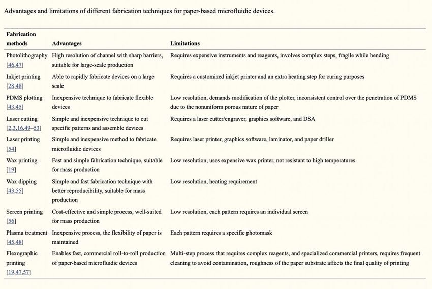
23Table1.
Note. Limitations and advantages of various fabrication procedures for microfluidic paper-
based analytical devices (μPAD’s). From "Paper-based analytical devices for clinical
diagnosis: Recent advances in the fabrication techniques and sensing mechanisms" by
Sher, 2017, Expert Review of Molecular Diagnostics, 17(4), 351-366.
We are aiming to identify the right paper to use for our microfluidic device since
transparency, as well as cellulose paper, has shown low sensitivity and decreased detection
rates. Among the many issues we have to address, special consideration will be given on
controlling evaporation and sample retention since it can lead to significant challenging as
it concerns the transportation of our urine or blood samples. All of our reagents should be
tested and engineered to withstand harsh environmental conditions during the
transportation process of our devices. Before moving to scale up our operation, it is
24required that our testing devices pass successfully clinical validations and the need to have
nonexistent variation in the sensitivity and specificity of our μPADs (Sher, 2017).
We must choose a fabrication technique that will fit our operation perfectly since
almost all of them have their own sets of limitations, table 1. Our goal is to use the process
of elimination for all the current fabrication procedures and pick one that will not require
expensive reagents or instrumentation. We want a high-resolution assay in which the
roughness of our paper will not interfere with the final printing quality of our substrate and
have a uniform surface design. Our ultimate device will be user-friendly, reliable, with
consistent specificity and detectability rates, inexpensive with high-resolution capabilities,
and biodegradable (Sher, 2017).
This research aims to accelerate further the latest progress accompanying the design
and fabrication of μPADs. Various sensing and new techniques are being researched
throughout the world; there is still space for innovation and further advancements in regard
to sensitivity, specificity, and reliability before mass adaptation by the scientific
community. Mass production and widespread acceptance in the diagnostics community for
paper-based microfluidic devices will lead to more patients accessing the technology
cheaply from the comfort of their homes.
Sample Processing and Analysis: Our paper based microfluidic diagnostics device
requires the correlation and integration of various multistep processes that work in
harmony with each other. Traditional methods include the drowning of blood via syringe
and its processing in various microtubes via pipettes and other laboratory consumables for
their safe usage and then proper safety disposal upon protocol completion. Our approach
is to bioengineer simplified versions of existing techniques and replace the workflow with
25innovative paper-based strategies that lead to a 3-1 model, meaning the incorporation of
sample collection, arrangement, and analysis all in our single usage μPAD (Gong, 2017).
Patient Sample collection:
The objective is to integrate simplicity in sample collection and eliminate the extra
step of using syringes and vials for the transfer of blood to μPAD’s. The ultimate goal is
simplicity and a μPAD that is practical and reliable for providing real time accurate data.
We also want to design a microneedle patch that with a simple adjustable mechanism it
opens and closes before and after administering the test. (Li, 2015).
Figure 3. The microneedle is integrated on the μPAD to enable efficient sample collection.
From "Advancing Paper-Based Microfluidics for Broad Diagnostic Application." by Gong,
2017, Analytical Chemistry, 117(12), 8447-8480.
We are incorporating on our design a microneedle that is activated by pressing the finger
on it (Fig. 3). This will enable us with a simplistic method of performing sample collection,
processing and real time analysis.
26Sample Preparation and Analysis:
Blood components composed of molecular, biochemical and immunological
biomarkers, when introduced to engineered μPAD enables the sample processing to be
initiated instantaneously. Our paper diagnostics device utilizes a significant lesser blood
sample volume that can be in a range of few milliliters and enable in field sample
processing and real time analysis. Capillary tubes within channels on μPAD, allow blood
transport from finger prick to the analysis zone. Red blood cell filtration on a milliliter
scale for size exclusion is enabled by designing a larger surface paper microfluidic area.
On detection zones colorimetric analysis will determine the presence human
immunoglobulin levels in serum, its intensity will indicate they abundance and
concentration (Gong, 2017).
Fabrication of paper-based analytical device:
Our process is utilizing wax printing techniques that uses graphical software for the
design of our paper geometry. Channel barriers are being developed by melting wax at 179
degrees census via the usage of a hot plate. Using adhesive tape, we are preventing leakage
and keeping our device intact, thus creating regions for sample droppage and channels for
its transport and analysis.
27Figure 4. Our layout concept for the μPAD used for human immunoglobulin colorimetric
assays with samples being blood or urine. From “Paper-Based Colorimetric Biosensor for
Tear Glucose Measurements.” By Gabriel, 2017, Micromachines (Basel), 8(4), 104.
Our μPAD is composed of three main areas, the sample inlet being square and with a
specified parameter and two circular zones, one of them being the control area and the
other the testing site respectively, demonstrated in Figure 4 (Gabriel, 2017).
Engineering enzymatic and colorimetric reactions and their appropriate detection:
IgG sensitized solution are being implied in the control zone area and used as a
negative control; the detection zone as well is being coated with Anti-Human Globulin
reagent and IgG sensitized red blood cells to confirm the presence of IgG when blood
sample is being introduced. Administering patient blood aliquots at the insert location and
with the help of lateral flow allows the sample pass through the control and detection zone.
Our goal is to use the blood samples in their raw state without the need of diluting them or
28performing any pretreatment techniques on them. The colorimetric changes must be
obvious with the naked eye and not require additional instrumentation for the readout
(Gabriel, 2017).
29Chapter III.
Results
Multiple Myeloma is a form of cancer that effects the immune system, the
antibodies produced to defend us from various infections are malfunctional in these
patients. There are numerous types of antibodies on the heavy chain as well as light chain.
The heavy chain antibodies are IgA, IgM, IgE, IgD, and IgG, and the ones on the light
chain being Kappa and Lambda. The goal of this research is to construct easy to use at
home paper microfluidic devices that will monitor the progress of the disease along with
the effectiveness of treatment. Our research focuses on measuring quantitatively the levels
of immunoglobulins present in a blood or urine sample. The myeloma protein we are
researching and are most interested on is IgG, and its levels will detect the course of the
disease in our patient’s population.
Table 2.
The Visualized Quantitation of Human Immunoglobulin
Human
IgG
mg/dl
>2000
400-1600The paper construct is designed with sample insert, control and three layers on IgG
detection levels, starting with 2000 mg/dl,
showed in Table 2.
The lower level indicates that the patient is doing well, a level above that it means
that patient needs to closely monitor their progress and it’s the first sign of concern.
Anything above >2000 will be a good indicator that the patient needs to contact their doctor
immediately and proceed with further tests and closer monitoring. Light blue color
indicates normal levels of IgG, and the darker the color the higher the chances that the
patient is relapsing and requires medical attention. Our experiment grasps the coated
antibody and via adding on the solution known quantities of protein we then proceed to
follow the washing and incubate at room temperature. The protein along with the detection
antibody which is conjugated with the horse-radish peroxidase is measured and then
washed after the incubation period. Tetramethylbenzidine and hydrogen peroxide is added
to develop blue color which is then turned into yellow when acid stop solution is applied.
Our laboratory preliminary experiments and demo demonstrate that there is a good
chance of our device to achieving commercialization and deliver this technology to users
in resource scarce areas and in homes. Thus, we aim to demonstrate in this experiment that
users will be able to operate the system and Interpret the results via the naked eye without
the need of extra instrumentation for readouts.
31You can also read