Craniofacial implants in a failed autologous reconstruction of microtia: a case report - International Journal of Implant Dentistry
←
→
Page content transcription
If your browser does not render page correctly, please read the page content below
Frias International Journal of Implant Dentistry
https://doi.org/10.1186/s40729-021-00337-8
                                                          (2021) 7:55
                                                                                                                              International Journal of
                                                                                                                                    Implant Dentistry

 CASE REPORT                                                                                                                                       Open Access

Craniofacial implants in a failed autologous
reconstruction of microtia: a case report
Vladimir Frias

  Abstract
  Plastic surgical reconstruction is considered to be the gold standard for the repair of microtia as the results are
  permanent and constructed from the patient’s own tissue; however, the multiple surgeries required and the
  difficulty in attaining adequate cosmetic results often result in patients choosing a prosthesis as a long-term
  rehabilitation. Advances in osseointegration in the craniofacial region have improved the outcomes with auricular
  prosthetics by providing a reliable method of attachment of the prosthesis and increasing patient acceptance. A
  case presentation illustrates the results of both treatment modalities and examines the outcomes on the same
  patient.
  Keywords: Microtia, Craniofacial implants, Auricular prosthesis

Introduction                                                                          Nagata [8]. The Brent technique is based on the ori-
Although the word microtia (micro-otia) literally                                     ginal surgical approach used by Tanzer but uses four
translates as “small ear,” the clinical condition pre-                                surgical stages instead of the original six. The proce-
sents as anything from an ear that presents with                                      dures include the fabrication of the auricular frame-
minor deformities but with all major landmarks                                        work with costal cartilage followed by transposition of
present, to a severely malformed ear that presents                                    the lobule, elevation of the framework, and recon-
with few identifiable landmarks [1]. The remnants of                                  struction of the tragus. The number of stages needed
the auricle may be displaced, and the condition is                                    for reconstruction has often been cited as a deficiency
often associated with aural atresia, hearing loss, and                                of the technique as, in practice, the number of surgi-
craniofacial syndromes [2]. Risk factors for developing                               cal procedures including revision procedures can
microtia include embryonic vascular disruption; envir-                                often reach seven or eight. Although the Nagata tech-
onmental factors such as maternal age, illness, or                                    nique also uses autogenous rib cartilage, it differs
medication; or genetic pathways [3].                                                  from the Brent technique by proposing two stages
  Options for the rehabilitation of microtia have in-                                 which combine framework harvesting and contouring,
cluded plastic surgical reconstruction and craniofacial                               tragus reconstruction, and lobule transposition in one
prosthetics with or without the use of osseointegrated                                procedure followed by framework elevation at the sec-
implant retention mechanisms [4]. The use of au-                                      ond stage. This reduces the number of surgeries sig-
togenous rib cartilage for the reconstruction was de-                                 nificantly; however, the procedure has been shown to
scribed by Tanzer [5], and his method has formed the                                  result in an increased rate of complications including
basis for most current surgical options. Two widely                                   flap necrosis, framework extrusion or resorption, and
used and successful techniques based on autogenous                                    increased donor site complications. Even in success-
rib grafts have been proposed by Brent [6, 7] and                                     fully treated cases with either technique, there is
                                                                                      often an esthetic compromise resulting from the lack
                                                                                      of definition of the concha and surrounding
Correspondence: vladimir.frias@roswellpark.org
Department of Oral Oncology, Roswell Park Comprehensive Cancer Center,
                                                                                      structures.
Elm & Carlton Streets, Buffalo, NY 14203, USA

                                      © The Author(s). 2021 Open Access This article is licensed under a Creative Commons Attribution 4.0 International License,
                                      which permits use, sharing, adaptation, distribution and reproduction in any medium or format, as long as you give
                                      appropriate credit to the original author(s) and the source, provide a link to the Creative Commons licence, and indicate if
                                      changes were made. The images or other third party material in this article are included in the article's Creative Commons
                                      licence, unless indicated otherwise in a credit line to the material. If material is not included in the article's Creative Commons
                                      licence and your intended use is not permitted by statutory regulation or exceeds the permitted use, you will need to obtain
                                      permission directly from the copyright holder. To view a copy of this licence, visit http://creativecommons.org/licenses/by/4.0/.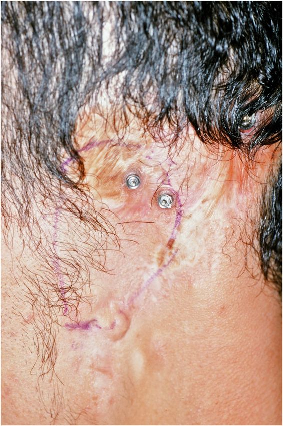
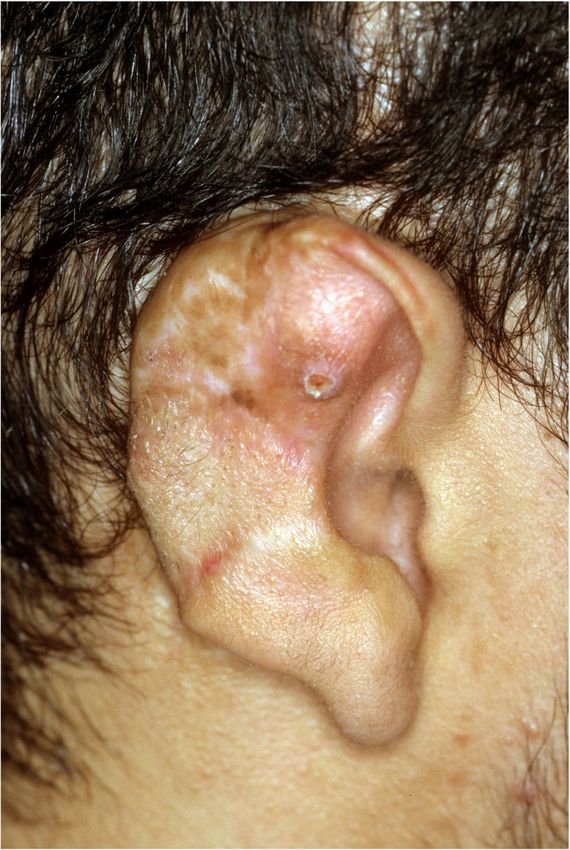
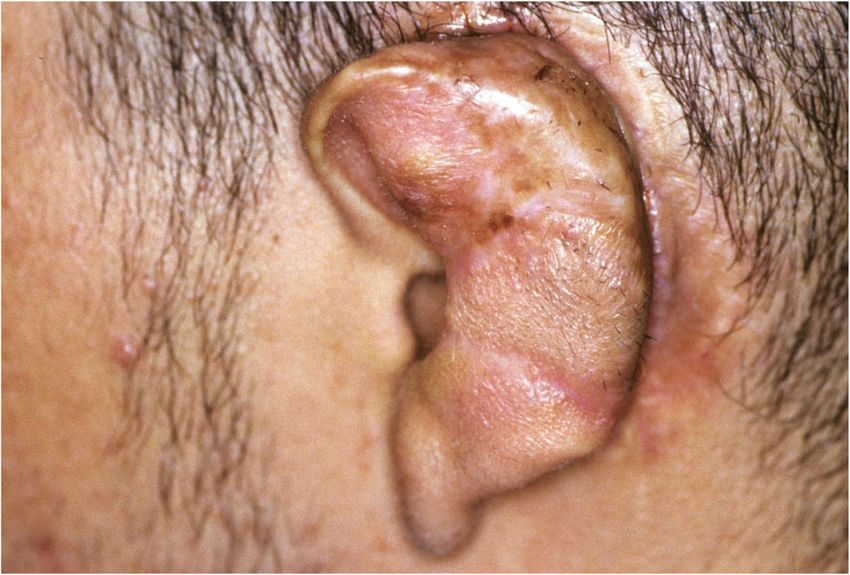
Frias International Journal of Implant Dentistry   (2021) 7:55                                                    Page 2 of 7

   Advances in biomedical engineering may eliminate
some of the problems with the surgical reconstruction of
auricular defects. Current experiments are focused on
the creation of tissue-engineered cartilage that has im-
proved elasticity compared to harvested rib cartilage [9].
The advantage of this technique is that it allows a pre-
cise framework to be created in the laboratory rather
than sculpting the cartilage in the operating room, and it
also reduces the surgical invasiveness of the reconstruc-
tion [10].
   An alternative approach to the treatment of microtia
has been to use a prosthetic material to replace the miss-
ing or malformed portions of the ear. The use of artifi-
cial prostheses to restore facial structures has been             Fig. 2 Left auricle, post-reconstruction
recorded since ancient times [11], and the use of leather,
fabrics, clay, and metal as prosthetic materials and reten-
tion mechanisms have all been reported [12]. The im-             of prosthetic attachment have improved the issues with
provement in dental materials in the twentieth century           durability, cosmetics, and retention. Because of the fa-
allowed for increasingly realistic prosthetics; however,         vorable conditions at the mastoid region, the success
many of the newer materials did not possess the durabil-         rate of osseointegrated implants retaining auricular pros-
ity required of a long-term prosthetic restoration, nor a        theses or bone-anchored hearing aids has been excep-
reliable method to attach it to a defect. The evolution of       tionally high with success rates above 95% [14].
advanced silicone elastomers and the introduction of               The use of craniofacial prosthetics for the rehabilita-
osseointegrated craniofacial implants [13] as a method           tion of microtia often results in superior esthetic results

 Fig. 1 Right auricle, post-reconstruction                        Fig. 3 Implants with abutments attached

Frias International Journal of Implant Dentistry       (2021) 7:55                                                          Page 3 of 7

with a minimal number of surgical procedures; however,               of rehabilitation centers, the cost, and the time involved
there are several marked deficiencies with this approach,            in either reconstructive method, many patients choose
chief amongst which is the removable nature of the re-               their treatment options by what is available locally rather
construction. The daily maintenance procedures which                 than explore all available treatments. A careful consider-
include careful debridement of the supporting structures             ation of the position and size of the auricular remnants
with a brush and detergent can also be complicated for               and the long-term needs and desires of the patient
patients with limited mobility [15]. Despite the high suc-           should be included in the decision-making process prior
cess rates of auricular implants, there are multiple issues          to the selection and implementation of a therapeutic
that arise with the tissue and the prostheses themselves.            protocol. The following case presentation illustrates the
Local tissue reactions from erythema through granula-                different results attained in a patient who had both a
tion tissue have been noted [16]. Studies have also                  plastic surgical reconstruction followed by an osseointe-
shown that the remake rate of an auricular prosthesis                grated craniofacial prosthesis.
due to poor fit or discoloration is approximately 14
months. Other complications that may arise are loss of               Clinical report
retention of the attachment clips, loosening of bar screw            A 19-year-old man presented with a history which in-
or abutments, separation of the retention clips or acrylic           cluded bilateral congenital defects that had been surgi-
base from the silicone, and rupture of the silicone [17].            cally reconstructed over the past 6 years. Surgical
Also important is the fact that the removal of residual              intervention was begun on the right ear at age 13, and
tissues for the placement of osseointegrated craniofacial            the surgical procedures were deemed complete after six
prostheses often eliminates the possibility of future plas-          procedures (Fig. 1). Surgical intervention for the left ear
tic surgery reconstruction.                                          was begun, and after a total of seven procedures, the
   In many cases, the ultimate choice of rehabilitation is           surgical course of treatment was completed (Fig. 2). The
determined by the experiences and the expertise of the               patient was especially dissatisfied with the appearance of
treating physicians, and because of the limited number

 Fig. 4 Pickup of the impression copings in silicone                  Fig. 5 Abutment analogs connected to the impression
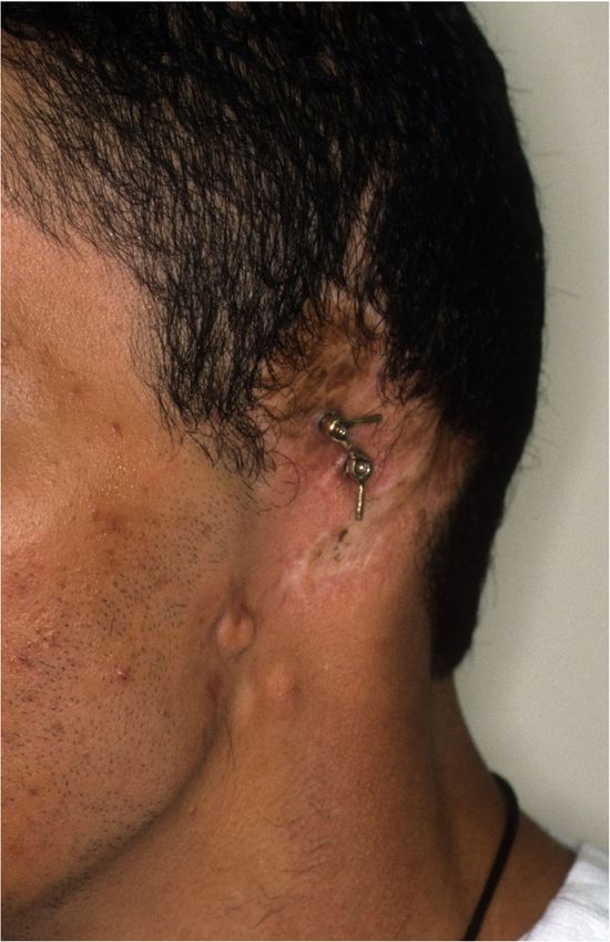
Frias International Journal of Implant Dentistry    (2021) 7:55                                                       Page 4 of 7

his left ear, and his plastic surgeon gave him the option         and 4mm with a diameter of 4.5mm. The length of im-
for an implant-retained craniofacial prosthesis.                  plant chosen is determined from a pre-operative medical
   An impression of the reconstructed ear was made with           CT which allows bone measurement as well as clinical
irreversible hydrocolloid [Jeltrate, Dentsply Caulk, Mil-         confirmation of osseous housing during surgery. Cover
ford DE], and a wax pattern of an idealized auricle was           screws were placed, and the implants were submerged
created and approved by the patient. In cases where a             and allowed to heal for 3 months. On completion of
normal ear is present, a copy of the opposing auricle can         healing, the implants were uncovered and multi-unit
be created in a mirror image from digital data; however,          abutments were attached. The outline of the wax pattern
since the patient was not satisfied with the esthetics of         was traced to confirm positioning of the abutments and
his contra-lateral ear, the pattern for the new auricle was       for framework design (Fig. 3). Impression copings were
created by hand. The wax pattern was replicated in                attached and connected with pattern resin (GC pattern
acrylic to create a surgical guide for implant placement.         resin, GC America, Alsip, IL). The impression was made
Since the meatus would be the only remaining anatom-              with injectable low-viscosity vinyl polysiloxane (Reprosil,
ical landmark after removal of the reconstructed ear, the         Dentsply Caulk, Milford DE) and reinforced with putty
guide was scored along the inferior ala-superior meatal           silicone (Figs. 4 and 5). The impression was boxed and
line to allow for ease of orientation. The removal of the         poured according to standard techniques. A pattern for
auricular reconstruction and placement of the implants            the retention bar was created by connecting plastic bar
were performed at the same surgical appointment. Two              patterns (CBS bar system, Attachments Intl, San Mateo,
VXI 300 implants [Vistafix 3, Cochlear Americas, Cen-             CA) to the prefabricated gold cylinders screwed to the
tennial, CO] were inserted into the mastoid area at posi-         abutments (Fig. 6). A silicone index for the helix and
tions which would allow the placement of the                      anti-helical portions of the auricle was created, and the
framework under the antihelix. The Vistafix 3 implant             bar extensions were adjusted to fit under the contours of
system is specifically designed for craniofacial applica-         the wax pattern of the auricle. The bar pattern was
tions, and the implants are available in lengths of 3mm

 Fig. 6 Framework designed to fit under the helix                  Fig. 7 Retention bar cast in type III gold alloy
Frias International Journal of Implant Dentistry (2021) 7:55 Page 5 of 7
invested and cast in type III dental gold (Fig. 7), then
polished and verified (Fig. 8). Matching retention clips
were placed on the bar and connected with acrylic resin
(Fig. 9). Retention nodules were placed on the acrylic
framework to aid in adhesion to the silicone overlay.
The wax pattern for the final prosthesis was created
using the original wax pattern as a guide, and the bar
and wax pattern were tried in and verified. The wax an-
terior to the tragus was extended anteriorly to prevent a
gap from developing between the prosthesis and skin
surface on jaw opening (Fig. 10). The patient’s skin
shade was matched using the FI-SK skin coloration sys-
tem (Factor II Inc., Lakeside, AZ). The wax pattern was
finished and invested, and the prosthesis was processed
in silicone (A2000, Factor II Inc., Lakeside, AZ) using
conventional techniques. The prosthesis was extrinsically
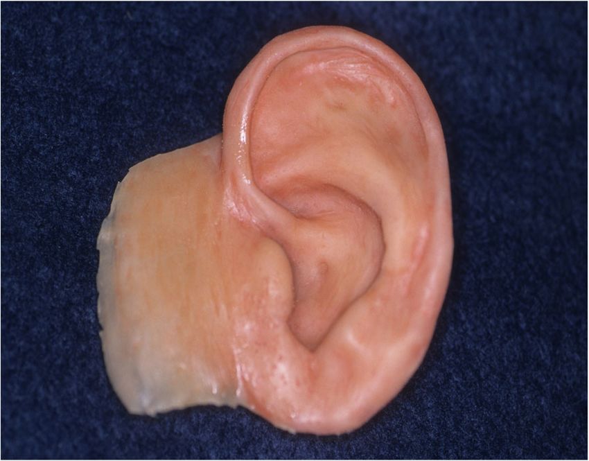
colored and sealed before delivery to the patient (Fig.
11). The sequence of procedures is delineated in Table
1.
The patient was instructed in home care, and a recall
system was set up. He was followed up annually for 3
years prior to moving away. Mild erythema and buildup Fig. 9 Retention clips blocked out on the bar
of sebum were noted at the first recall which was
eliminated with improved hygiene of the area using a
soft bristle brush and 3% hydrogen peroxide. The issue
was evaluated by visual and tactile examination. No evi-
dence of swelling, bleeding, or suppuration was noted.
The prosthesis was remade at 18 months due to the de-
terioration of the silicone flap. No complications were
noted with the implants, abutments, or bar and clip
framework. The tissue reaction and remake rates are
similar to published studies [18].
Fig. 8 Bar framework attached to implants Fig. 10 Processed intrinsically colored prosthesis with tissue flange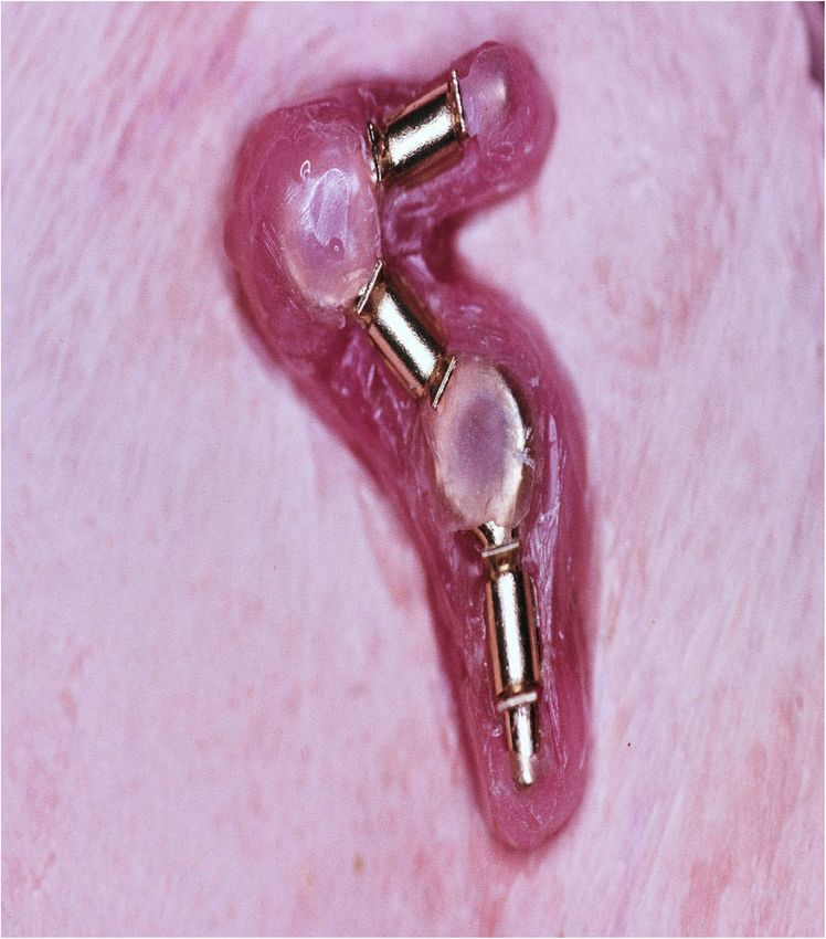
Frias International Journal of Implant Dentistry    (2021) 7:55                                                                       Page 6 of 7

                                                                  final reason for the use of osseointegrated prostheses is
                                                                  the failure of a surgical reconstruction due to extrusion,
                                                                  resorption, or a poor cosmetic outcome. The clinical
                                                                  presentation shows how even multiple surgeries by an
                                                                  experienced surgical team can result in less than accept-
                                                                  able cosmetic results and an osseointegrated prosthesis
                                                                  can serve as a viable alternative.

                                                                  Acknowledgements
                                                                  Not applicable.

                                                                  Author’s contributions
                                                                  The author read and approved the final manuscript.

                                                                  Funding
                                                                  Not applicable.

                                                                  Availability of data and materials
                                                                  Not applicable.

                                                                  Declarations

 Fig. 11 Lateral view of the completed prosthesis                 Ethics approval and consent to participate
                                                                  Consent for treatment and use of pictures were obtained and on the file.

                                                                  Consent for publication
                                                                  Consent for publication was obtained and on the file.
Conclusions
A clinical case presentation illustrates the treatment al-        Competing interests
ternatives that face the patient suffering from microtia.         Vladimir Frias declares that there are no competing interests.
Since the surgical reconstruction of microtia is often
                                                                  Received: 10 February 2021 Accepted: 30 March 2021
begun at a young age, parents are usually faced with a
decision to undergo a series of operations for the recon-
struction of the auricle or to have craniofacial implants         References
placed. The use of one or the other treatment modality            1. Weerda H. Classification of congenital deformities of the auricle. Facial Plast
                                                                      Surg. 1988;5(05):385–8. https://doi.org/10.1055/s-2008-1064778.
is often dependent on the skills and training of the surgi-       2. Bly RA, Bhrany AD, Murakami CS, Sie KCY. Microtia Reconstruction. Facial
cal staff as auricular reconstruction is a highly special-            Plast Surg Clin North Am. 2016;24(4):577–99. https://doi.org/10.1016/j.fsc.201
ized procedure which is not available at all medical                  6.06.011.
                                                                  3. Gendron C, Schwentker A, Van Aalst JA. Genetic advances in the
centers. Although in cases amenable to surgical recon-                understanding of microtia. J Pediatr Genet. 2016;5(4):189–97 (comment k).
struction, it is the option that should be considered first,      4. Wilkes GH, Wong J, Guilfoyle R. Microtia reconstruction. Plast Reconstr Surg.
some parents may decide not to subject their children to              2014;134(3):464–79 (comment k).
                                                                  5. Tanzer RC. Total reconstruction of the external ear. Plast Reconstr Surg.
the multiple surgeries involved in auricular reconstruc-              1959;23(1):1–15. https://doi.org/10.1097/00006534-195901000-00001.
tion. Other reasons for choosing an osseointegrated               6. Brent B. The correction of microtia with autogenous cartilage grafts: I. The
prosthesis are a significant deformity, burns, or a med-              classic deformity. Plast Reconstr Surg. 1980;66(1):1–12. https://doi.org/10.1
                                                                      097/00006534-198007000-00001.
ical condition that precludes surgical reconstruction. In         7. Brent B. The correction of microtia with autogenous cartilage grafts: II.
other cases, the placement of implants for a bone-                    Atypical and complex deformities. Plast Reconstr Surg. 1980;66(1):13–21.
anchored hearing aid is a good opportunity to insert                  https://doi.org/10.1097/00006534-198007000-00002.
                                                                  8. Nagata S. A new method for total reconstruction of the auricle for microtia.
additional implants for a craniofacial prosthesis. The                Plast Reconstr Surg. 1993;92(2):187–201. https://doi.org/10.1097/00006534-1
                                                                      99308000-00001.
Table 1 Sequence of treatment procedures                          9. Pappa AK, Caballero M, et al. Biochemical properties of tissue-engineered
                                                                      cartilage. J Craniofac Surg. 2014;25(1):111–5 (comment k).
Impression of auricular remnants
                                                                  10. Hwang CM, Lee BK, et al. Auricular reconstruction using tissue-engineered
Creation of ideal auricular contours
                                                                      alloplastic implants for improved clinical outcomes. Plast Reconstr Surg.
Creation of surgical guide
                                                                      2014;1333(3):360–9 (comment k).
Placement of implants
                                                                  11. Reisberg DJ, Habakuk SW. A history of facial and ocular prosthetics. Adv
Abutment connection
                                                                      Opth Plast Reconstr Surg. 1990;8:11–24.
Pickup of abutment copings
                                                                  12. Schaaf NG. Materials in maxillofacial prosthetics. Dent Clin Nort Am. 1975;19:
Fabrication of retentive bar
                                                                      347–56.
Fabrication of clip housing
                                                                  13. Tjellstrom A, Yontchev E, et al. Five years’ experience with bone-anchored
Final auricular wax pattern
                                                                      auricular prostheses. Otolaryngol Head Neck Surg. 1985;93(3):366–72.
Mold creation and silicone process
                                                                      https://doi.org/10.1177/019459988509300315.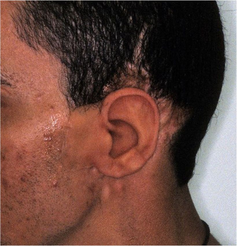
Frias International Journal of Implant Dentistry             (2021) 7:55             Page 7 of 7

14. Jacobsson M, Tjellstrom A, et al. A retrospective study of osseointegrated
    skin-penetrating titanium fixtures used for retaining facial prostheses. Int J
    Oral Maxillofac Implants. 1992;7:523–8.
15. Allen PF, Watson G, et al. Peri-implant soft tissue maintenance in patients
    with craniofacial implant retained prostheses. Int J Oral Maxillofac Surg.
    2000;29(2):99–103 (comment f).
16. Nishimura RD, Roumanas E. Auricular prostheses and osseointegrated
    implants: UCLA experience. J Prosthet Dent. 1995;73(6):553–8
    (comment m).
17. Karacoca S, Aydin C, et al. Retrospective study of treatment outcomes
    with implant-retained extraoral prostheses: survival rates and
    prosthetic complications. J Prosthet Dent. 2010;103(2):118–26
    (comment m).
18. Aydin C, Karakoca S, et al. Implant retained auricular prostheses: an
    assessment of implant success and prosthetic complications. Int J
    Prosthodont. 2008;21:241–4 (Comment b).

Publisher’s Note
Springer Nature remains neutral with regard to jurisdictional claims in
published maps and institutional affiliations.
You can also read