Detection and phylogeny of Wolbachia in field-collected Aedes albopictus
←
→
Page content transcription
If your browser does not render page correctly, please read the page content below
European Journal of Molecular & Clinical Medicine
                                                   ISSN 2515-8260        Volume 09, Issue 03, 2022

 Detection and phylogeny of Wolbachia in field-collected Aedes albopictus and
                Aedes aegypti from Manila City, Philippines
           Maria Angenica F. Regilme1,2, Tatsuya Inukai1, and Kozo Watanabe1,2,*
  1
     Center for Marine Environmental Studies (CMES), Ehime University, Matsuyama, Ehime,
                                             Japan
  2
    Ehime University - De La Salle University International Collaborative Research Laboratory,
                                  Laguna Province, Philippines
                       *Correspondence: watanabe.kozo.mj@ehime-u.ac.jp

Abstract: Wolbachia is the most common bacterial endosymbiont of arthropods, such as the
medically important Aedes albopictus. Recent reports also detected in Aedes aegypti. This study
collected 12 adults Ae. albopictus and 359 Ae. aegypti from 183 households in a dengue-prone
area, Manila, Philippines, between June and September 2017. Aedes larvae (n = 509) were also
collected from 17 water containers from 11 households. The DNA of the Aedes larvae and adults
were screened for the presence of Wolbachia using the wsp and 16S markers, following
optimized polymerase chain reaction (PCR) conditions, and sequenced. Our results showed that
12 out of 12 (100%) adult Ae. albopictus and3 out of 359 (0.84%) adult Ae. aegypti were
Wolbachia positive, whereas all larvae tested negative for Wolbachia (0/509; 0%). The wsp
marker revealed six Wolbachia-positive Ae. albopictus belonging to supergroups A (n = 2) and B
(n = 4). The 16S marker showed Wolbachia in ten Ae. albopictus and 3 Ae. albipictus,most
sequences were in supergroup A (n = 11) and two in supergroup B. Our results revealed
Wolbachia supergroups in field-collected Ae. albopictus and Ae. aegypti with implications for a
successful Wolbachia mass release programs.

Keywords: Wolbachia, dengue, Aedesaegypti, Aedes albopictus

1. Introduction
Wolbachia is a maternally inherited endosymbiotic bacteria, infecting 40% of arthropod species
(Werren et al. 2008; Zug et al. 2012). Wolbachia can alter the reproductive mechanisms of its
hosts, such as cytoplasmic incompatibility, feminization, parthenogenesis, and male-killing
(Fraser et al. 2017). Maternal transmission permits the rapid spread of Wolbachia in mosquito
populations (Fraser et al. 2017). At present, the identified supergroups or major clades are 17 (A-
Q) that infects arthropods such as insects (Kaur et al. 2021). Wolbachia is naturally present in
medically important mosquito species, including Aedes albopictus (Albuquerque et al. 2011;
Joanne et al. 2015; Kitrayapong et al. 2002) and Aedes aegypti(Balaji et al. 2019; Carvajal et al.
2019; Kulkarni et al. 2019 and Teo et al. 2017) that may inhibit the replication of arboviral
pathogens (Iturbe-Ormaetxe et al. 2011). Natural populations of Ae. aegypti were considered
negative for Wolbachia, but recent studies have reported both positive (Balaji et al. 2019;
Carvajal et al. 2019; Kulkarni et al. 2019; Teo et al. 2017; Coon et al. 2016; Thongsripong et al.
2017; Hegde et al. 2018; Bennett et al. 2019) and negative (Gloria-Soria et al. 2018 and Ross et
al. 2019) results for Wolbachia in field-collected Ae. aegypti among different countries. Due to
these varying results, further studies of natural infection of Wolbachia in field-collected Ae.
aegypti is needed.

                                                                                              3060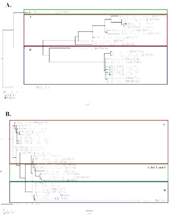
European Journal of Molecular & Clinical Medicine
                                                   ISSN 2515-8260        Volume 09, Issue 03, 2022

The presence of natural Wolbachia strains in field-collected Ae. aegypti may affect the current
release programs. For example, mating of a transfected Wolbachia infected Ae. aegypti with
naturally occurring Wolbachia infected Ae. aegypti may cause reduced or no viable offspring due
to the different Wolbachia strains of the parents thereby, halting the successful invasion of
Wolbachia strains from transfected mosquitoes into the natural population. Most studies have
found that naturally occurring Wolbachia in Ae. aegypti were strains that were phylogenetically
close to the wAlbB strain, which also infects Ae. albopictus(Balaeji et al. 2019;Carvajal et al.
2019;Kulkarni et al. 2019; and Coon et al. 2016). In the study of (Coon et al. 2016) wAlbA and
wAlbB strains were found in Ae. aegypti. Thus, additional Wolbachia detection studies on
natural populations of Ae. aegypti are needed to support the presence of wAlbA strongly. Ae.
albopictus is known to be infected with both wAlbA and wAlbB strains, which belong to the
supergroups A and B, respectively (Zhou et al. 1998).The phylogenetic relationship of the
Wolbachia strains of Ae. albopictus and Ae. aegypti may suggest the possibility of post-lateral
transfer between these two species.
A high Wolbachia detection rate has always been reported in Ae. albopictus(Joanne et al. 2015
and Kitrayapong et al. 2002)because of the reproductive manipulations of Ae. albopictus that
have enabled Wolbachia to spread within the mosquito populations (Nugapola et al.
2017).Wolbachia has been detected in Ae. aegypti natural populations (Balaji et al.
2019;Carvajal et al. 2019; Kulkarni et al 2019, Teo et al. 2017; Coon et al. 2016 Thongsripong et
al. 2017; Hegde et al. 2018 and Bennett et al 2019), but some studies showed negative for
Wolbachia in Ae. aegypti (Gloria-Soria et al. 2018 and Ross et al. 2019). The absence of
Wolbachia in Ae. aegypti and the low detection rate might be because Wolbachia is not well
adapted to Ae. aegypti that makes it difficult to have stable Wolbachia infection and proliferation
in the Ae. aegypti populations. In the study of Teo et al. (2017) they compared the Wolbachia
detection rate in larval Ae. albopictus and Ae. aegypti from Malaysia. Their results revealed
71/284 (25%) Wolbachia infected Ae. albopictus and 4/16 (25%) in Ae. aegypti. However, the
sample size for Ae. aegypti was small to have a solid validation about the comparison of the
detection rate of these two species. Other adult studies also found a high Wolbachia detection
rate in Ae. albopictus (Wiwatanaratanabutr et al. 2013; Nugapola et al. 2017 and Kittayapong et
al. 2000) but did not detect Wolbachia in Ae. aegypti.
Wolbachia load in adult mosquitoes is commonly found in the thoracic muscles, malphigian
tubules, thoracic ganglia, head, ovaries, and salivary glands (Moreira et al. 2009). However,
these organs are still not yet fully developed during the immature stage of mosquitoes which may
cause a lower Wolbachia detection rate of larval Aedes sp. as compared to adult mosquitoes.
Previous studies also revealed that mosquitoes' larval stage has lower Wolbachia density than the
adult stage (Coon et al. 2016 and Tolley et al. 2019) because of the influence of the larval
crowding and temperature changes. The reduced Wolbachia density during the immature stage
can cause failure in the maternal transmission in Wolbachia (Zhou et al. 1998). Thus, Wolbachia
detection rate might be lower in larval stage than the adult mosquitoes.
Here, we studied the Wolbachia supergroups found in field-collected Ae. albopictus and Ae.
aegypti simultaneously collected in the same site using phylogenetic analysis of wsp and 16S
sequences. We tested three hypotheses. The first hypothesis was that both supergroups A and B
can be observed in both Ae. albopictus and Ae. aegypti. The second hypothesis was that Ae.
albopictus has a higher Wolbachia detection rate than Ae. aegypti. The third hypothesis was that

                                                                                              3061
European Journal of Molecular & Clinical Medicine
ISSN 2515-8260 Volume 09, Issue 03, 2022
larval Aedes sp. mosquitoes have a lower Wolbachia detection rate than adultAedes sp.
mosquitoes.
2. Materials and Methods
2.1. Study site, adult mosquitoes, larvae collection, and identification
We selected the study site in Manila City, the capital city of the Philippines. It is a highly
populated and urbanized area that connects two major cities, Manila City and Quezon City. It
consists of residential, commercial, and industrial infrastructure. We collected adults and larvae
between June and September 2017, the rainy season and the peak time for dengue cases.
Adult mosquitoes were collected using commercially available mosquito light traps (Jocanima
Corporation, Manila, Philippines) placed inside or outside each randomly selected household.
The target sample size was calculated based on the two-stage cluster systematic sampling design.
An estimate of p (0.23) was used with the maximum tolerable error of 10%. An additional 15%
allowance was added as a buffer resulting in 472 households as summarized in the recent study
of (Regilme et al. 2021). We set the mosquito trap for 48 hours inside or outside the surveyed
households. The mosquito trap attracts the host-seeking and blood-fed mosquitoes to enter a
capture net via the heat and CO2 transmitted by a strong current from the ventilator (Regilme et
al. 2021 and Balingit et al. 2020). We also surveyed water containers in each household (n =
428). The collected adult and immature mosquitoes were morphologically identified as Aedes sp.
using a stereomicroscope using the keys published by (Rueda et al. 2004). The larval stages (L1-
L4) of the collected larvae were not identified. The adult and larval samples were preserved in
RNAlater (Ambion, Invitrogen, CA) solution to keep their RNA and DNA intact and stored at
−200C before nucleic acid extraction.
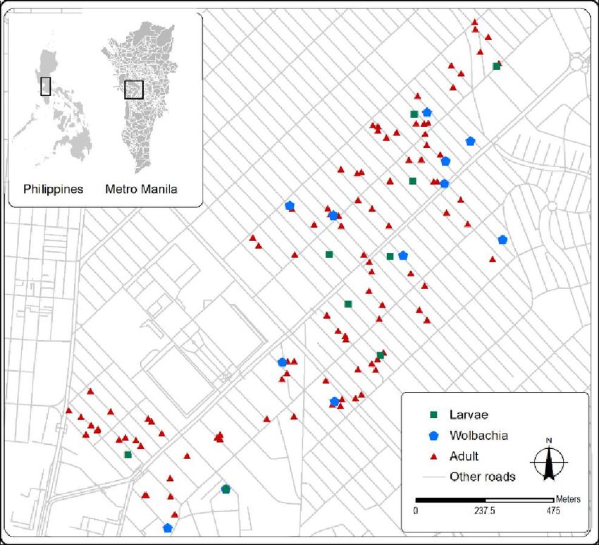
Figure 1. Spatial distribution of Wolbachia-positive households (n = 11) out of 428 households
surveyed in a highly urbanized area in Manila, Philippines
To confirm the species of the collected adult and immature mosquitoes, we used microsatellite
data from a recent study of Regilme et al. (2021) that used species-specific primers for Ae.
3062European Journal of Molecular & Clinical Medicine
ISSN 2515-8260 Volume 09, Issue 03, 2022
aegypti(Chambers et al. 2007 and Slotman et al. 2007). One representative immature mosquito
from water containers was tested for species identification using microsatellite primers. The
DNA of 359 adult Ae. aegypti were also used for the population genetics analysis (Regilme et al.
2021) and the remaining DNA of the 359 adult Ae. aegypti and 12 adult Ae. albopictus were
used for this study of Wolbachia detection. Using cox1 sequences compared with reference
sequences from GenBank using BLASTn, we confirmed the species identification of the
Wolbachia positive adult mosquitoes as Ae. albopictus or Ae. aegypti.
2.2. DNA extraction, molecular identification, PCR amplification, and sequencing
We extracted DNA using Qiagen AllPrep DNA/RNA micro kit and Qiagen DNA Blood and
Tissue DNeasy Kits© (Qiagen, Hilden, Germany) from adult (n = 371) and larval (n = 509)
samples, respectively. In this study, the extracted DNA was only used while the extracted RNA
was stored in -800C for future studies. DNA concentration and quality were measured using a
NanoDrop 2000 Spectrophotometer (Thermo Fisher Scientific, Waltham, MA, USA).
Wolbachia was detected using two molecular markers: wsp (610 base pairs) using the primer
pairs wsp 81F (5′-TGGTCCAATAAGTGATGAAGAAAC-3′) and wsp 691R (5′-
AAAAATTAAACGCTACTCCA-3′) (Zhou et al. 1998) and 16S specific for Wolbachia (850
base pairs) with the primer pairs WolbF (5′-GAAGATAATGACGGTACTCAC-3′) and Wspecr
(5′-AGCTTC GAGTGAAACCAATTC-3′) (Folmer et al. 1994).
For both wsp and 16Sgene amplification, we followed the protocol published in (Carvajal et al.
2019), in a final volume of 10 µl with 1 µl of the genomic DNA. We used the following
components for the PCR reaction for both markers: 10x Ex Taq buffer, 25 mM MgCl2, 2.5 mM
dNTP, 10 µM forward and reverse primers, 10% dimethyl sulfoxide (Sigma-Aldrich, St. Louis,
MO, USA), and five units/µl of Takara Ex Taq™ (Takara Bio Inc., Shiga, Japan). The wsp PCR
amplification was as follows: initial denaturation at 95℃ for 3 minutes, denaturation at 95℃ for
1 minute, annealing at 55℃ for 1 minute, an extension at 72℃ for 1 minute for 40 cycles, and a
final extension at 72℃ for 3 minutes. The 16Samplification followed these conditions: initial
denaturation at 95℃ for 2 minutes, denaturation at 95℃ for 2 minutes, annealing at 60℃ for 1
minute, an extension at 72℃ for 1 minute for two cycles, another 35 cycles of denaturation at
95℃ for 30 seconds, annealing at 60℃ for 1 minute, an extension at 72℃ for 45 seconds, and a
final extension at 72℃ for 10 minutes. We included a positive control of a Wolbachia-positive
Culex sp. and negative control of water in each PCR run.
PCR products were analyzed in 1.5% agarose gel stained with Midori Green Advance DNA stain
at 100 V for 30 minutes. To validate the presence of Wolbachia in each sample, we performed
PCR amplification twice per marker. The criteria for a positive Wolbachia test were based on
two successful amplifications per molecular marker, wsp, and 16S.
We also amplified the cox1 mitochondrial gene of Wolbachia-positive samples using the primer
pairs LCO-1490 (5′-GGTCAACAAATCATAAAGATATTGG-3′) and HCO1-2198 (5′-
AAACTTCAGGGTGACCAAAAAATCA-3′) (Folmer et al. 1991). We used the following PCR
amplification profile: initial denaturation at 95℃ for 5 minutes, denaturation at 95℃ for 30
seconds, annealing at 48℃ for 45 seconds for 35 cycles, and a final extension at 72℃ for 7
minutes. The amplified PCR products were purified using QIAquick (Qiagen, Hilden, Germany)
PCR Purification kits and sequenced by Eurofin Genomics Inc. Tokyo, Japan.
We assembled the forward and reverse sequences for each marker using the CodonCode Aligner
version 1.2.4 software (https://www.codoncode.com/aligner/). We aligned the sequences using
3063European Journal of Molecular & Clinical Medicine
ISSN 2515-8260 Volume 09, Issue 03, 2022
the online program MAFFT version 7 with the default settings
(https://mafft.cbrc.jp/alignment/software/). We checked the sequence quality of the aligned
sequences in Mesquite version 3.5 (Maddison et al. 2019) by confirming the absence of stop
codons. Finally, we checked all generated sequences for similarities with reference sequences
from GenBank (NCBI, 2016) using Basic Local Alignment Search Tool–Nucleotide BLAST
(https://blast.ncbi.nlm.nih.gov/Blast.cgi?PAGE_TYPE=BlastSearch).
2.3. Identification of Wolbachia strains, haplotypes, and phylogenetic analysis
All wsp, 16S, and cox1 sequences were separately analyzed using DnaSP version 6.12.03 (Rozas
et al. 2017) to determine the number of haplotypes. We assessed the relationship of the
Wolbachia strains of our study with representative sequences from different insect hosts by
constructing a phylogenetic tree for the wsp and 16S sequences using PhyML 3.1 (Lefort et al.
2017) using the default settings. We applied the GTR + G model for wsp and the GTR + G + 1
model for 16S. The model per marker was selected using the SMART model selection method
(Guindon et al. 2003). We used Brugia pahangi (AY527207) and Rickettsia sp. (U11021)as the
outgroups for the wsp and 16S, respectively.
We acknowledge the uncertainties of Wolbachia-positive Ae. aegypti due to the 16S (Ma et al.
2017) and the conventional PCR method, e.g., the false-positive rate. We were careful to
ascertain positive Wolbachia in adult Ae. aegypti in light of this information. Thus, we used the
primers of Simoes et al. (2011) which are known to produce fewer false-positive results and
negative detections of Wolbachia. PCR amplication of individual samples were performed twice
per marker. We also performed repeated PCR tests of our Wolbachia-positive samples to ensure
successful Wolbachia detection as defined in the previous study by (Carvajal et al.2019). To
address the issue of the sensitivity of the conventional PCR method, such as the false-positive
detection, we combined the sequencing analysis of the PCR amplicon.
3. Results
3.1. Detection of Wolbachia infection in adult and larval mosquitoes
A total of ten (10/12; 83.33%) adult Ae. albopictus were Wolbachia positive based on the 16S
marker while six adult Ae. albopictus (6/12; 50.00%) were positive based on wsp marker. Four
(4/12; 33.33%) adult Ae. albopictus were Wolbachia positive both in wsp and 16S markers. A
total of three (3/359; 0.83%) were positive out of the 359 adult Ae. aegypti based on the 16S
marker. However, we did not detect Wolbachia using wsp marker in the 359 adults Ae. aegypti.
Larval Aedes samples (n = 509) showed no evidence of Wolbachia from either marker. The
species identification of 12 Ae. albopictus and 3 Ae. aegypti adults with Wolbachia infection
detected by wsp or 16S markers or both markers was confirmed by BLAST analysis using cox1
marker. It was confirmed that there was no error in species identification with 100% identity
match.
The phylogenetic tree built using the wsp marker(Figure 2 A) revealed two major clades:
supergroup A and supergroup B (Zhou et al. 1998). Two Ae. albopictus were infected with
Wolbachia belonging to supergroup A and four with Wolbachia belonging to supergroupB. The
haplotype sequences from supergroup A were grouped with the Wolbachia type strain A (wAlb
A), identified in an Ae. albopictus host in the USA (Zhou et al. 1998). In contrast, the sequences
from supergroup B clustered with the Wolbachia-type strains from Ae. aegypti (wAegB) in the
Philippines (Carvajal et al. 2019) and Ae. albopictus (wAlbB) in the USA (Zhou et al. 1998).
3064European Journal of Molecular & Clinical Medicine
ISSN 2515-8260 Volume 09, Issue 03, 2022
Figure 2. The maximum likelihood phylogenetic tree of Wolbachia sequences found from
mosquitoes based on the (A) wsp sequences with reference sequences from supergroups A, B,
and C and an outgroup of Brugia pahangi(B) 16S sequences with reference sequences from
supergroups A, B, C, D, E, F, and J and an outgroup of Rickettsia sp. The colored boxes
represent the Wolbachia supergroups of the sequences, red for supergroup A, green for
supergroups C, D,E, F and J and blue represents supergroup B. The codes in parenthesis are the
GenBank Accession number. The phylogenetic trees are redrawn for better resolution in Adobe
InDesign.
In the phylogenetic tree constructed using 16Ssequences, 8 out of 10 (80%) Ae. albopictus and
three Ae. aegypti (100%)in this study were clustered within supergroup A (Figure 2 B). The
remaining two Ae. albopictus sequences (20%) in this study were clustered together with the
supergroup B with reference sequences of Ae. albopictus and Ae. aegypti.
3.2. Wolbachia and mitochondrial cox1 haplotypes
We identified six cox1, three wsp, and nine 16S haplotypes among the 15 adult samples positive
with Wolbachia (Table 1). We found that three Ae. albopictus samplescollected from one
household (NH149) were all Wolbachia positive, based on the wsp and 16S markers (Table 1).
An identical cox1 haplotype (sequence), C1, was observed among all these three Ae. albopictus.
The identical haplotypes of wsp (W2) and 16S (S6) also occurred among these three Ae.
albopictus (Table 2). Another household (NH56) had three Wolbachia-positive Ae. albopictus
with the same cox1 haplotype (C1). Two out of these three Ae. albopictus (Individual codes 177
3065European Journal of Molecular & Clinical Medicine
ISSN 2515-8260 Volume 09, Issue 03, 2022
and 178)were Wolbachia positive according to the wsp sequences, while two Ae. albopictus
(Individual codes 95 and 177) were positive,according to the 16S sequence. We observed
different haplotypes in each wsp (W1 and W2)and 16S(S8 and S9)marker. Other than these two
households, no other households had more than one Wolbachia-positive mosquito.
Table 1.Summary of sampling data and Wolbachia detection results
Total
Surveyed households 428
Households with adult Aedes mosquitoes 183
Households with larval Aedes 11
Collected Aedes mosquitoes 371
Aedes albopictus 12
Aedes aegypti 359
Collected Aedes larvae 509
Wolbachia positive
Larva 0
Adult Ae. albopictus 12
Adult Ae. aegypti 3
Households with Wolbachia-positive
11
mosquitoes
Table 2.A detailed summary of the results of the Wolbachia infection in selected Aedes
mosquitoes using wsp and 16S markers
cox1
HH MSI IC wsp(S) wsp(H) 16S(H)
(H)
1 SH9 Ae. 40 C1 S7
2 SH27 Ae. 306 C5 S3
3 SH134 Ae. aegypti 384 C6 S1
4 SH214 Ae. 425 C1 S6
5 NH43 Ae. 172 C4 S3
A W1 S8
095,
Ae.albopictus (177); (177); (095);
6 NH56 177, C1
(n=3) B W2 S9
178
(178) (178) (177)
7 NH107 Ae. 218 C5 S4
8 NH130 Ae. aegypti 116 C2 S1
9 NH131 Ae. 240 C5 A W3 S5
B W2
Ae. 258, S6
(258, (258,
10 NH149 albopictus 259, C1 (258,
259, 259,
(n=3) 260 259)
260) 260)
11 NH181 Ae. aegypti 121 C3 S2
Total number of haplotypes 6 3 9
3066European Journal of Molecular & Clinical Medicine
ISSN 2515-8260 Volume 09, Issue 03, 2022
Abbreviations: HH households; MSI molecular species identification; IC individual code; H
haplotype; S supergroup;
4. Discussion
We found that the adult Ae. albopictus were infected with both supergroups A and B of
Wolbachia using the wsp, widely used for Wolbachia strain identification and phylogeny studies
(Zhou et al. 1998). A previous study (Carvajal et al. 2019) found that field-collected Ae. aegypti
positive for Wolbachia from Manila, Philippines were clustered into A and B supergroups.
However, Ae. albopictus were not collected in the study of (Carvajal et al. 2019).We found more
Wolbachia-positive Ae. albopictus in the Wolbachia supergroup B (4/6; 66.66%) than in
supergroup A (2/6; 33.33%), as observed in a previous study (Hu et al. 2020) (China; 631/693;
91.05%). In this study, we observed two Wolbachia supergroups (A and B) in wsp sequences of
Ae. albopictus and supergroups A and B in the 16S phylogenetic tree of Ae. albopictus and Ae.
aegypti. Our results were consistent with the previous studies of (Zhou et al. 1998) in Ae.
albopictus that found both Wolbachia supergroups A and B while the clustering of Wolbachia
infected Ae. aegypti in supergroup A supports the study of (Coon et al. 2016). The presence of
two Wolbachia supergroups is common in Ae. albopictus populations (Zhou et al. 1998 and
Kittayapong et al. 2002) but the observance of two supergroups in two co-occurring adult
mosquito species of Ae. albopictus and Ae. aegypti from the same site is not yet studied to the
best of our knowledge. Our result is significant because it can give information about the
phylogenetic relationship of Wolbachia and the supergroups present in the Ae.albopictus and Ae.
aegypti collected from Manila, Philippines. However, the limited number of Wolbachia infected
Ae. aegypti in this study may not strongly show the relationship of Wolbachia found in both
Aedes species from the same site. Despite that, our results can be a starting information to know
the phylogenetic relationship between Wolbachia supergroups A and B of both Aedes species.
The molecular phylogeny based on the 16S marker revealed that most of the Wolbachia positive
Ae. albopictus (n=8) and three Wolbachia Ae. aegypti were clustered in Wolbachia supergroup
A. Most studies found that Wolbachia in Ae. aegypti were usually closely related to wAlbB
infection of supergroup B that is also common to Ae. albopictus(Balaji et al. 2019; Carvajal et al
2019 and Kulkarni e al. 2019). However, in the study of (Coon et al. 2016) both supergroups A
and B were present in the Wolbachia infected Ae. aegypti. The presence of supergroup A in Ae.
aegypti in our study supports the previous study of (Coon et al. 2016). Our results show that
supergroup A detected in Ae. aegypti are related to Wolbachia we identified in the Ae. albopictus
samples, which might suggest the possibility of post-lateral transfer because of the sharing of
habitat ranges between Ae. albopictus and Ae. aegypti (Coon et al. 2016). At present, there are no
studies about the transfer of Wolbachia between hosts in the field, thus in future studies we
recommend investigaiting the possible post-lateral transfer of Wolbachia from Ae. albopictus to
Ae. aegypti in field populations.
We found a low detection rate of Wolbachia in adult Ae. aegypti, which isconcordant with the
previous results of Kulkarni et al. (2019) obtained from field-collected Ae. aegypti in Florida,
USA, and Carvajal et al. (2019) conducted in Metro Manila, Philippines. In contrast, a high
(>50%) detection rate was observed in the studies of Teo et al. (2017) conductedin Malaysia and
Kulkarni et al. (2019) conductedin New Mexico, USA. The low Wolbachia detection rate in Ae.
3067European Journal of Molecular & Clinical Medicine
ISSN 2515-8260 Volume 09, Issue 03, 2022
aegypti in our study and Carvajal et al. (2019).might be due to the low density of Wolbachia that
cannot be detected by conventional PCR (Iturbe-Ormaetxe et al. 2011). Previous metabarcoding
studies on Ae. aegypti found a low number of Wolbachia sequence reads in the Ae. aegypti
midgut (Kulkarni et al. 2019; Thongsripong et al. 2017 and Hegde et al. 2018), implying low
Wolbachia density. Although our results are limited since we did not quantitatively measure
Wolbachia density, our 40-cycle PCR amplification method adapted from Carvajal et al. (2019)
could infer that Wolbachia-positive Ae. aegypti might be present in Metro Manila, Philippines.
Further Wolbachia detection studies are thus needed to affirm natural Wolbachia infection in
field Ae. aegypti.
Our research discovered a higher detection rate of Wolbachia in Ae. albopictus (12/12; 100%)
than in Ae. aegypti (3/359; 0.84%). A previous study of Wolbachia detection in field mosquitoes
also found a higher Wolbachia detection rate in Ae. albopictus than in other mosquito species
(Wiwataratanabutr et al. 2013). Previous studies found a low detection rate of Wolbachia in
Anopheles sp. and Ae. aegypti compared with Ae. albopictus because of the lack of a symbiotic
relationship between Wolbachia and its hosts (Baldini et al. 2018; Jeffries et al. 2018 and
Sawasdichai et al. 2019). Therefore, Wolbachia and Ae. aegypti might have a weak stable
symbiotic relationship that makes it difficult for Wolbachia to spread in the Ae. aegypti
populations. In contrast, Ae. albopictus displayed a higher Wolbachia detection rate because of
the host's ability to utilize better reproductive manipulations, enabling the Wolbachia to spread
further in the mosquito populations.
Our study found eight out of 10 (80.00%) Wolbachia infected Ae. albopictus in the 16S marker,
but no bands were amplified using the wsp marker. On the other hand, three Ae. aegypti were
positive using the 16S marker but negative for wsp marker. In the study of (Wolfgang et al.
2009), repeated failures in Wolbachia detection using the standard PCR amplifying the wsp gene
were encountered due to the mismatch in the primer and DNA template which may also explain
our negative results in the wsp marker.
Our results were negative for Wolbachia in all the collected Aedes larvae(n = 509). One possible
reason is that we found only 17 water containers during our field sampling. Since Wolbachia is
maternally transmitted, all of its offspring would also be negative for Wolbachia if the mother is
negative. Wolbachia might be detectable in larval samples if we increase the number of water
containers by setting up ovitraps. Another possible reason for obtaining negative results for
Wolbachia in larvae can be linked to a lower Wolbachia density during the larval stage of
mosquitoes. Previous studies (Coon et al. 2016 and Stevanovic et al. 2015) have found that the
Wolbachia density is much lower in the larval stage than in the adult stage. Wiwatanaratanbutr et
al. (2013) discovered that temperature and larval crowding may affect the relative Wolbachia
densities in Ae. aelbopictus. Stevanovic et al. (2015) found a lower Wolbachia density in
Drosophila melanogaster larvae than in adult females. We presume that this is due to the
immature organs of larva, such as the thoracic ganglia and muscles, ovaries, head, and salivary
glands. In the present study, we could not quantify the Wolbachia density in Wolbachia-positive
individuals because of low DNA quantity.
The same haplotype of Wolbachia (wsp and 16S)and mitochondrial DNA (cox1) among the three
Wolbachia-positive Ae. albopictus collected from household NH149 may infer the maternal
transmission of the Wolbachia occurring in the field, which may be different from laboratory-
controlled conditions. The maternal transmission of Wolbachia in mosquitoes is usually
confirmed by detecting Wolbachia in the offspring of infected mothers(Balaji et al. 2019 and
3068European Journal of Molecular & Clinical Medicine
ISSN 2515-8260 Volume 09, Issue 03, 2022
Kulkarni et al. 2019). For example, Wolbachia-microinjected Ae. aegypti embryos were reared
into adults and then tested using PCR for the presence of Wolbachia(McMeniman et al. 2009).
We assumed that using haplotype inference of cox1 sequences of field-collected Wolbachia
infected mosquitoes may eliminate the bias of laboratory-controlled conditions, including
temperature, light, and feeding times, during mosquito rearing.
However, in another household (NH56), an unexpected pattern was also observed: although three
Wolbachia-infected mosquitoes had an identical cox1 haplotype, the haplotypes of Wolbachia
wsp and 16S sequences were not identical among the three individuals. Based on the number of
cox1 haplotypes [i.e., 4 (C1–C4)] found in the 12 Wolbachia-positive Ae. albopictus, we
calculated a Poisson probability of 0.149 for obtaining the same cox1 haplotype among 3
randomly selected individuals. The mitochondrial genome reflects the long-term maternal
lineage (Folmer et al. 1994). Therefore, although the mothers of these three individuals were
different, they can have a common ancestor in their maternal lineage, although how many
generations ago cannot be estimated. One of the limitations of this study is the few number of
Wolbachia-positive mosquitoes. In future studies, we suggest increasing the sample size to
obtain more Wolbachia-positive mosquitoes from one household for a stochastically strong
validation of the maternal transmission through whole genome sequencing or double-digest
restriction site-associated DNA.
5. Conclusions
Overall, our study displayed Wolbachia supergroups A and B in the wsp sequences of Ae.
albopictus and in the 16S sequences of Ae. albopictus and Ae. aegypti. The result suggests that
Wolbachia supergroups A and B are currently infecting the natural populations of Ae. albopcitus
and Ae. aegypti in Manila, Philippines. Understanding the Wolbachia supergroups of these two
species collected simultaneously in the same site can give insights into their phylogenetic
relationship in the Culicidae phylogenetic tree and can give information about the possibility of
post-lateral transfer of Wolbachia. A higher Wolbachia detection rate was observed in the adult
Ae. albopictus (12/12;10%) than the Ae. aegypti (3/349; 0.84%). This result may suggest the
higher reproductive manipulations ability of Ae. albopictus that may have contributed to the
higher Wolbachia detection rate. We found no Wolbachia-infected larval samples in our study,
thus supporting our hypothesis of a lower Wolbachia detection rate in larva than in adults. This
might be due to the lower Wolbachia density during the immature stages of Aedes sp. than the
adult stage. Our findings emphasize the need to further detect Wolbachia infection in field-
collected Ae. albopictus and Ae. aegypti and to characterize the existing Wolbachia strains
present. This information is vital in the successful Wolbachia deployment programs to ensure the
compatibility of the Wolbachia strain that may already be present in the natural mosquito
populations.
Reproducibility: All data generated and/or analyzed during this study are included in this
published article and its additional files. All newly generated sequences are available in the
GenBank database under the Accession Numbers OM131675-OM131689, OM169215 –
OM169223 and OM304377 – OM304379.
Acknowledgments: We are thankful for all the households that participated in our sampling
collection of mosquitoes and larvae and the village officials of Sampaloc, Manila, for their
3069European Journal of Molecular & Clinical Medicine
ISSN 2515-8260 Volume 09, Issue 03, 2022
assistance and support. We appreciate the help of Johanna Beulah T. Sornillo of the Research
Institute for Tropical Medicine, Philippines, for her technical assistance in the household
sampling design. We want to thank Katherine Viacrusis for her service in the fieldwork. Our
deepest thanks to Dr. Thaddeus M. Carvajal, Dr. Divina Amalin, Dr. Mary Jane Flores, Dr.
Joeselle M. Serrana, Jerica Isabel Reyes and Micanaldo Francisco for their valuable suggestions
and technical assistance. The authors would like to thank Enago (www.enago.jp) for the English
language review. MAFR is a recipient of the Japanese Government (Monbukagakusho)
Scholarship from the Ministry of Education, Science, Sport, and Culture of Japan.
Funding: This study was supported in part by the Japan Society for the Promotion of Science
(JSPS) Grant-in-Aid Fund for the Promotion of Joint International Research [Fostering Joint
International Research (B)] under grant number 19KK0107, JSPS Grant-in-Aid for Scientific
Research (B) under grant number 21H02206, the JSPS Core-to-Core Program B Asia-Africa
Science Platforms under grant number JPJSCCB20190008, and the Endowed Chair Program of
the Sumitomo Electric Industries Group Corporate Social Responsibility Foundation.
Competing interests: The authors declare no conflict of interest.
Author Contributions: Conceptualization, MAFR and KW; methodology, MAFR and TI;
software, MAFR; validation, MAFR and KW; formal analysis, MAFR; investigation, MAFR;
resources, KW; data curation, MAFR and TI; writing—original draft preparation, MAFR;
writing—review and editing, MAFR and KW visualization, MAFR; supervision, KW; project
administration, MAFR and KW; funding acquisition, KW All authors have read and agreed to
the published version of the manuscript.
References
Albuquerque, A.L.D., Magalhães, T. and Ayres, C.F.J., 2011. High prevalence and lack of
diversity of Wolbachia pipientis in Aedes albopictus populations from Northeast
Brazil. Memórias do Instituto Oswaldo Cruz, 106(6), pp.773-776.
Balaji, S., Jayachandran, S. and Prabagaran, S.R., 2019. Evidence for the natural occurrence of
Wolbachia in Aedes aegypti mosquitoes. FEMS microbiology letters, 366(6), p.fnz055.
Baldini, F., Rougé, J., Kreppel, K., Mkandawile, G., Mapua, S.A., Sikulu-Lord, M., Ferguson,
H.M., Govella, N. and Okumu, F.O., 2018. First report of natural Wolbachia infection in the
malaria mosquito Anopheles arabiensis in Tanzania. Parasites & vectors, 11(1), pp.1-7.
Balingit, J.C., Carvajal, T.M., Saito-Obata, M., Gamboa, M., Nicolasora, A.D., Sy, A.K.,
Oshitani, H. and Watanabe, K., 2020. Surveillance of dengue virus in individual Aedes aegypti
mosquitoes collected concurrently with suspected human cases in Tarlac City,
Philippines. Parasites & vectors, 13(1), pp.1-13.
Bennett, K.L., Gómez-Martínez, C., Chin, Y., Saltonstall, K., McMillan, W.O., Rovira, J.R. and
Loaiza, J.R., 2019. Dynamics and diversity of bacteria associated with the disease vectors Aedes
aegypti and Aedes albopictus. Scientific reports, 9(1), pp.1-12.
Carvajal, T.M., Hashimoto, K., Harnandika, R.K., Amalin, D.M. and Watanabe, K., 2019.
Detection of Wolbachia in field-collected Aedes aegypti mosquitoes in metropolitan Manila,
Philippines. Parasites & vectors, 12(1), pp.1-9.
Chambers, E.W., Meece, J.K., McGowan, J.A., Lovin, D.D., Hemme, R.R., Chadee, D.D.,
McAbee, K., Brown, S.E., Knudson, D.L. and Severson, D.W., 2007. Microsatellite isolation and
3070European Journal of Molecular & Clinical Medicine
ISSN 2515-8260 Volume 09, Issue 03, 2022
linkage group identification in the yellow fever mosquito Aedes aegypti. Journal of
Heredity, 98(3), pp.202-210.
Coon, K.L., Brown, M.R. and Strand, M.R., 2016. Mosquitoes host communities of bacteria that
are essential for development but vary greatly between local habitats. Molecular ecology, 25(22),
pp.5806-5826.
Fraser, J.E., De Bruyne, J.T., Iturbe-Ormaetxe, I., Stepnell, J., Burns, R.L., Flores, H.A. and
O'Neill, S.L., 2017. Novel Wolbachia-transinfected Aedes aegypti mosquitoes possess diverse
fitness and vector competence phenotypes. PLoS pathogens, 13(12), p.e1006751.
Folmer, R., 1994. DNA primers for amplification of mitochondrial cytochrome c oxidase subunit
I from diverse metazoan invertebrates. Mol Mar Biol Biotechnol, 3(5), pp.294-9.
Gloria-Soria, A., Chiodo, T.G. and Powell, J.R., 2018. Lack of evidence for natural Wolbachia
infections in Aedes aegypti (Diptera: Culicidae). Journal of medical entomology, 55(5), pp.1354-
1356.
Guindon, S. and Gascuel, O., 2003. A simple, fast, and accurate algorithm to estimate large
phylogenies by maximum likelihood. Systematic biology, 52(5), pp.696-704.
Hegde, S., Khanipov, K., Albayrak, L., Golovko, G., Pimenova, M., Saldaña, M.A., Rojas,
M.M., Hornett, E.A., Motl, G.C., Fredregill, C.L. and Dennett, J.A., 2018. Microbiome
interaction networks and community structure from laboratory-reared and field-collected Aedes
aegypti, Aedes albopictus, and Culex quinquefasciatus mosquito vectors. Frontiers in
microbiology, p.2160.
Hu, Y., Xi, Z., Liu, X., Wang, J., Guo, Y., Ren, D., Wu, H., Wang, X., Chen, B. and Liu, Q.,
2020. Identification and molecular characterization of Wolbachia strains in natural populations
of Aedes albopictus in China. Parasites & vectors, 13(1), pp.1-14.
Iturbe-Ormaetxe, I., Walker, T. and O'Neill, S.L., 2011. Wolbachia and the biological control of
mosquito-borne disease. EMBO reports, 12(6), pp.508-518.
Jeffries, C.L., Lawrence, G.G., Golovko, G., Kristan, M., Orsborne, J., Spence, K., Hurn, E.,
Bandibabone, J., Tantely, L.M., Raharimalala, F.N. and Keita, K., 2018. Novel Wolbachia
strains in Anopheles malaria vectors from sub-Saharan Africa. Wellcome open research, 3.
Joanne, S., Vythilingam, I., Yugavathy, N., Leong, C.S., Wong, M.L. and AbuBakar, S., 2015.
Distribution and dynamics of Wolbachia infection in Malaysian Aedes albopictus. Acta
tropica, 148, pp.38-45.
Kaur, R., Shropshire, J.D., Cross, K.L., Leigh, B., Mansueto, A.J., Stewart, V., Bordenstein, S.R.
and Bordenstein, S.R., 2021. Living in the endosymbiotic world of Wolbachia: a centennial
review. Cell Host & Microbe, 29(6), pp.879-893.
Kittayapong, P., Baimai, V. and O'Neill, S.L., 2002. Field prevalence of Wolbachia in the
mosquito vector Aedes albopictus. American Journal of Tropical Medicine and Hygiene, 66(1),
pp.108-111.
Kittayapong, P., Baisley, K.J., Baimai, V. and O'Neill, S.L., 2000. Distribution and diversity of
Wolbachia infections in Southeast Asian mosquitoes (Diptera: Culicidae). Journal of medical
entomology, 37(3), pp.340-345.
Kittayapong, P., Mongkalangoon, P., Baimai, V. and O'Neill, S.L., 2002. Host age effect and
expression of cytoplasmic incompatibility in field populations of Wolbachia-superinfected Aedes
albopictus. Heredity, 88(4), pp.270-274.
Kulkarni, A., Yu, W., Jiang, J., Sanchez, C., Karna, A.K., Martinez, K.J., Hanley, K.A.,
Buenemann, M., Hansen, I.A., Xue, R.D. and Ettestad, P., 2019. Wolbachia pipientis occurs in
3071European Journal of Molecular & Clinical Medicine
ISSN 2515-8260 Volume 09, Issue 03, 2022
Aedes aegypti populations in New Mexico and Florida, USA. Ecology and evolution, 9(10),
pp.6148-6156.
Lefort, V., Longueville, J.E. and Gascuel, O., 2017. SMS: smart model selection in
PhyML. Molecular biology and evolution, 34(9), pp.2422-2424.
Ma, Y., Chen, W.J., Li, Z.H., Zhang, F., Gao, Y. and Luan, Y.X., 2017. Revisiting the phylogeny
of Wolbachia in Collembola. Ecology and evolution, 7(7), pp.2009-2017.
Maddison, W.P. and Maddison, D.R., 2019. Mesquite: a modular system for evolutionary
analysis, v. 3.61. See http://mesquiteproject. org.
McMeniman, C.J., Lane, R.V., Cass, B.N., Fong, A.W., Sidhu, M., Wang, Y.F. and O'Neill,
S.L., 2009. Stable introduction of a life-shortening Wolbachia infection into the mosquito Aedes
aegypti. Science, 323(5910), pp.141-144.
Moreira, L.A., Iturbe-Ormaetxe, I., Jeffery, J.A., Lu, G., Pyke, A.T., Hedges, L.M., Rocha, B.C.,
Hall-Mendelin, S., Day, A., Riegler, M. and Hugo, L.E., 2009. A Wolbachia symbiont in Aedes
aegypti limits infection with dengue, Chikungunya, and Plasmodium. Cell, 139(7), pp.1268-
1278.
Nugapola, N.N.P., De Silva, W.P.P. and Karunaratne, S.P., 2017. Distribution and phylogeny of
Wolbachia strains in wild mosquito populations in Sri Lanka. Parasites & vectors, 10(1), pp.1-8.
Regilme, M.A.F., Carvajal, T.M., Honnen, A.C., Amalin, D.M. and Watanabe, K., 2021. The
influence of roads on the fine-scale population genetic structure of the dengue vector Aedes
aegypti (Linnaeus). PLoS neglected tropical diseases, 15(2), p.e0009139.
Ross, P.A., Ritchie, S.A., Axford, J.K. and Hoffmann, A.A., 2019. Loss of cytoplasmic
incompatibility in Wolbachia-infected Aedes aegypti under field conditions. PLoS neglected
tropical diseases, 13(4), p.e0007357.
Rozas, J., Ferrer-Mata, A., Sánchez-DelBarrio, J.C., Guirao-Rico, S., Librado, P., Ramos-
Onsins, S.E. and Sánchez-Gracia, A., 2017. DnaSP 6: DNA sequence polymorphism analysis of
large data sets. Molecular biology and evolution, 34(12), pp.3299-3302.
Rueda, L.M., 2004. Pictorial keys for the identification of mosquitoes (Diptera: Culicidae)
associated with dengue virus transmission. Walter Reed Army Inst Of Research Washington Dc
Department Of Entomology.
Sawasdichai, S., Chaumeau, V., Dah, T., Kulabkeeree, T., Kajeechiwa, L., Phanaphadungtham,
M., Trakoolchengkaew, M., Kittiphanakun, P., Akararungrot, Y., Oo, K. and Delmas, G., 2019.
Detection of diverse Wolbachia 16S rRNA sequences at low titers from malaria vectors in Kayin
state, Myanmar. Wellcome open research, 4.
Simões, P.M., Mialdea, G., Reiss, D., Sagot, M.F. and Charlat, S., 2011. Wolbachia detection: an
assessment of standard PCR protocols. Molecular Ecology Resources, 11(3), pp.567-572.
Slotman, M.A., Kelly, N.B., Harrington, L.C., Kitthawee, S., Jones, J.W., Scott, T.W., Caccone,
A. and Powell, J.R., 2007. Polymorphic microsatellite markers for studies of Aedes aegypti
(Diptera: Culicidae), the vector of dengue and yellow fever. Molecular Ecology Notes, 7(1),
pp.168-171.
Stevanovic, A.L., Arnold, P.A. and Johnson, K.N., 2015. Wolbachia-mediated antiviral
protection in Drosophila larvae and adults following oral infection. Applied and Environmental
Microbiology, 81(23), pp.8215-8223.
Teo, C.H.J., Lim, P., Voon, K. and Mak, J.J.T.B., 2017. Detection of dengue viruses and
Wolbachia in Aedes aegypti and Aedes albopictus larvae from four urban localities in Kuala
Lumpur, Malaysia. Trop Biomed, 34, pp.583-597.
3072European Journal of Molecular & Clinical Medicine
ISSN 2515-8260 Volume 09, Issue 03, 2022
Thongsripong, P., Chandler, J.A., Green, A.B., Kittayapong, P., Wilcox, B.A., Kapan, D.D. and
Bennett, S.N., 2018. Mosquito vector-associated microbiota: Metabarcoding bacteria and
eukaryotic symbionts across habitat types in Thailand endemic for dengue and other
arthropod-borne diseases. Ecology and evolution, 8(2), pp.1352-1368.
Tolley, S.J., Nonacs, P. and Sapountzis, P., 2019. Wolbachia horizontal transmission events in
ants: what do we know and what can we learn?. Frontiers in Microbiology, 10, p.296.
Werren, J.H., Baldo, L. and Clark, M.E., 2008. Wolbachia: master manipulators of invertebrate
biology. Nature Reviews Microbiology, 6(10), pp.741-751.
Wiwatanaratanabutr, I., 2013. Geographic distribution of wolbachial infections in mosquitoes
from Thailand. Journal of invertebrate pathology, 114(3), pp.337-340.
Wolfgang, A., Markus, R., Dimitrios N, A. and Christian, S., 2009. Evidence for low-titre
infections in insect symbiosis: Wolbachia in the bark beetle Pityogenes chalcographus
(Coleoptera, Scolytinae). Environmental Microbiology, 11(8), pp.1923-1933.
Zhou, W., Rousset, F. and O'Neill, S., 1998. Phylogeny and PCR–based classification of
Wolbachia strains using wsp gene sequences. Proceedings of the Royal Society of London. Series
B: Biological Sciences, 265(1395), pp.509-515.
Zug, R. and Hammerstein, P., 2012. Still a host of hosts for Wolbachia: analysis of recent data
suggests that 40% of terrestrial arthropod species are infected. PloS one, 7(6), p.e38544.
3073You can also read