Differentiating novel coronavirus pneumonia from general pneumonia based on machine learning
←
→
Page content transcription
If your browser does not render page correctly, please read the page content below
Differentiating novel coronavirus pneumonia from
general pneumonia based on machine learning
Chenglong Liu
University of Shanghai for Science and Technology https://orcid.org/0000-0003-1392-4600
Xiaoyang Wang
Ruian City People's Hospital
Chenbin Liu
Chinese Academy of Medical Sciences & Peking Union Medical College Institute of Radiation Oncology,
Shenzhen
Qingfeng Sun
Ruian City People's Hospital
Wenxian Peng ( pengwx@sumhs.edu.cn )
Research
Keywords: Machine learning, Novel coronavirus pneumonia, Common pneumonia, Chest CT
DOI: https://doi.org/10.21203/rs.3.rs-31313/v1
License: This work is licensed under a Creative Commons Attribution 4.0 International License.
Read Full License
Page 1/16Abstract
Background
Chest CT screening as supplementary means is crucial in diagnosing novel coronavirus pneumonia
(COVID-19) with high sensitivity and popularity. Machine learning was adept in discovering intricate
structures from CT images and achieved expert-level performance in medical image analysis.
Methods
To develop and validate an integrated machine learning framework on chest CT images for
differentiating COVID-19 from common pneumonia (CP). Seventy-three con rmed COVID-19 cases were
consecutively enrolled together with twenty-seven con rmed common pneumonia patients from Ruian
People’s Hospital, from January 2020 to March 2020. Statistical textual features of COVID-19 and CP
images were extracted. After feature selection, the reserved features were applied to the ensemble of
bagged tree (EBT) and four other machine learning classi ers with 10-fold cross-validation.
Results
The classi cation accuracy, precision, sensitivity, speci city and F1 score of our proposed method are
91.66%, 97.91%, 85.26%, 98.15% and 91.15% respectively. The AUC of its receiver operating characteristic
is 0.98.
Conclusions
The experimental results indicate that the EBT algorithm with statistical textural features on chest CT for
differentiating COVID-19 from common pneumonia achieved high transferability, e ciency, speci city,
and impressive accuracy.
Background
Since the rst COVID-19 case was discovered in 2019, more than 4.78 million cases of novel coronavirus
pneumonia have been diagnosed worldwide, with 318,789 deaths recently according to World Health
Organization Coronavirus disease (COVID-2019) situation report − 121. Currently, the detection of COVID-
19 mainly relies on nucleic acid testing. However, many infected patients with obvious typical symptoms
were received multiple nucleic acid tests but diagnosed positive consequently [1], resulting in delayed
treatment and even aggravating the spread of the epidemic. On February 5, the Chinese National Health
and Health Commission launched the "Novel Coronavirus Pneumonia Diagnosis and Treatment Program
(Trial Version 5)", which updated the diagnostic criteria for novel coronavirus pneumonia with adding CT
imaging examinations as one of the main basics for clinical diagnosis of COVID-19. CT screening is
Page 2/16considerably popular, easy to operate and sensitive to COVID-19, which is critical for both early diagnosis
and epidemic control.
Nevertheless, in uenza virus pneumonia and other types of pneumonia might occur as well. In some
aspects, especially according to clinical features, it is troublesome to differentiate COVID-19 from
common pneumonia. For instance, the main manifestations of COVID-19 in the early stage were fever,
fatigue, dry cough, and expiratory dyspnea while patients with common pneumonia have similar
symptoms [2]. Pneumonia places a huge burden on the health care system because of its high morbidity
and mortality. Therefore, early diagnosis and isolation of CP patients and COVID-19 patients can better
prevent the spread of the epidemic and optimize the allocation of medical resources. However, except for
the overlapping symptoms and detection abnormalities, CT manifestations of CP and COVID-19 were
similar, causing instability and uncertainty for distinguishing them [3, 4].
In the current shortage of medical resources, some countries even recruited medical interns who have not
graduated yet and aged retired doctors to return to the hospital for treatment of COVID-19 patients,
including radiologists. Nonetheless, the accuracy of CT diagnosis varied from houseman to experts while
analyzing thousands of CT images would be no doubt an extravagant challenge for inexperienced
doctors.
To tackle this problem, we developed a machine learning method, particularly focusing on differentiating
COVID-19 from CP, demonstrating high e ciency in the identi cation of COVID-19 and CP, helping to
reduce misdiagnosis and control pandemic transmission.
Results
After feature selection by T-test results (p < 0.05), twenty- ve features showed the signi cant difference
between COVID-19 and CP groups were included. Table 1 shows the feature names.
Table 1 Twenty- ve features with signi cant difference between COVID-19 and CP groups (p < 0.05).
Page 3/16Comparison of EBT and other algorithms
The classi cation accuracies of EBT, SVM, LR, DT and KNN are 91.66%, 85.90%, 80.98%, 88.66% and
85.99% respectively. The AUCs of EBT, SVM, LR, DT and KNN are 0.98, 0.93, 0.88, 0.91, and 0.86
respectively. Notably, EBT-1 means the EBT algorithm without feature selection. The accuracy of it is
90.58% and the AUC is 0.97. Under most circumstances, EBT achieves the best performance among
others. Table 2 shows more speci c results of ve algorithms. Figure 1 shows the ROCs of ve classi ers.
Table.2 Precision, recall and F1-score of body parts based on different classi ers
Comparison of COVID-19 and CP
As is shown in Table 3, no matter what algorithm it is, the number of correctly classi ed CP images is
slightly larger than that of COVID-19. The results demonstrated that approximately from 15.12% (93 out
of 615) to 21.46% (132 out of 615) COVID-19 samples are wrongly classi ed while the maximum error
rate of CP is 14.21% (83 out of 584).
Page 4/16Table 3
TP & TN of ve classi ers
TP TN
LR 467 501
SVM 490 528
KNN 475 554
DT 519 555
EBT-1 515 585
EBT 526 585
Discussion
Because of the absence of CP images, we conducted data augmentation on the ROIs of CP images to
balance with the amount of COVID-19 images. Compared with the diverse features of COVID-19 CT
images, it is much uncomplicated to discriminate CP samples. It caused superior classi cation result
than that of COVID-19. Whereas without data augmentation, the total accuracy rate of the algorithms
would decline. More CP cases should be accumulated for further study.
In CT images of COVID-19, there were multiple small patches, obvious stromal changes in the lung
exudate, and the damage to the alveolar epithelium was very obvious [5–8]. To be more speci c, many
patients presented with ground-glass opacity (GGO), consolidation, GGO plus a reticular pattern, vacuolar
sign, microvascular dilation sign, brotic streak, pleural thickening, pleural retraction sign, and pleural
effusion [9–12]. Patients with common pneumonia generally have aky high-density shadows in the
lung, with relatively few ground-glass changes and alveolitis manifestations which are rather grievous in
COIVD-19 CT images [13]. Multiple ground-glass shadows and consolidation in double lungs with a small
amount of pleural effusion appears in common pneumonia CT images as well [14–16]. The differences
between GGO change, consolidation, density, and other distinct characteristics can be extremely well
distributed in statistical textural features. The pronouncedly superior performance of the proposed
method bene ts overwhelmingly from this advantage.
At present, many scholars have been working on the prediction and diagnosis COVID-19, and most of the
algorithms they developed are based on deep learning, which obtained impressive results [17, 18]. For
example, the deep learning model (COVNet) developed by Li et al.[19] can accurately detect COVID-19 and
distinguish it from lung diseases such as community-acquired pneumonia (CAP). The sensitivity and
speci city for detecting COVID-19 in the independent test group were 114 (90% [95% CI: 93%, 98%]) out of
127 (90% [95% CI: 83%, 94%]) ]) and 307 (96% [95% CI: 93%, 98%]) of 294 (90% [95% CI: 93%, 98%]), the
AUC was 0.96 (p-value < 0.001). The sensitivity and speci city for detecting the CAP in the independent
test set were 87% (152 (175)) and 92% ((239 (259)), and the AUC was 0.95 (95% CI: 0.93, 0.97). However,
Page 5/16development of the practical deep learning diagnostic systems for epidemic response is rather different
from the development of traditional deep learning diagnostic systems due to deep learning-based COVID-
19 diagnostic systems is time-consuming to some extent, while initially the COIVD-19 samples are in
shortage, thus not enough to train deep learning algorithms that often require a large scale of training
data. Furthermore, deep neural networks require high-end Graphics Processing Units (GPUs), which are
extremely expensive, to be trained within a reasonable amount of time. It is not practical to train deep
neural networks to achieve high performance without GPUs. To effectively leverage such high-end GPUs,
fast-access Central Processing Units (CPUs), Solid State Disk (SSD) and large-capacity random-access
memory (RAM) are also required. In contrast, machine learning is a relatively fabulous choice. Machine
learning can achieve relatively considerable results in a small data set. At the same time, machine
learning does not need high-performance hardware, and can try a variety of different machine learning
algorithms in a short time to choose the best one. Traditional machine learning algorithms are based on
feature engineering which is easier to explain and understand than deep networks that lack transparency
and interpretability (e.g., it is impossible to determine what imaging features are being used to determine
the output). Our proposed machine learning method in combination with statistical textural features
accomplished the accuracy of 91.66% for distinguish COVID-19 from CP and AUC of 0.98. It also has a
high sensitivity and speci city of 85.26% and 98.15% respectively.
Limitations
1. The ROI is manually delineated which is time-consuming especially when doctors are racing against
time to save lives. 2. The CT images, particularly CP images, are not adequate. Despite that the proposed
model attained state-of-the-art performance, more clinical images are required to test the generalizability
of the machine learning model to other patients. 3. Instead of distinguishing CP from COVID-19, the result
of our established model didn’t determine which speci c pneumonia it was, such as viral or bacterial,
mainly due to insu cient data and quiet over-lapping CT manifestations. Prognosis of CPs will be
considered in our future study.
Conclusions
This study explored an ensemble of bagged tree (EBT) algorithm with statistical textural features for
differentiating novel coronavirus pneumonia from common pneumonia. The experimental results show
that, as compared to the other four classi ers and EBT without feature selection, the proposed method
achieved pronouncedly superior performance with a small amount of CT images.
Methods
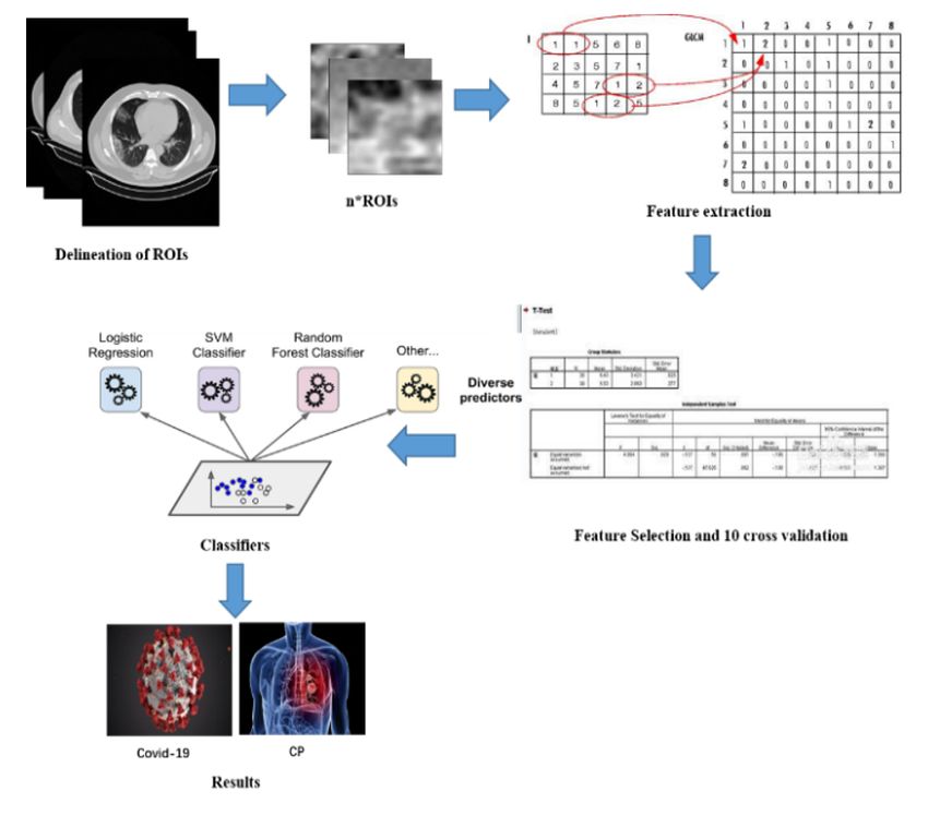
Figure 2 illustrates the scheme of this study. Machine learning algorithms integrated with statistical
textural features are leveraged to differentiate COVID-19 from CP. Firstly, manually delineation of the ROIs
of both COVID-19 and CP would be performed. Then we wrote a Matlab algorithm to crop the ROIs from
DICOM images. Secondly, input the ROIs to feature extraction algorithms. Thirdly, select features with
Page 6/16signi cant difference for time-saving and reducing over tting. Finally, input the selected features with
labels to ve classi ers and then the result is the prediction of the classi ers.
Patient selection
From January 2020 to March 2020, there were 73 COVID-19 cases con rmed by nucleic acid test positive
and 27 common pneumonia cases enrolled in this study (age ranges from 14 to 72 years). Both COVID-
19 and CP patients who had undergone chest CT scans were retrospectively reviewed by two senior
radiologists. Of the COVID-19 cases, twelve patients without obvious characteristics on CT images were
excluded (negative rate 16.4%, 12/73). Finally, sixty-one con rmed COIVD-19 cases, and 27 common
pneumonia cases were enrolled in this study.
Delineation of ROIs
The images were independently assessed by two radiologists. If the radiologists disagreed with each
other, a senior radiologist would be invited to review the pulmonary CT images and make the nal
examination. All the CT images were generated from the Siemens Sensation 16-layer spiral CT (Siemens,
Erlangen, Germany). The image format was Digital Imaging and Communications in Medicine (DICOM).
The scan parameters were: tube voltage 120 kV; tube current automatic regulation; 1–2 millimeters cross-
sectional thickness; 1–2 millimeters cross-sectional distance; scan pitch 1.3; and 16 × 0.625 millimeters
collimation.
The software of MRIcro 1.4 was used to extract the rectangle ROI of COVID-19 and CP. ROIs were
sketched from CT images based on features of COVID-19 and CP, such as multiple ground-glass opacity
in both lungs in the progression stage for COVID-19 and individual ground glass nodule for CP. The main
processes of ROI delineation: 1) A rectangular region as large as possible, which is the ROI, was
delineated within the features and export the whole image with delineation to a PNG image; 2) Binarize
the PNG image to get the ROI boundary and ll the rectangular region to get the ROI template; 3) Using
the ROI template to extract the ROI in the original DICOM image; 4) Convert the gray level of the ROI
image to 256 gray levels and resize the image to 32 × 32 pixels.. Figure 3 demonstrates the owchart of
ROI delineation. Consequently, 615 COVID-19 CT images and 146 CP ROIs were cropped. Because COVID-
19 images were four times larger than CP images which could reduce the classi cation accuracy, we
rotated the CP images by 90, 180, 270 degrees. Ultimately, the number of CP images were augmented to
584. Typical ROIs cropped from COVID-19 and CP cases were shown in Fig. 4.
Sub-image from number 1 to number 8 are the ROIs of COVID-19, sub-image from a to h are the ROIs of
CP.
Feature extraction
The statistical texture feature is based on the gray-scale feature parameters extracted by a certain image
processing algorithm based on pixels and their neighborhood. The gray level co-occurrence matrix
Page 7/16(GLCM) [20] and the gray level-gradient co-occurrence matrix (GLGCM) [21] are the second-order
statistical texture features of the gray-scale variation of the image and the basic functions to describe the
characteristics of texture structure[22], which have been widely used in medical image processing[23, 24].
In this paper, 13-dimensional GLCM features and 15-dimensional GLGCM features are applied. In the gray
level co-occurrence matrix, supposing that the distance d of the pixel is 1, rstly, calculate the gray level
co-occurrence matrix in four directions of 0, 45, 90 and 135 degrees, and secondly, calculate the texture
features based on the matrix in each direction. Eventually, the average of the texture features in 4
directions was taken as the nal feature. Studies have shown that the statistical texture features
extracted by GLCM and GLGCM can achieve effective identi cation of medical images. Also, the
histological characteristics of COVID-19 and CP can be well re ected in the gray mode, and the gray
histogram is an intuitive statistical method [25]. It is a one-dimensional function of the gray level and
belongs to the rst-order statistical method. A total of 34-dimensional features including 6-dimensional
rst-order histogram features, 13-dimensional GLCM features, and 15-dimensional GLGCM features of
second-order statistical texture features are utilized in this paper. After obtaining all texture feature data,
due to the different calculation methods of each feature, the numerical value changes in a wide range.
Therefore, to facilitate calculation, all data are normalized to [0, 1] based on their respective dimensions,
the normalized Eq. (1) is as follows:
X* = (X MIN) / (MAX MIN)
Where X is the original data of the Nth dimension, MIN is the minimum value in the Nth dimension, MAX is
the maximum value in the Nth dimension, X* is the normalized feature.
Feature selection & Classi cation
Feature selection can usually improve classi cation performance and reduce over tting, especially for
problems of small sample size. T-test, also known as Student's T-test, is mainly used for normal
distribution data with a small sample size and unknown population standard deviation σ. It uses the T-
distribution theory to infer the probability of the difference [26], to determine whether the difference
between the two samples is signi cant. Feature with p < 0.05 indicates that it has a signi cant difference,
which matches the inclusion criteria.
Ensemble of bagged tree (EBT) algorithm adopts the idea of self-help method while it forms multiple
training samples and generates numerous tree models [27]. Besides, the result was predicted by majority
voting. EBT uses the same algorithm to train the samples multiple times with various independent
classi ers. As for classi cation, the nal output is the voting result of each classi er [28, 29]. It improves
the generalization ability, and each prediction function of EBT can be generated in parallel, thereby saving
a lot of time.
Different feature classi cation algorithms have their advantages. For comparison with the performance
of the EBT algorithm, support vector machine (SVM), logistic regression (LR), decision tree (DT), K-nearest
neighbor with Minkowski distance equal weight (KNN) are implemented with the same texture feature
Page 8/16extraction methods and the same feature selection method. To superiorly identify the differences of the
results, a 10-fold cross-validation method is adopted. All image data are divided into ten equal parts. Nine
of them are employed as the training set, and the remaining one is applied as the validation set to
calculate the corresponding accuracy. Take turns until every part is utilized as the validation set. The
average value of 10 accuracies is taken as the nal accuracy of the classi cation algorithm.
Statistics
The classi cation metrics used included area under the receiver operating characteristic curve (AUC),
sensitivity, speci city, accuracy, precision, and F1 score. Let TP (true positive) denote the number of
samples belonging to class positive and correctly classi ed; FN (false negative) denote the number of
samples belonging to class negative but misclassi ed; FP (false positive) denote the number of samples
not belonging to class positive but misclassi ed as class positive; FN (false negative) denote the number
of samples not belonging to class negative but misclassi ed as class negative [30]. Classi cation
accuracies are reported in terms of accuracy, sensitivity, speci city, precision and F1 score as
Accuracy = (TP+TN) ⁄ (TP + TN + FP + FN)
Sensitivity = Recall = TP ⁄ (TP+FN)
Precision=TP ⁄ (TP+FP)
Speci city = TN ⁄ (TN+FP)
F1score = 2 × Precision × Recall / (Precision + Recall)
Abbreviations
CT: Computed Tomography; COVID-19: novel coronavirus pneumonia; CP: common pneumonia; EBT:
ensemble of bagged tree; DICOM: digital imaging and communications in medicine; kV: kilovolt; GLCM:
gray level co-occurrence matrix; GLGCM: gray level-gradient co-occurrence matrix; SVM: support vector
machine; LR: logistic regression; DT: decision tree; KNN: K-nearest neighbor with Minkowski distance
equal weight; ROC: receiver operating characteristic curve; AUC: the receiver operating characteristic curve;
GPU: Graphics Processing Unit; CPU: central processing units; SSD: solid state disk; RAM: random-access
memory.
Declarations
Availability of data and materials
The dataset analyzed during the current study was derived from the following public domain resources:
https://pan.baidu.com/s/1Ux9dpa1wtquNee4hEh1OWQ, code: k23c.
Ethics approval and consent to participate
The Institutional Review Board of Ruian People’s Hospital, Ruian city, Zhejiang province of China
approved the retrospective study (YJ202014), and the requirement for written informed consent was
Page 9/16waived.
Consent for publication
All authors consent for the publication of this manuscript.
Competing interests
The authors declare that they have no competing interests.
Funding
This work was supported by the funding of Ruian Science and Technology Bureau (MS2020023,
MS2020025).
Authors’ contributions
CL and XW are equally contributed as the rst authors. Corresponding Author: WP. CL, XW, and WP
accomplished the manuscript writing, data analysis, and machine learning model developing. CL and QS
accomplished data collecting, clinical expertise providing, and manuscript revising. All authors read and
approved the nal manuscript.
Acknowledgements
We would like to acknowledge the funding agencies for the support of the work. The content is solely the
responsibility of the authors and does not necessarily represent the o cial views of Ruian Science and
Technology Bureau.
Author details
1
School of Medical Instrument and Food Engineering, University of Shanghai for Science and
Technology, Shanghai 200093, China. 2 College of Medical Imaging, Shanghai University of Medicine and
Health Sciences, Shanghai 201318, China. 3 Department of Radiology, Ruian People’s Hospital, Zhejiang,
325200, China. 4 Department of Radiation Oncology, National Cancer Center/National Clinical Research
Center for Cancer/Cancer Hospital & Shenzhen Hospital, Chinese Academy of Medical Sciences and
Peking Union Medical College, Shenzhen, 518117, China. 5 Infectious Disease Department, Ruian People’s
Hospital, Zhejiang, 325200, China.
References
Page 10/161. Li D, Wang D, Dong J, Wang N, Huang H, Xu H, Xia C. "False-Negative Results of Real-Time Reverse-
Transcriptase Polymerase Chain Reaction for Severe Acute Respiratory Syndrome Coronavirus 2:
Role of Deep-Learning-Based CT Diagnosis and Insights from Two Cases". Korean J Radiol.
2020;21(4):505–8.
2. Cheng Z, Lu Y, Cao Q, Qin L, Pan Z, Yan F, Yang W. "Clinical Features and Chest CT Manifestations of
Coronavirus Disease 2019 (COVID-19) in a Single-Center Study in Shanghai, China" AJR. American
journal of roentgenology, pp. 1–6, 2020.
3. Chung M, Bernheim A, Mei X, Zhang N, Huang M, Zeng X, Cui J, Xu W, Yang Y, Fayad ZA, et al., "CT
Imaging Features of 2019 Novel Coronavirus (2019-nCoV)," Radiology, vol. 295, no. 1, pp. 202–207,
2020.
4. Li CX, Wu B, Luo F, Zhang N, "[Clinical Study and CT Findings of a Familial Cluster of Pneumonia
with Coronavirus Disease 2019 (COVID-19)]," Sichuan da xue xue bao. Yi xue ban = Journal of
Sichuan University. Medical science edition, vol. 51, no. 2, pp. 155–158, 2020.
5. Dai H, Zhang X, Xia J, Zhang T, Shang Y, Huang R, Liu R, Wang D, Li M, Wu J, et al., "High-resolution
Chest CT Features and Clinical Characteristics of Patients Infected with COVID-19 in Jiangsu, China"
International journal of infectious diseases: IJID : o cial publication of the International Society for
Infectious Diseases, 2020.
6. Mungmungpuntipantip R, Wiwanitkit V. "Clinical Features and Chest CT Manifestations of
Coronavirus Disease (COVID-19)" AJR. American journal of roentgenology, pp. W1, 2020.
7. Li X, Zeng W, Li X, Chen H, Shi L, Li X, Xiang H, Cao Y, Chen H, Liu C, et al. "CT imaging changes of
corona virus disease 2019(COVID-19): a multi-center study in Southwest China". Journal of
translational medicine. 2020;18(1):154.
8. Zhu T, Wang Y, Zhou S, Zhang N, Xia L. "A Comparative Study of Chest Computed Tomography
Features in Young and Older Adults With Corona Virus Disease (COVID-19)" Journal of thoracic
imaging, 2020.
9. Liu M, Song Z, Xiao K. "High-Resolution Computed Tomography Manifestations of 5 Pediatric
Patients With 2019 Novel Coronavirus" Journal of computer assisted tomography, 2020.
10. Zhong Q, Li Z, Shen X, Xu K, Shen Y, Fang Q, Chen F, Liang T, "[CT imaging features of patients with
different clinical types of coronavirus disease 2019 (COVID-19)]," Zhejiang da xue xue bao. Yi xue
ban = Journal of Zhejiang University. Medical sciences, vol. 49, no. 1, pp. 0, 2020.
11. Guan CS, Lv ZB, Yan S, Du YN, Chen H, Wei LG, Xie RM, Chen BD, "Imaging Features of Coronavirus
disease 2019 (COVID-19): Evaluation on Thin-Section CT," Academic radiology, 2020.
12. Salehi S, Abedi A, Balakrishnan S, Gholamrezanezhad A. "Coronavirus Disease 2019 (COVID-19): A
Systematic Review of Imaging Findings in 919 Patients" AJR. American journal of roentgenology,
pp. 1–7, 2020.
13. Cao MS, Sheng J, Wang TZ, Qiu XH, Wang DM, Wang Y, Xiao YL, Cai HR. "Acute exacerbation of
idiopathic pulmonary brosis: usual interstitial pneumonitis vs. possible usual interstitial
pneumonitis pattern". Chin Med J. 2019;132(18):2177–84.
Page 11/1614. Ryu AJ, Navin PJ, Hu X, Yi ES, Hartman TE, Ryu JH. "Clinico-radiologic Features of Lung Disease
Associated With Aspiration Identi ed on Lung Biopsy," Chest, vol. 156, no. 6, pp. 1160–1166, 2019.
15. Kloth C, Thaiss WM, Beck R, Haap M, Fritz J, Beer M, Horger M. "Potential role of CT-textural features
for differentiation between viral interstitial pneumonias, pneumocystis jirovecii pneumonia and
diffuse alveolar hemorrhage in early stages of disease: a proof of principle". BMC Med Imaging.
2019;19(1):39.
16. Koo HJ, Lim S, Choe J, Choi SH, Sung H, Do KH, "Radiographic and CT Features of Viral Pneumonia,"
Radiographics: a review publication of the Radiological Society of North America, Inc, vol. 38, no. 3,
pp. 719–739, 2018.
17. Alimadadi A, Aryal S, Manandhar I, Munroe PB, Joe B, Cheng X. "Arti cial intelligence and machine
learning to ght COVID-19" Physiological genomics, vol. 52, no. 4, pp. 200–202, 2020.
18. Santosh KC. "AI-Driven Tools for Coronavirus Outbreak: Need of Active Learning and Cross-
Population Train/Test Models on Multitudinal/Multimodal Data". Journal of medical systems.
2020;44(5):93.
19. Li L, Qin L, Xu Z, Yin Y, Wang X, Kong B, Bai J, Lu Y, Fang Z, Song Q, et al., "Arti cial Intelligence
Distinguishes COVID-19 from Community Acquired Pneumonia on Chest CT," Radiology, pp. 200905,
2020.
20. Xu X, Zhang X, Tian Q, Zhang G, Liu Y, Cui G, Meng J, Wu Y, Liu T, Yang Z, et al. "Three-dimensional
texture features from intensity and high-order derivative maps for the discrimination between bladder
tumors and wall tissues via MRI". Int J Comput Assist Radiol Surg. 2017;12(4):645–56.
21. Jiang S, Mao H, Ding Z, Fu Y. "Deep Decision Tree Transfer Boosting" IEEE transactions on neural
networks and learning systems, 2019.
22. Haralick RM, Shanmugam K, Dinstein I, "Textural Features for Image Classi cation," IEEE
Transactions on Systems, Man, and Cybernetics, vol. SMC-3, no. 6, pp. 610–621, Nov. 1973.
23. Lam SW. Texture feature extraction using gray level gradient based co-occurence matrices. Presented
at IEEE International Conference on Systems,1996,1:267–271.
24. Yang Q, Gao F, Nie Q. "Analysis of rotation invariance in texture image recognition". Computer
Engineering Applications. 2010;46:205–7.
25. Langner T, Wikstrom J, Bjerner T, Ahlstrom H, Kullberg J. "Identifying morphological indicators of
aging with neural networks on large-scale whole-body MRI" IEEE transactions on medical imaging,
2019.
26. Motz L, Weaver JH. "The story of mathematics" Princeton University Press Princeton Nj, vol. 9, 2005.
27. Al-Barazanchi KK, Al-Neami AQ, Al-Timemy AH, "Ensemble of bagged tree classi er for the diagnosis
of neuromuscular disorders," Presented at 2017 Fourth International Conference on Advances in
Biomedical Engineering (ICABME), Beirut, 2017, pp. 1–4.
28. Tan AC, Gilbert D. "Ensemble machine learning on gene expression data for cancer classi cation.
Applied bioinformatics. 2003;2(3 Suppl):S75–83.
Page 12/1629. Pei C, Sun Y, Zhu J, Wang X, Zhang Y, Zhang S, Yao Z, Lu Q. "Ensemble Learning for Early-Response
Prediction of Antidepressant Treatment in Major Depressive Disorder" Journal of magnetic
resonance imaging: JMRI, 2019.
30. Yan Z, Zhan Y, Peng Z, Liao S, Shinagawa Y, Zhang S, Metaxas DN, Zhou XS. "Multi-Instance Deep
Learning: Discover Discriminative Local Anatomies for Bodypart Recognition". IEEE Trans Med
Imaging. 2016;35(5):1332–43.
Figures
Figure 1
ROCs of ve classi ers.
Page 13/16Figure 2
The owchart of this study
Page 14/16Figure 3
The owchart of ROI Delineation
Page 15/16Figure 4
The ROI of COVID-19 and CP.
Page 16/16You can also read