DNA EXTRACTION METHODS FOR FIN OF MACKEREL IN THAILAND
←
→
Page content transcription
If your browser does not render page correctly, please read the page content below
International Journal of GEOMATE, March, 2020, Vol.18, Issue 67, pp. 45-50
ISSN: 2186-2982 (P), 2186-2990 (O), Japan, DOI: https://doi.org/10.21660/2020.67.5660
Special Issue on Science, Engineering and Environment
DNA EXTRACTION METHODS FOR FIN OF MACKEREL
IN THAILAND
*Walaiporn Makkapan, Patcharaporn Narkthewan and Kaewalee Viboonkit
King Mongkut’s Institute of Technology Ladkrabang Prince of Chumphon Campus,
Pathiu, Chumphon, Thailand
*Corresponding Author, Received: 14 June 2019, Revised: 20 Nov. 2019, Accepted: 16 Dec. 2019
ABSTRACT: The quality and quantity of DNA are crucial aspects for fish studies based on molecular
techniques. The extraction method of genomic DNA is depended on tissue types and organism species. This
research aimed to identify an appropriate extraction method for gDNA from the fin of three popular mackerel
species in Thailand; short-bodied mackerel (Rastrelliger brachysoma), island mackerel (R. faughni) and
Indian mackerel (R. kanagurta). Four different methods for gDNA extraction were compared based on time,
quality and quantity of extractable gDNA for PCR technique. Method III showed the highest quantity of
gDNA in R. brachysoma and R. kanagurta. Nevertheless, the highest purity of gDNA for both species was
obtained by method II and IV, respectively. The gDNA from method IV was successful to amplify the
intense band of β-actin fragment. The highest concentration and purity of gDNA from R. faughni were
received using method II. However, β-actin gene fragment amplified from gDNA of method IV showed
intense bands. These results indicated that method IV was suitable for gDNA extraction from the fin of three
mackerel species because of the fastest, high quality and quantity for PCR amplification.
Keywords: DNA extraction, Fin, Mackerel, Fish
1. INTRODUCTION
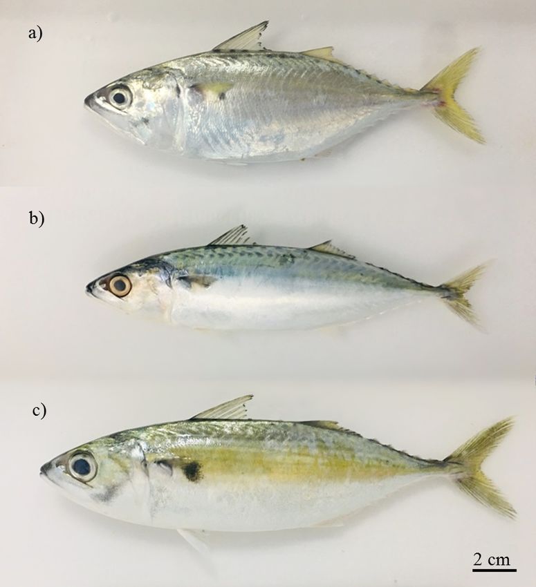
R. brachysoma, R. faughni and R. kanagurta
Extraction of genomic DNA is an important (Fig.1) were obtained from the port in Chumphon
step of fish studies based on molecular techniques. province, Thailand. Caudal fin was isolated and
Several reports related to DNA extraction from subjected to gDNA extraction using four different
various fish suggested quality and quantity of methods composed of method I-V.
extracted gDNA were depended on fish species, Method I was modified from urea treatment
tissues types and extraction methods [1-5]. method [7]. Fin tissue (20 mg) was mixed with
Nowadays, attempt to extract gDNA from fish 300 µl of extraction buffer (10 mM Tris-HCl pH
tissue without or less invasive sampling method 8.0, 10 mM EDTA, 0.5% SDS, 4 M Urea) with
was investigated in many fish. Several reports 5 µl of RNase (10 mg/ml). The mixture was
demonstrated the different protocols of DNA homogenized and incubated at 42°C with shaking
extraction from the external organs of fish, such as at 200 rpm. After incubation for 1 h, the mixture
fins [1, 5], and scales [3]. The mackerel of the was gently mixed with 10 µl of Proteinase K
genus Rastrelliger are pelagic fish species, (10 mg/ml) and then incubated at 42°C with
belonging to the Scombridae family. The shaking at 200 rpm for 16-18 h. An equal volume
Rastrelliger mackerel comprise three species, of phenol:chloroform:isoamyl alcohol (25:24:1)
namely, R. brachysoma, R. faughni and R. was then added into the mixture and inverted
kanagurta. Their distribution is widely in the several times. The mixture was centrifuged at
Indo-Pacific Ocean [6]. There is little information 11,000 rpm for 15 min. The upper phase was
related to molecular studies in these popular collected to a new tube, and mixed with 1 M NaCl
mackerel species in Thailand. Therefore, the and an equal volume of absolute ethanol. The
objective of this study was to identify an mixture was centrifuged at 11,000 rpm for 15 min.
appropriate method for gDNA extraction from the The supernatant was discarded. The 70% ethanol
fin of three mackerel species for PCR-based was added to DNA pellet and then centrifuged at
techniques. The result data will be as a beneficial 11,000 rpm for 5 min. The DNA pellet was air-
knowledge for further mackerel research such as dried and re-suspended in sterile water.
genomic analysis, biomarker study, and genetic Method II was modified from the report of [1].
species identification, etc. Fin tissue (20 mg) was homogenized in 300 µl
of extraction buffer (50 mM Tris-HCl pH 8.0,
2. MATERIALS AND METHODS 100 mM NaCl, 50 mM EDTA, 1% SDS) with
7.5 µl of Proteinase K (10 mg/ml). The mixture
2.1 Fish Sample and Extraction Methods was incubated in the shaking incubator
45International Journal of GEOMATE, March, 2020, Vol.18, Issue 67, pp. 45-50
at 55°C for 16-18 h with oscillation of The supernatant was taken in a new tube and
200 rpm. Subsequently, 300 µl of 5 M NaCl inversely mixed with an equal volume of
was mixed with the mixture and centrifuged phenol:chloroform:isoamyl alcohol (25:24:1).
at 11,000 rpm for 10 min. The supernatant was After centrifugation at 12,000xg for 10 min,
collected and inversely mixed with an equal the upper phase was collected and mixed with
volume of phenol:chloroform:isoamyl alcohol an equal volume of isopropanol, and then
(25:24:1). The mixture was then centrifuged centrifuged at 12,000xg for 10 min. The
at 11,000 rpm for 10 min. The upper phase supernatant was decanted and the DNA pellet was
was collected and mixed with an equal volume washed with 70% ethanol. The DNA pellet was
of absolute ethanol for DNA precipitation. air-dried and re-suspended in sterile water.
The mixture was centrifuged at 11,000 rpm for 15 Method IV was modified from the report
min. The supernatant was discarded. The 70% of [5, 8]. Fin sample (20 mg) was homogenized
ethanol was added to DNA pellet and then in 400 µl of extraction buffer (200 mM Tris-HCl
centrifuged at 11,000 rpm for 5 min. The DNA pH 8.0, 100 mM EDTA, 250 mM NaCl, 1.2%
pellet was air-dried and re-suspended in sterile SDS, 200 µg/ml Proteinase K) and incubated
water. in the shaking incubator at 48°C for 3 h.
Method III was followed by Rapid MT An equal volume of phenol:chloroform:isoamyl
method [4]. The fish fin (20 mg) was homogenized alcohol (25:24:1) was added into the mixture
in 660 µl of extraction buffer (100 mM Tris-HCl and mixed by inversion several times. After
pH 8.0, 5 mM EDTA, 0.2% SDS, 200 mM centrifugation at 11,000 rpm for 15 min, the
NaCl) and then 1 µl of Proteinase K (10 mg/ml) upper layer was taken into a new tube.
was added. The mixture was incubated in An equal volume of isopropanol and 0.2 volume
the shaking incubator at 55°C for 24 h with of 10 M ammonium acetate was added and
oscillation of 200 rpm. After incubation, the mixed. The sample was centrifuged at 11,000 rpm
sample was centrifuged at 12,000xg for 15 min. for 15 min. The supernatant was removed and
the DNA pellet was washed in 70% ethanol,
air-dried and re-suspended in sterile water.
Fig. 1 Mackerel species of this study a) R. brachysoma, b) R. faughni, and c) R. kanagurta
46International Journal of GEOMATE, March, 2020, Vol.18, Issue 67, pp. 45-50
2.2 Quality and Quantity of gDNA Analysis The gDNA extraction method should be rapid
and efficient for achieving the high quantity and
The quality and quantity of gDNA from each quality of gDNA [9, 10]. The fin gDNA was
method were determined by agarose gel extracted from four different methods: method
electrophoresis and spectrophotometer. The DNA I-IV. For the extraction time, the method I-III
sample was visualized in 1% agarose gel stained needed more than 20 h for the whole process,
with SYBR Safe DNA Gel Stain. The whilst the time requirement for method IV was not
concentration and purity were evaluated using more than 6 h. Extraction buffer of all methods
UV/visible spectrophotometer with absorbance of was generally used SDS and proteinase K as a
260 and 260/280 nm, respectively. The data were detergent for tissue digestion process, which was
presented as a mean ± standard deviation varied the concentration of those. Moreover, urea
(mean±SD; n=5). The statistical significance was and RNase were added into the extraction buffer of
examined using the One-Way Analysis of method I for protein and RNA denaturation [5, 7],
Variance followed by a Tukey's HSD test at a respectively. The extraction buffer of method II-IV
significance level of ≤ 0.05. was additionally mixed with NaCl used for
extracting protein [4]. The temperature in tissue
2.3 Polymerase Chain Reaction lysis step in all methods was carried out at 42-
55°C. Subsequently, phenol:chloroform: isoamyl
The quality of gDNA was further confirmed by alcohol was required to remove the proteins and
amplification in the conserved region of β-actin DNA was precipitated by either absolute ethanol
gene, consisting of 1244 bp, using specific or isopropanol. Interestingly, DNA precipitation
primers: Forward 5’ ATGAAATCGCCGCACT step of all did not take additional time.
GG 3’ and Reverse 5’ TGGATGGCAACGTACA A characteristic pattern of DNA bands
TGGC 3’. PCR reaction was performed in the total observed by 1.0% agarose gel electrophoresis
volume of 50 µl mixture consisting of 100 ng of was shown in Fig. 2. The gDNA bands of each
gDNA, 1X Standard Taq Reaction buffer, 1 mM species were distinct among different methods.
MgCl2, 0.2 mM dNTPs mixture, 0.4 µM of The quantity and quality of gDNA from the
forward and reverse specific primers, and 1.25 U fin of Rastrelliger sp. obtained from different four
Taq DNA polymerase. Amplification conditions methods was summarized in Table 1. The
were 95°C for 3 min followed by 30 cycles at extractable gDNA concentration from different
95°C for 30 s, 53°C for 30 s, 72°C for 30 s and methods varied in each species. The concentration
then subjected to a final extension of 72°C for 3 of gDNA from R. brachysoma was shown in
min. The amplified products were analyzed a range of 0.45-0.98 µg/µl. The highest
through 1.5% agarose gel stained with SYBR Safe oncentration of gDNA from R. brachysoma was
DNA Gel Stain. found from method III (0.98±0.10 µg/µl), followed
by method IV (0.74±0.10 µg/µl), method II
3. RESULTS AND DISCUSSION (0.64±0.12 µg/µl), and method I (0.45±0.18 µg/µl),
respectively. The concentration of gDNA of R.
This study identified the appropriate extraction faughni was obtained in a range of 0.62-1.10 µg/µl.
method for gDNA from the fin tissue of three The gDNA of R. faughni was shown
popular mackerel species in Thailand based the highest concentration obtained from
on extraction time, quality and quantity of gDNA. method II (1.10±0.07 µg/µl). This was followed by
Table 1 Quantity and quality of extracted DNA from different extraction methods
R. brachysoma R. faughni R. kanagurta
Extraction
Method Concentration Purity Concentration Purity Concentration Purity
(µg/µl) (A260/A280) (µg/µl) (A260/A280) (µg/µl) (A260/A280)
Method I 0.45±0.18a 1.48±0.03a 0.62±0.08a 1.49±0.07a 0.49±0.03a 1.67±0.08a
Method II 0.64±0.12a,b 1.61±0.07b 1.10±0.07b 1.77±0.05b 0.70±0.08b 1.75±0.06a,b
Method III 0.98±0.10c 1.54±0.02a,b 0.94±0.12b 1.66±0.09b 0.84±0.07c 1.70±0.02a
Method IV 0.74±0.10b 1.59±0.03b 1.05±0.14b 1.74±0.08b 0.70±0.07b 1.83±0.04b
Note: Concentration and purity were represented as mean±SD (n=5). The different letters indicated the
significant difference between extraction methods within same species. The significant difference was
analyzed by Tukey's HSD at P≤0.05.
47International Journal of GEOMATE, March, 2020, Vol.18, Issue 67, pp. 45-50
Fig. 2 Genomic DNA extracted from fin of a) R. brachysoma, b) R. faughni, and c) R. kanagurta by using
different methods (method I-IV).
method IV (1.05±0.14 µg/µl), method III gDNA attained from method IV (1.59±0.03),
(0.94±0.12 µg/µl), and method I (0.62±0.08 µg/µl), method III (1.54±0.02), and method I (1.48±0.03),
respectively. The concentration of gDNA of R. respectively. The gDNA of R. faughni showed the
kanagurta was extractable in a range of 0.49-0.84 purity values in a range of 1.49-1.77. The gDNA
µg/µl. The highest concentration of gDNA from R. obtained from method II showed the highest purity
kanagurta was found in method III (0.84±0.07 for R. faughni (1.77±0.05), following by method
µg/µl), followed by method IV (0.70±0.07 µg/µl) IV (1.74±0.08), method III (1.66±0.09), and
and method II (0.70±0.08 µg/µl). The lowest method I (1.49±0.07), respectively. Meanwhile,
concentration of extracted gDNA was observed the gDNA of R. kanagurta showed the purity
from method I (0.49±0.03 µg/µl). This study found values in a range of 1.67-1.83. The highest purity
the significantly lower values were attained from in this species was shown in the extracted DNA
method I supplemented with urea in extraction obtained by method IV (1.83±0.04), following by
buffer, suggesting this method did not suitable for method II (1.75±0.06), method III (1.70±0.02), and
DNA extraction from the fin of three mackerel method I (1.67±0.08), respectively. These results
species. suggested the method II produced the highest
The quality of the gDNA was indicated by quality of gDNA for R. brachysoma and
the A260/A280 ratio. The purity of extracted R. faughni compared to other methods. The
DNA is generally accepted a yield of A260/A280 method IV was able to extract the highest quality
ratio in a range of 1.8-2.0 [11]. The gDNA of R. of gDNA for R. kanagurta. Corresponding with
brachysoma from all extraction methods expressed quantity result, the quality of gDNA extracted
a low purity, which had a value in a range of 1.48- from method I was lowest for all species.
1.61. The highest purity in this species was Report of [1] revealed the purity of the fin DNA
exhibited in the extracted DNA obtained by extracted from modified salt method showed
method II (1.61±0.07). This was followed by the in a range of 1.83-2.19. These results showed
48International Journal of GEOMATE, March, 2020, Vol.18, Issue 67, pp. 45-50
Fig. 3 β-Actin fragments amplified from gDNA extracted from fin of a) R. brachysoma, b) R. faughni, and
c) R. kanagurta by using different methods (method I-IV). M = 100 bp DNA ladder marker and
N = negative control.
similar results of reports from [4, 5] demonstrated The facilities were supported by King Mongkut’s
the purity of fish DNA was in a range of 1.6-2.0. Institute of Technology Ladkrabang Prince of
Subsequently, the quality of gDNA was Chumphon Campus, Thailand.
considered by using as a template for PCR
amplification of β-actin gene fragment. The 6. REFERENCES
amplified products of β-actin gene analyzed
by 1.5% agarose gel electrophoresis were shown [1] Lopera-Barrero N. M., Povh J. A., Ribeiro R.
in Fig. 3. The PCR products in R. brachysoma P., Gomes P. C., Jacometo C. B., and Lopes, T.
as well as R. kanagurta were achieved from S., Comparison of DNA Extraction Protocols
gDNA obtained from all methods. The intense of Fish Fin and Larvae Samples: Modified
band of PCR products was represented from the Salt (NaCl) Extraction. Ciencia e
gDNA obtained from method I and IV. Whereas, Investigación Agraria, Vol. 35, Issue 1, 2008,
some of gDNA obtained from method II and pp.65-74.
III showed the light band of the amplified PCR [2] Mukhopadhyay T., and Bhattacharjee, S.,
fragment. The PCR amplification in R. faughni Standardization of Genomic DNA Isolation
was attained by using gDNA extracted from all from Minute Quantities of Fish Scales and
methods. The PCR products of gDNA from Fins Amenable to RAPD-PCR. Proceedings of
method IV showed the intense band. The gDNA the Zoological Society, Vol. 67, Issue 1, 2014,
from method I, II and III were amplified the PCR pp.28-32.
products with a light band. [3] Li Y., Gul Y., Cui L., Cao X., and Wang, W.,
Comparative Analysis of Different Protocol
4. CONCLUSION for Extraction of DNA from Fish Scales of
Cyprinus carpio. Indian Journal of
The quality and quantity of gDNA from the fin Biotechnology, Vol. 14, Issue 3, 2015, pp.
varied according to fish species and extraction 382-387.
methods. The method IV was the appropriate [4] Chowdhury M. M., Rahman A. S. M. S.,
method for DNA extraction from the fin of three Nahar L., Rahman M., Reza H. A., and
mackerel species in Thailand. It indicated that the Ahmed Md. S., Efficiency of Different DNA
advantage of method IV was rapid, good quantity Extraction Methods for Fish Tissues: A
and quality of gDNA for PCR amplification. Comparative Analysis. IOSR Journal of
Pharmacy and Biological Sciences, Vol. 11,
5. ACKNOWLEDGMENTS Issue 3, 2016, pp. 11-15.
[5] Muhammad H., Iqbal Z., Iqbal M. U., Younas
This work was financially supported by the T., and Bashir Q., An Efficient Method for
National Research Council of Thailand, Thailand. DNA Isolation from Fish Fin. Pakistan Journal
49International Journal of GEOMATE, March, 2020, Vol.18, Issue 67, pp. 45-50
of Agricultural Sciences, Vol. 53, Issue 4, Baptiste L. J., Kotconi S. O., and Gachomo E.
2016, pp. 843-850. W., A Simple and Efficient Genomic DNA
[6] Collette B. B., and Nauen C. E., Systematic Extraction Protocol for Large Scale Genetic
catalogue, FAO species catalogue, Scombrids Analyses of Plant Biological Systems. Plant
of the world. An annotated and illustrated Gene, Vol. 1, 2015, pp. 43-45.
catalogue of tunas, mackerels, bonitos and [10] Shafi N., Rauf A., Akhtar T., Minhas R. A.,
related species known to date, Vol. 2, FAO, Ulfat, and Bibi S., Comparative Isolation and
1983, pp. 46-49. Amplification of Cytochrome oxidase 1
[7] Wasko A. P., Martins C., Oliveira C., and DNA from Oncorhynchus mykiss
Foresti F., Non-destructive Genetic Sampling (Rainbow Trout) of Azad Jammu & Kashmir.
in Fish. An Improved Method for DNA International Journal of Fisheries and Aquatic
Extraction from Fish Fins and Scales. Studies, Vol. 4, Issue 6, 2016, pp. 196-199.
Hereditas, Vol. 138, Issue 3, 2003, pp.161-165. [11] Boesenberg-Smith K. A., Pessarakli M. M.,
[8] Kumar R., Singh P. J., Nagpure N. S., and Wolk D. M., Assessment of DNA Yield
Kushwaha B., Srivastava S. K., and Lakra, W. and Purity: an Overlooked Detail of PCR
S., A Non-invasive Technique for Rapid Troubleshooting. Clinical Microbiology
Extraction of DNA from Fish Scales. Indian Newsletter, Vol. 34, Issue 1, 2012, pp. 1, 3-6.
Journal of Experimental Biology, Vol. 45,
Copyright © Int. J. of GEOMATE. All rights reserved,
Issue 11, 2007, pp. 992-997.
including the making of copies unless permission is
[9] Sika K. C., Kefela T., Adoukonou-Sagbadja
obtained from the copyright proprietors.
H., Ahoton L., Saidou A., Baba-Moussa L.,
50You can also read