Effectiveness of Yoga on the Interrectus Distance in Early Postpartum Women: A High-Frequency Ultrasound Study - Hindawi.com
←
→
Page content transcription
If your browser does not render page correctly, please read the page content below
Hindawi
BioMed Research International
Volume 2022, Article ID 8908095, 8 pages
https://doi.org/10.1155/2022/8908095

Research Article
Effectiveness of Yoga on the Interrectus Distance in Early
Postpartum Women: A High-Frequency Ultrasound Study

          Qunfeng Li ,1 Siman Lei ,2 Yanhong Liu ,3 Qiongzhu Liu ,3 Ying Chen ,4 Xin Lin ,4
          and Xinling Zhang 4
          1
            Guangdong Polytechnic of Science and Technology, Zhuhai, 510640 Guangdong, China
          2
            Faculty of Education, University of Macau, Taipa, 999078 Macao SAR, China
          3
            Zhuhai Women and Children’s Hospital, Zhuhai, 519001 Guangdong, China
          4
            Third Affiliated Hospital of Sun Yat-Sen University, Guangzhou, 510630 Guangdong, China

          Correspondence should be addressed to Xinling Zhang; zhxinl@mail.sysu.edu.cn

          Received 17 February 2022; Revised 13 April 2022; Accepted 23 April 2022; Published 20 May 2022

          Academic Editor: Yuvaraja Teekaraman

          Copyright © 2022 Qunfeng Li et al. This is an open access article distributed under the Creative Commons Attribution License,
          which permits unrestricted use, distribution, and reproduction in any medium, provided the original work is properly cited.

          To investigate the effects of ultrasonic evaluation of a progressive yoga exercise program on reducing the inter-recti distance (IRD)
          among women in the early postpartum period. Postpartum women (n = 116), free of obstetric complications and in recovery
          following vaginal delivery between weeks 1 and 12, were recruited. Participants were randomly assigned to the control and
          yoga exercise group in the pre- and post-intervention design. The control group received no treatment, while the yoga exercise
          group participated in a guided 12-week progressive yoga exercise program started at postpartum week 1. The IRD was
          examined using high-frequency ultrasound at postpartum weeks 6 and 12. The results showed that the supraumbilical,
          umbilical, and subumbilical IRD were significantly decreased in the yoga exercise group after the 12-week progressive yoga
          exercise intervention compared with the first (week 6) and second (week 12) measurements. The differences in IRD at
          supraumbilical, umbilical, and subumbilical intervals between weeks 6 and 12 significantly increased in the yoga exercise
          group. Progressive yoga exercises are effective program that reduce IRD among women in the early postpartum period through
          ultrasound evaluation. In conclusion, women should advocate combined yoga exercise in the early postpartum days following a
          supervised program.

1. Introduction                                                          overweight and obesity. Thus, DRA impacts the quality of
                                                                         life of postpartum women, increases the risk in childcare,
Diastasis recti abdominis (DRA) is a common postpartum                   and has become a public health concern. Moreover, the dete-
syndrome occurring in 30%~70% of women [1]. The diasta-                  riorated physical function related to DRA may fall under the
sis is caused by the excessive pressure that leads to stretching         responsibility of the public healthcare system.
and thinning of the white line connecting the rectus abdom-                   Appropriate and effective assessment of DRA is essential
inis, that is, the increase in the inter-recti distance (IRD) and        for clinicians and researchers to evaluate the effects of treat-
the width of the white line [2]. This issue is closely related to        ment or rehabilitation training. Owing to the barriers of cost
pelvic floor dysfunction and the development of chronic                   and access, both computed tomography (CT) and magnetic
back pain. Long-term nonunion of DRA may even lead to                    resonance imaging (MRI) scans are not feasible and have lim-
organ displacement [3], hernia, and other complications                  ited utility as a common measure for postpartum women. The
[4] and reduce the stability of the spine [5]. It was reported           general evaluation of various treatments used in clinical prac-
that 60.0% of primiparas suffered from DRA at week 6,                     tice, such as finger-width palpation or dial caliper examina-
45.5% at month 6, and 32.6% at month 12 after delivery                   tion, is unreliable for accurate assessment of IRD [7]. A
[6]. DRA seriously affects body image acceptance, is a risk               recent review on DRA management suggested that two-
factor predisposing women to mental illness, and leads to                dimensional ultrasound imaging is a reliable method for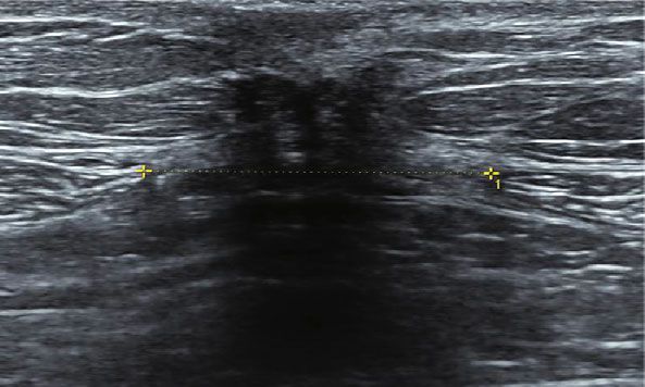
2                                                                                                  BioMed Research International

accurate diagnosis of women during the postnatal period [8].        2.2. Instrumentation and Examiners. A digital color doppler
In addition, the intuitive ultrasound imaging feedback for          ultrasound diagnostic instrument was used to perform the
IRD of different sites can improve the potential positive effect      IRD assessment. The ultrasound system, a product of volu-
by strengthening correct actions in the postpartum period, for      son series E10 (General Electric Company, GE Healthcare,
rehabilitation training guidance. However, a systematic review      Milwaukee, WI, USA) with a high-frequency linear array
[9] revealed that varying design and quality of DRA measure-        probe (ML6-15-D), was used with a working frequency of
ment could not provide conclusive evidence, even though             5-14 MHz. The probe condition was chosen as the muscle
noninvasive exercise treatment could reduce the IRD.                condition. All assessments and scans were performed by
    Studies [7, 10] examining the effectiveness of exercise          three doctors, each with 12 years of clinical experience. In
programs present uncertainty regarding the measurement              addition, all ultrasound imaging was adjusted by one exam-
of IRD. Also, studies [11, 12] have shown that core exercise        iner who was specifically trained in the investigation and
is most often involved in treatment to reduce IRD. One              assessment of IRD images for 12 years.
study [13] indicated that core muscle group exercise and                The inspection method included the participants lying in
abdominal breathing could relieve a series of clinical symp-        the supine position and fully relax their abdominal muscles.
toms caused by DRA. Moreover, the total effective rate of            Subsequently, the IRD was measured in this resting state.
early postpartum self-rehabilitation exercise is 100% [14].         During measurement, the probe plane was perpendicular
Nevertheless, no consensus protocol for abdominal exercises         to the long axis of the white abdomen. Therefore, the IRD
has been proposed and carried out in quality randomized             investigation was located at the umbilicus and 3 cm above
controlled trials.                                                  and below the umbilicus. Ultrasound image of rectus
    In this study, progressive yoga exercises are suggested to      abdominis separation was seen Figure 2.
resume function of muscles that influence the rehabilitation
process in DRA. However, only a few studies examined the            2.3. Exercise Intervention. The progressive yoga exercise pro-
impact of exercise intervention showing clear evidence on           gram is divided into two-level and combined with core
the intervention combined with yoga and core breath for             breathing and yoga postures, such as cat style, tiger style, half
early postpartum women. In this study, a progressive exer-          ship style, simple side plate style, antiquadruped style, and
cise program that combined yoga postures and breathing              other yoga postures that enhance core muscle strength.
was performed by postpartum women from week 1 to week               The exercise group has to participate in the 12-week exercise
12 after delivery. And the IRD was measured by ultrasound           intervention consisted of 60 minutes once a week and guided
imaging at fixed points to explore the effects of the rehabili-       by an experienced instructor. The yoga exercise group was
tation. The purpose of this study was to examine the effects         intervened by progressive yoga rehabilitation exercise.
of progressive yoga exercise interventions in early postpar-        According to the physical condition of postpartum women
tum women with IRD.                                                 and the fitness degree of exercise intensity, the intervention
                                                                    group was divided into two periods: (1) postpartum week 1
                                                                    to week 6 (1-1, 1-2, 1-3, and 1-4); (2) postpartum week 6
2. Materials and Methods                                            to week 12 (2-1, 2-2, 2-3, and 2-4). No activities were
                                                                    arranged for the control group. Flow chart of the partici-
2.1. Participants. Participants were recruited through              pants’ diagram is seen in Figure 3.
purposive-convenience sampling. Seven postpartum rehabil-
itation centers in Guangzhou and Zhuhai city agreed to pro-         2.4. Data Processing and Statistical Analysis. All data were
mote and assist seeking primipara or pluripara patients who         analyzed using SPSS 26.0 (IBM, New York, USA) and
underwent vaginal delivery and were willing to enroll in the        expressed as mean ± standard error of mean (SEM). An
study. A total of 129 participants consented to participate in      independent sample t-test was used to compare baseline
two checkup and test sessions (at postpartum weeks 6 and            data and ultrasound indices between the control and yoga
12) and practiced two levels of combined yoga exercise for          exercise group. In addition, paired sample t-test was used
12 weeks. In addition, a general health check was conducted         to compare the pre- and post-intervention differences in
to ensure that the participants were able to work out. Partic-      ultrasound indexes between the control and yoga exercise
ipants who had undergone rectus abdominis separation                group, which was normally distributed. The calculated data
therapy and those with active exercise habits were excluded         that did not obey the normal distribution was represented
(3 times per week, intensity > 70% HRmax per time). The             by M (P25 , P75 ). The comparison between groups was per-
flow chart of participate diagram was seen Figure 1.                 formed using the Mann–Whitney U test. Statistical signifi-
     The study was conducted between June 2020 and Decem-           cance was set at P < 0:05.
ber 2020. A total of 116 participants were assigned to either the
yoga exercise group (n = 63) or the control group (n = 53). All     3. Results
participants provided written informed consent, and partici-
pants’ rights were protected in accordance with the Declara-        Of the 129 participants who completed all sessions and
tion of Helsinki. The ethics committee approved this                assessments, 13 were excluded because six were unable to
prospective randomized study of the Institutional Review            schedule a testing time, and two withdrew from the study
Board of the Third Affiliated Hospital, Sun Yat-Sen University        for unknown reasons. Another five women were excluded
(Zhongda Fushan Yilun [2020] 02-150-01).                            because they missed at least one testing session due to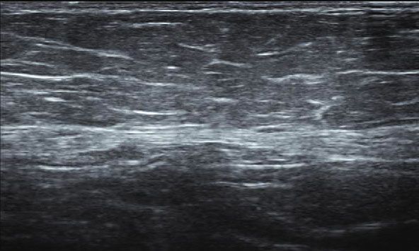
BioMed Research International                                                                                                               3

                Enrollment                                        129 patients met the inclusion criteria (n = 129)

                                                                                    Randomization

                                                    Exercise group (n = 65)                                 Control group (n = 64)
                 Allocation
                                                       1 session weekly                                          No exercise

                                    Week 1                    Week 6                  Week 12

                 Follow-up                      Level 1
                                                                            Level 2
                                              Yoga Asana
                                                                          Yoga Asana
                                             and breathing

                               Unable to             Missed 1 test                 Missed 1 test            Unable to
                                                                                                                                Unable to
                 Drop          schedule a           session due to                session due to            schedule a
                                                                                                                                follow-up
                 out          testing time          personal issues               personal issues            test time
                                                                                                                                  (n = 2)
                                 (n = 1)                (n = 1)                       (n = 4)                 (n = 5)

                Analysis                Week 1                                Week 6                              Week 12
                                        survey                              Ultrasound                           Ultrasound

                                                         Assessed and                         Assessed and
                                                           analyzed                             analyzed
                                                         exercise group                       control group
                                                            (n = 63)                            (n = 53)

                                                   Figure 1: Flow chart of participate diagram.

                              Above the umbilicus
                                   3.11 cm                                        Umbilicus 2.39 cm

                                                   (a)                                                (b)

                                                         Below the umbilicus
                                                              0.96 cm

                                                                            (c)

Figure 2: Ultrasound image of rectus abdominis separation. Note: (a) ultrasonography of 3 cm above the umbilicus; (b) ultrasonography of
umbilicus; (c) ultrasonography of 3 cm below the umbilicus.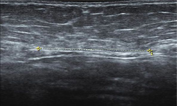
4 BioMed Research International
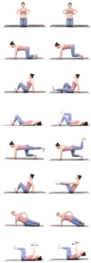
No. Yoga Asana Image signal
Yoga core breathing
1-1 method
1-2 Cat style (variant)
1-3 Sitting leg extension
1-4 Supine leg lift
2-1 Tiger (variant)
2-2 Half ship type
2-3 Simple side plate style
2-4 Reversing four feet style
Figure 3: The program of progressive yoga rehabilitation exercise. (1) postpartum week 1 to week 6 (1-1, 1-2, 1-3, and 1-4); (2) postpartum
week 6 to week 12 (2-1, 2-2, 2-3, and 2-4).
personal issues. The average age of participants in the con- respectively) and week 12 (P < 0:001 and P < 0:001, respec-
trol and exercise groups was 30 and 31 years, respectively tively), but no significant change was reported in the subum-
(Table 1). The baseline data showed no difference between bilical IRD.
the two groups. The differences in IRD at the supraumbilical, umbilical,
A comparison of the supraumbilical, umbilical, and sub- and subumbilical intervals of the control and yoga exercise
umbilical IRD in the control and yoga exercise group at group were compared (Table 4 and Figure 5). Results of
postpartum weeks 6 and 12 is shown in Table 2. Results the independent sample t-test revealed that compared with
from paired sample t-test revealed a significant decrease in the control group, the differences in IRD at the supraumbi-
IRD at supraumbilical (P < 0:001), umbilical (P < 0:001), lical (P < 0:001), umbilical (P < 0:001), and subumbilical
and subumbilical (P < 0:001) in the yoga exercise group at (P < 0:01) intervals at postpartum weeks 6 and 12 increased
week 12, compared with week 6. However, in the control significantly in the yoga exercise group.
group, the significant decrease in the IRD was reported at
only supraumbilical (P = 0:024) and subumbilical 4. Discussion
(P = 0:006) at postpartum week 12.
The results of supraumbilical, umbilical, and subumbili- This study is aimed at measuring IRD at different sites
cal IRD were compared for the control and yoga exercise among early postpartum women. High-frequency ultrasonic
group at postpartum weeks 6 and 12 (Table 3 and imaging can lead to precise, institutive, and accurate mea-
Figure 4). Results from the independent sample t-test surements, making it valuable to discuss this evaluation
revealed that compared with the control group, the method of IRD. This study found that a 12-week progressive
supraumbilical and umbilical IRD in the yoga exercise group yoga rehabilitation exercise intervention effectively short-
decreased significantly at week 6 (P < 0:001 and P < 0:01, ened the supraumbilical, umbilical, and subumbilical IRD.BioMed Research International 5
Table 1: Baseline data of control group and yoga exercise group.
Variable Control groups Experiment group t P
Number of cases 53 63
Age 30 31 1.76 0.08
Parity 1 1 0.99 0.32
Progestation BMI (kg/m2) 20:58 ± 2:06 20:34 ± 1:90 0.65 0.52
2
Week 6 postpartum BMI (kg/m ) 22:17 ± 4:76 22:02 ± 1:96 0.22 0.83
Week 12 postpartum BMI (kg/m2) 21:71 ± 2:09 21:38 ± 1:99 0.87 0.39
Table 2: Data of IRD at postpartum weeks 6 and 12 in control group and yoga exercise group.
Groups Check point Week 6 postpartum Week 12 postpartum P
3 cm above the umbilicus 2:45 ± 0:82 2:25 ± 0:80 0.024
Control group (n = 53) Umbilicus 2.9 (2.15, 3.67) 2.58 (2.1, 3.45) 0.098
3 cm below the umbilicus 0.52 (0.2, 0.95) 0.4 (0.1, 0.98) 0.006
3 cm above the umbilicus 1:93 ± 0:76 1:38 ± 0:616 BioMed Research International
Table 4: The difference of IRD at postpartum weeks 6 and 12 between the control group and the yoga exercise group.
Location Control group (n = 53) Experiment group (n = 53) t P
Interval difference above the umbilicus 0:19 ± 0:60 0:56 ± 0:45 3.674BioMed Research International 7
reported by previous research [29, 30], showing that various Conflicts of Interest
exercises with abdominal contractions such as supine head
up and twisting forward flexion are significantly effective in The authors declare that there is no conflict of interest with
narrowing the IRD. However, few studies provide quality any financial organizations regarding the material reported
randomized controlled trials to prove that exercise can in this manuscript.
reduce IRD in the early postpartum [30].
To our knowledge, this study is the first trial that exer-
cise intervenes at the first week after delivery and com- References
bines the core breathing method with progressive
[1] N. Kimmich, C. Haslinger, M. Kreft, and R. Zimmermann,
exercise, which is different from the traditional abdominal
“Diastasis recti abdominis and pregnancy,” Praxis, vol. 104,
breathing method of yoga. Based on the anatomical struc- pp. 803–806, 2015.
ture principle of the rectus abdominis, when we inhale,
[2] M. Hsia and S. Jones, “Natural resolution of rectus abdominis
our chest expands, and our abdomen relaxes without
diastasis. Two single case studies,” The Australian Journal of
upliftment, and it can reduce excessive abdominal uplift Physiotherapy, vol. 46, no. 4, pp. 301–307, 2000.
and aggravate DRA. Upon exhaling, the intercostal mus-
[3] D. R. Benjamin, H. C. Frawley, N. Shields, A. T. M. van de
cles contract. The ribs sink and adduct. The pubis slightly Water, and N. F. Taylor, “Relationship between diastasis of
lifts upward, causing abdominal wall tension, shortening the rectus abdominis muscle (DRAM) and musculoskeletal
the distance between the starting and ending points of dysfunctions, pain and quality of life: a systematic review,”
the rectus abdominis to achieve the effects of contracting Physiotherapy, vol. 105, no. 1, pp. 24–34, 2019.
rectus abdominis and reducing DRA. However, eight stud- [4] W. Ha, S. Y. Song, C. S. Yoon, and K. N. Kim, “Severe irrevers-
ies were included in the systematic review by Benjamin ible diastasis recti abdominis and abdominal hernia in post-
et al. [3] which summarized that postpartum exercise partum women: rare case report,” International Surgery,
results in reducing IRD were inconsistently caused by het- vol. 105, no. 1-3, pp. 10–13, 2021.
erogeneity in exercise dose (i.e., duration, frequency, and [5] M. Eriksson Crommert, K. Petrov Fieril, and C. Gustavsson,
intensity) and the study design. Therefore, it could not “Women's experiences of living with increased inter-recti dis-
draw plausible assumptions. In this study, the intervention tance after childbirth: an interview study,” BMC Women's
was instructed by an experienced postpartum yoga exercise Health, vol. 20, no. 1, pp. 260–260, 2020.
coach, and the program was based on the physical fitness [6] J. B. Sperstad, M. K. Tennfjord, G. Hilde, M. Ellström-Engh,
level of postpartum women and divided into two stages: and K. Bø, “Diastasis recti abdominis during pregnancy and
from week 1 to week 6 and from week 6 to week 12. 12 months after childbirth: prevalence, risk factors and report
The program has been planned with different postures of lumbopelvic pain,” British Journal of Sports Medicine,
and movements with detailed guidance and intensities. vol. 50, no. 17, pp. 1092–1096, 2016.
Another reason for selecting exercise as a possible nonsur- [7] A. Carlstedt, S. Bringman, M. Egberth et al., “Management of
gical solution for DRA might be its effectiveness adopted diastasis of the rectus abdominis muscles: recommendations
in the week 1 early postnatal period and related studies for Swedish national guidelines,” Scandinavian Journal of Sur-
generally began to intervene from postpartum week 6 gery, vol. 110, no. 3, pp. 452–459, 2021.
and seldom found to enroll postpartum women in the trial [8] N. Keshwani, N. Hills, and L. McLean, “Inter-rectus distance
before week 3 in the previous study protocol [31]. measurement using ultrasound imaging: does the rater mat-
There are some limitations of this study. As separation of ter?,” Physiotherapy Canada, vol. 68, no. 3, pp. 223–229, 2016.
the rectus abdominis has only been a concern in recent years [9] D. R. Benjamin, A. T. M. van de Water, and C. L. Peiris,
because of lack of data supporting long-term exercise inter- “Effects of exercise on diastasis of the rectus abdominis muscle
vention effects, it should be noted that yoga practitioners in the antenatal and postnatal periods: a systematic review,”
Physiotherapy, vol. 100, pp. 1–8, 2014.
need professional guidance at first. Without guidance or
nonprofessional guidance, this effect may be biased. [10] W. Reinpold, F. Köckerling, R. Bittner et al., “Classification of
rectus diastasis-a proposal by the German Hernia Society
(DHG) and the International Endohernia Society (IEHS),”
Frontiers in Surgery, vol. 6, p. 1, 2019.
5. Conclusions
[11] A. A. Thabet and M. A. Alshehri, “Efficacy of deep core stabil-
ity exercise program in postpartum women with diastasis recti
Early postpartum progressive yoga exercise improved the abdominis: a randomised controlled trial,” Journal of Musculo-
positive effect of rehabilitation on IRD. High-frequency skeletal & Neuronal Interactions, vol. 19, no. 1, pp. 62–68,
ultrasound improved the accuracy of evaluation of effect 2019.
on yoga exercise. Therefore, it is necessary to encourage [12] P. Gerber, C. Anderin, and A. Thorell, “Weight loss prior to
postpartum women to start progressive yoga rehabilitation bariatric surgery: an updated review of the literature,” Scandi-
exercises as soon as possible. navian Journal of Surgery, vol. 104, no. 1, pp. 33–39, 2015.
[13] N. Keshwani, S. Mathur, and L. McLean, “The impact of exer-
cise therapy and abdominal binding in the management of dia-
Data Availability stasis recti abdominis in the early post-partum period: a pilot
randomized controlled trial,” Physiotherapy Theory and Prac-
No data were used to support this study. tice, vol. 37, no. 9, pp. 1018–1033, 2021.8 BioMed Research International
[14] A. Michalska, W. Rokita, D. Wolder, J. Pogorzelska, and [28] M. Fukano, Y. Tsukahara, S. Takei, S. Nose-Ogura, T. Fujii,
K. Kaczmarczyk, “Diastasis recti abdominis - a review of treat- and S. Torii, “Recovery of abdominal muscle thickness and
ment methods,” Ginekologia Polska, vol. 89, no. 2, pp. 97–101, contractile function in women after childbirth,” International
2018. Journal of Environmental Research and Public Health,
[15] N. Keshwani and L. McLean, “Ultrasound imaging in postpar- vol. 18, no. 4, p. 2130, 2021.
tum women with diastasis recti: intrarater between-session [29] S. B. Gluppe, M. E. Engh, and K. Bø, “Immediate effect of
reliability,” The Journal of Orthopaedic and Sports Physical abdominal and pelvic floor muscle exercises on interrecti dis-
Therapy, vol. 45, no. 9, pp. 713–718, 2015. tance in women with diastasis recti abdominis who were par-
[16] P. Mota, A. G. Pascoal, F. Sancho, and K. Bø, “Test-retest and ous,” Physical Therapy, vol. 100, no. 8, pp. 1372–1383, 2020.
intrarater reliability of 2-dimensional ultrasound measure- [30] P. Mota, A. G. Pascoal, A. I. Carita, and K. Bø, “The immediate
ments of distance between rectus abdominis in women,” The effects on inter-rectus distance of abdominal crunch and
Journal of Orthopaedic and Sports Physical Therapy, vol. 42, drawing-in exercises during pregnancy and the postpartum
no. 11, pp. 940–946, 2012. period,” The Journal of Orthopaedic and Sports Physical Ther-
[17] S. L. Gluppe, G. Hilde, M. K. Tennfjord, M. E. Engh, and K. Bø, apy, vol. 45, no. 10, pp. 781–788, 2015.
“Effect of a postpartum training program on the prevalence of [31] N. Acharry and R. K. Kutty, “Abdominal exercise with bracing,
diastasis recti abdominis in postpartum primiparous women: a a therapeutic efficacy in reducing diastasis-recti among post-
randomized controlled trial,” Physical Therapy, vol. 98, no. 4, partal females,” International Journal of Physiotherapy
pp. 260–268, 2018. Research, vol. 3, no. 2, pp. 999–1005, 2015.
[18] J. Bellido Luque, A. Bellido Luque, J. Valdivia et al., “Totally
endoscopic surgery on diastasis recti associated with midline
hernias. The advantages of a minimally invasive approach.
Prospective cohort study,” Hernia, vol. 19, no. 3, pp. 493–
501, 2015.
[19] A. M. Rath, P. Attali, J. L. Dumas, D. Goldlust, J. Zhang, and
J. P. Chevrel, “The abdominal linea alba: an anatomo-
radiologic and biomechanical study,” Surgical and Radiologic
Anatomy, vol. 18, no. 4, pp. 281–288, 1996.
[20] N. Tahan, K. Khademi-Kalantari, M. A. Mohseni-Bandpei,
S. Mikaili, A. A. Baghban, and S. Jaberzadeh, “Measurement
of superficial and deep abdominal muscle thickness: an ultra-
sonography study,” Journal of Physiological Anthropology,
vol. 35, no. 1, pp. 1–5, 2016.
[21] J. Depledge, P. McNair, and R. Ellis, “Exercises, tubigrip and
taping: can they reduce rectus abdominis diastasis measured
three weeks post-partum?,” Musculoskeletal Science and Prac-
tice, vol. 53, article 102381, 2021.
[22] W. L. Gilleard and J. M. M. Brown, “Structure and function of
the abdominal muscles in primigravid subjects during preg-
nancy and the immediate postbirth period,” Physical Therapy,
vol. 76, no. 7, pp. 750–762, 1996.
[23] D. G. Lee, L. J. Lee, and L. McLaughlin, “Stability, continence
and breathing: the role of fascia following pregnancy and
delivery,” Journal of Bodywork and Movement Therapies,
vol. 12, no. 4, pp. 333–348, 2008.
[24] A. G. Pascoal, S. Dionisio, F. Cordeiro, and P. Mota, “Inter-
rectus distance in postpartum women can be reduced by iso-
metric contraction of the abdominal muscles: a preliminary
case-control study,” Physiotherapy, vol. 100, no. 4, pp. 344–
348, 2014.
[25] M. F. Sancho, A. G. Pascoal, P. Mota, and K. Bø, “Abdominal
exercises affect inter-rectus distance in postpartum women: a
two- dimensional ultrasound study,” Physiotherapy, vol. 101,
no. 3, pp. 286–291, 2015.
[26] M. Leopold, K. Santiago, J. Cheng et al., “Efficacy of a core
strengthening program for diastasis rectus abdominis in post-
partum women: a prospective observational study,” Physical
Therapy, vol. 45, no. 4, pp. 147–163, 2021.
[27] U. J. Hwang, S. H. Jung, H. A. Kim, J. H. Kim, and O. Y. Kwon,
“Effect of abdominal electrical muscle stimulation training
with and without superimposed voluntary muscular contrac-
tion on lumbopelvic control,” Journal of Sport Rehabilitation,
vol. 29, no. 8, pp. 1137–1144, 2020.You can also read