Effects of Feralization with Thalidomide Pre-treated Sperm on Sea Urchin
←
→
Page content transcription
If your browser does not render page correctly, please read the page content below
Proceedings of The National Conference
                                                                              On Undergraduate Research (NCUR) 2020
                                                                                Montana State University, Bozeman MT
                                                                                                    March 26-28, 2020

  Effects of Feralization with Thalidomide Pre-treated Sperm on Sea Urchin
                            Embryonic Development

                                      Kathryn Cebula and Abigail Paul
                                                 Biology
                                          Susquehanna University
                                          514 University Avenue
                                          Selinsgrove, PA 17870

                                 Faculty Advisor: Dr. Jan Reichard-Brown

                                                     Abstract

Thalidomide was released in the 1950s as a nonaddictive, nonbarbiturate sedative used to treat morning sickness in
pregnant women. Unfortunately, the drug had unforeseen teratogenic side effects. Numerous studies examined the
teratogenic mechanism of action for thalidomide, but only recently experiments on sperm exposure to thalidomide
and its effect on embryonic development have been undertaken. One specific thalidomide abnormality researchers
have examined is the disruption of serotoninergic development after embryonic exposure, which is thought to cause
autism. Sea urchin embryos serve as an acceptable model to observe thalidomide's effects on early development since
many highly conserved developmental pathways are shared between humans and sea urchins. Untreated Lytechinus
pictus eggs were fertilized with three types of sperm: untreated, pretreated with DMSO, and pretreated with
thalidomide (0.25nM) for one hour at 17ºC. One hundred embryos were evaluated from each of triplicate cultures
every 24 hours for the first 72 hours. Immunohistochemical staining for serotonin was performed on each treatment
and the swimming behavior observed at 72 hours using a light test. Embryos fertilized with thalidomide pretreated
sperm exhibited five times more abnormalities at 24, 48, and 72 hours than controls. The light test demonstrated that
the embryos fertilized with thalidomide pretreated sperm did not respond to the light stimulus in the same manner as
controls. We observed decreased expression of serotonin in experimental Strongylocentrotus purpuratus embryos
which implies thalidomide can cause serotonergic neurons to become dysfunctional. In addition, our findings suggest
that these embryos may develop abnormalities, potentially affecting neurogenesis. Historically, most studies only
focused on examining the effects of maternal exposure. Our results further suggest that paternal exposure to
thalidomide prior to conception may alter early development, indicating that it may be critical to view the effects of
thalidomide exposure on both parents before and after conception.

Keywords: Thalidomide, Sea Urchin, Sperm

1. Introduction

1.1 Creation of Thalidomide/congenital abnormalities

Thalidomide was first introduced globally in the late 1950s amid a post-war era when many people in the United
States and Europe experienced trouble sleeping and relied on sedatives. Initially synthesized and released in 1957 by
the German pharmaceutical company Chemie-Grunenthal, thalidomide quickly gained accepted use. Chemie-
Grunenthal marketed thalidomide as a nonaddictive, non-barbiturate, non-depressive sedative. Thalidomide posed
great appeal around the world after its release in Germany, being marketed as a very safe drug.1 Chemie-Grunenthal
made the statement that they “could not find a dose high enough to kill a rat”, noted by Fintel et al.2 Soon after its
release, thalidomide showed effectiveness in treating morning sickness in pregnant women. Doctors began to prescribe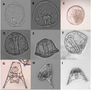
the drug to pregnant women as an off-label use. Vargesson also noted that thalidomide was eventually sold, under
various names and by various pharmaceutical companies, in 46 countries all over the world. However soon after the
release of thalidomide into the market, mothers who took thalidomide during their pregnancy gave birth to babies with
severe birth defects.1
  These birth defects were observed in many different body tissues and organs, often resulting in high mortality rates.3
Vargesson describes some of the most common birth defects, that includes short, flipper-like limbs known as
phocomelia, shortening of lower limb bones, polydactyly, craniofacial malformations, internal organ defects, as well
as others.1 It is also likely that thalidomide exposure during pregnancy resulted in miscarriages due to severe
malformations, but these numbers remain unknown. Noting the high prevalence of birth defects among mothers who
took thalidomide, clinicians Widukind Lenz and William McBride both reported in 1961 that thalidomide caused the
severe birth defects seen in newborn babies. After this claim, Germany and the United Kingdom took thalidomide off
the market in 1961 and eventually many countries withdrew thalidomide by 1962.1 In the six short years pregnant
women were taking thalidomide, Vargesson estimates that 10,000 children with severe congenital abnormalities were
associated with thalidomide use during their mother’s pregnancy.1
  Thalidomide is still used today and is FDA-approved to aid in the treatment of multiple myeloma as a
chemotherapeutic agent, HIV, Crohn's disease, leprosy, and a few other cancers.2,4 One type of cancer that thalidomide
can be used to treat is multiple myeloma either as a single-agent or in combination therapy. In the preliminary clinical
trials for cancers such as multiple myeloma, thalidomide is thought to inhibit the vascular endothelial growth factor
(VEGF) and angiogenesis. The multiple myeloma patients exhibit a different response rate to thalidomide, depending
on if they had previous treatment and are in relapse or if they are first-time patients for myeloma. Sudulaguntla et al.
noted that the patients who took thalidomide and had relapsed myeloma had a 32% response rate while the untreated
patients taking thalidomide as their first treatment had a 36% response rate.4
   Even though thalidomide is still used today, the drug is regulated by the Risk Evaluation and Mitigation Strategies
(REMS), which is mandated by the FDA and monitored by pharmacies that prescribe the medication. The REMS
program works to prevent the risk of embryo-fetal exposure to thalidomide. It also educates patients, prescribers, and
pharmacists about the risk of taking thalidomide.5
   Researchers Di Bernardo and Di Carlo first reported using sea urchins to study early embryonic development over
a century ago and continue to use urchins to study embryonic development.6 McClay declares that sea urchins serve
as good model systems to study teratogens during development since they have easily obtainable gametes that can be
collected in large quantities.7 Likewise, Ettensohn explains that, sea urchin embryos develop rapidly and can be
observed easily using light microscopy.8 Sea urchins, like humans, are deuterostomes, which as a group exhibit
evolutionarily conserved developmental pathways, making them a suitable model organism in which to study
teratogens during early development according to McClay.7 A study conducted by Reichard-Brown et al. using sea
urchins, Lytechinus pictus and Strongylocentrotus purpuratus, found that these species were susceptible to
thalidomide-induced teratogenesis during development, making them a good system to study thalidomide effects.9
   Early development of sea urchin embryos occurs very rapidly, with important developmental stages occurring within
the first 72 hours after fertilization. The embryo divides until it reaches the 128-cell blastula stage. From there, the
archenteron begins to form and elongates until it almost reaches the top of the blastocoel. As described by Gilbert, the
archenteron then attaches to the top on the blastocoel to form the mouth, while the opposite end of the tube is the
anus.10 Next skeletogenesis occurs and the sea urchin spicules are formed. Primary mesenchymal cells (PMCs) migrate
into the blastocoel and these cells derive the micromeres which form the skeleton of the sea urchin embryo.
  Embryonic exposure to thalidomide in pregnant rats, showed disruption of early serotonergic development which is
thought to be a cause of autism. The irregular distribution of serotonergic neurons in the dorsal raphe indicates an
abnormality of serotonergic neuronal differentiation and migration.11 In sea urchins, serotonergic neurons are
predictable in location, making them easy to locate along the dorsal margin of the animal pole domain and the neurons
base of the postural arms. Through Garner and others research looking at the neurons' position, scientists were able to
interpret data at different developmental stages of the same embryo.12 Yaguchi and others found that in sea urchins,
serotonergic neurons can be found at the lower lip of the larval mouth in the apical organ and the esophagus.13
   One study by Wei et al. found that in sea urchins Six3, SoxC, Brn1/2/4, and Z167 genes provide the framework for
a gene regulatory network for neurogenesis.14 This study showed how gene regulatory pathways lead to the
differentiation of SynB-positive and serotonergic neurons. Reichard-Brown et al. notes that sea urchins have similar
neuronal development as humans.9 The study by Wei and others also states that the neural network of a sea urchin has
40-50 peripheral neurons, including 10-12 serotoninergic neurons that develop thought the larval life.14
   An abundance of serotonergic neurons is present in the apical ganglion of the sea urchin. Another study by Yaguchi
and Katow suggests that the apical ganglion plays a role in swimming behavior of the sea urchins. 15 Katow et al.
believed an increase in serotonin enables the calcium levels to rise in the embryo, which allows for cilia movement to

                                                         1022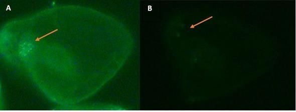
direct swim behavior.16 Abnormal swimming behavior can occur when sea urchins lack functional serotonergic
neurons. The movement and orientation of sea urchin embryos are directed by light. Studies by Yaguchi and Katow
show that spatial larval swimming is inhibited when serotonergic synthesis is inhibited. 15 Some studies demonstrate
the adding rate-limiting enzymes to alter serotonergic neurogenesis would potentially lead to abnormal swimming
behaviors.
   The fetal risk of maternal exposure to thalidomide during pregnancy was very well studied and remained the focal
point after the thalidomide tragedy observations. The role of paternal exposure to thalidomide has not been explored
or understood as much as maternal exposure. Lutwak-Mann et al., using rabbits as a model system, found that male
rabbits exposed to thalidomide fathered litters with congenital abnormalities and the litters experienced increased
mortality.17 Young from the litters presented malformations such as spina bifida, absence of both kidneys, and
paralysis of the limbs. This study also found thalidomide present in the sperm and seminal plasma of the male rabbits
just six hours after ingestion and remained for twelve days after ingestion. Another study observed the potential drug
carry-over effect of thalidomide when used on sperm. With the use of Gas Chromatography-Mass Spectrometry, all
detectable thalidomide in the sperm sample was accounted for after the sperm were washed, which showed no
significant binding of thalidomide to sperm after the wash. 18
  Thalidomide is currently being used as a therapy for human immunodeficiency virus (HIV). Another study examined
if thalidomide was present in human semen and plasma samples after oral administration of the drug to male patients
who were HIV positive. All patients dosed with thalidomide had detectable levels of thalidomide in both their semen
and plasma.19 This study used a 0.25nM thalidomide concentration in which the sperm incubated in, but therapeutic
doses are usually administered at 250 mg doses. Teo et al. also state the importance of evaluating if thalidomide
presence in bodily fluids increases in a dose dependent manner.19

2. Methodology

2.1 Fertilization of Sea Urchins

Sea urchins (L. pictus and S. purpuratus), purchased from Marinus Scientific, LLC, were both spawned and fertilized
using traditional spawning techniques. To obtain sperm and eggs, the sea urchins were rinsed with deionized water
and KCL (1mL, 0.5M) was injected into the membrane surrounding the mouth, called Aristotle’s Lantern. Sperm were
collected by placing a male urchin upside down over a dry beaker to exude sperm while the eggs were collected in a
beaker filled with artificial sea water. Once the urchins finished spawning, the maturity of the eggs was evaluated
using a light microscope (10X). If the eggs were mature, they were pipetted through a cheesecloth to remove the jelly
coat. Eggs were re-suspended in clean sea water.
   Before fertilization, the sperm were pretreated with thalidomide. The thalidomide (101.6 mg) was dissolved into
DMSO (4.0 mL) and was further diluted in DMSO to yield a final concentration 0.25nM of in which the sperm were
incubated. The three sperm treatments were separated into individual 1.5 mL microcentrifuge tubes, each tube filled
with 125 µL of sperm. Along with the sperm, the thalidomide treatment tube contained 2 µL of the thalidomide and
DMSO solution (0.25 nM), the DMSO treatment tube contained 2 µL of DMSO, and the control treatment only
contained the sperm. Sperm aliquots were incubated for an hour at 17°C. The washed eggs were placed into a 50mL
centrifuge tube containing fresh sea water while the sperm incubated.
   After the sperm incubation period, eggs were equally divided into three separate 50 mL centrifuge tubes, one tube
for each treatment group, and re-suspended in 30 mL of fresh sea water. Each aliquot of sperm was transferred to a
15 mL centrifuge tube that contained 15 mL of sea water. Portions of the sperm solutions (5 mL) were transferred to
the corresponding treatment tube filled with eggs and used to fertilize the eggs. The sperm were not washed since the
concentration of thalidomide within the tube while the eggs were being fertilized (0.0022nM) was below the threshold
to trigger a culture's response to thalidomide.9 Another study also showed there was no significant binding of
thalidomide to sperm after a wash.18 The three treatment groups were incubated at 17ºC for 15 minutes to allow
fertilization to occur. After 15 minutes, a 95% fertilization success rate for each treatment was confirmed using a light
microscope (10X). The fertilized eggs were allowed to settle to the bottom of the 50 mL centrifuge tube. The sperm
filled water was removed and the pelleted eggs re-suspended in 30 mL of sea water. Culture dishes filled with 91 mL
of sea water had 9 mL of fertilized eggs from one treatment group added. To ensure oxygen levels were similar within
each treatment dish, the same amount of embryos were added to each culture to avoid embryo crowding. We prepared
triplicate cultures for each treatment group and incubated all the cultures at 17ºC.

                                                         1023
Observations of each treatment group occurred at different developmental stages, the early gastrula stage (24 hours),
the late gastrula stage (48 hours), and the pluteus stage (72 hours). Samples of 100 embryos were observed from each
culture using a light microscope (10X), noting specific abnormalities observed at each point of development as well
as the number of embryos that presented developmental abnormalities. We replicated the experiment 6 times and, in
total, used 54 treatment dishes.
2.2 Fixing Sea Urchin Embryos
Sea urchin embryos (S. purpuratus) were preserved at 48 hours after fertilization to perform immunohistochemical
staining. Swimming embryos from each treatment group were transferred into individual 15 mL centrifuge tubes, the
sea water removed from each tube, and the embryos fixed using 4% paraformaldehyde. After overnight incubation,
the fixed embryos were washed three times with PBS buffer and finally re-suspended in sea water. Fixed embryos
from each treatment were divided into three corresponding 0.5 mL microcentrifuge tubes.
2.3 Staining of Sea Urchin Embryos
The anti-5-HT serotonin rabbit primary antibody (2µL) from ImmunoStar was used for serotonin staining at a dilution
of 1:1500. The antibody was added to each microcentrifuge tube and incubated at 17ºC for 18 hours. After the
incubation period with the primary antibody, each treatment of embryos was washed three times with PBS buffer
(10X). The Vector Laboratories’ VectaFluor Excel Amplified Fluorescent Staining System was used to visualize the
serotonin in the embryos. A drop of normal horse serum (2.5%) was added to all the embryos and incubated at 17ºC
for 40 minutes to prevent non-specific binding. The embryos were washed three times with PBS buffer after the
incubation period. A drop, 50 µL, of goat anti-rabbit IgG amplifier antibody was added to all the embryos and
incubated at 17ºC for 30 minutes. The embryos were washed three times with PBS buffer after incubation. One drop
of VectaFluor Dylight 488 horse anti-goat IgG was added to all embryos and incubated at 17ºC for 1 hour in the dark.
The embryos were washed twice more for five minutes each using PBS buffer. The embryos were viewed using a
fluorescence microscope with the DAPI and FITC filters. Pictures and observations were taken of embryos from each
treatment.
2.4 Swimming Behavior Test
The swimming behavior of the L. pictus embryos were observed at 72 hours after fertilization. Embryos taken from
the middle of the water column in the dish were pipetted into a new culture dishes with fresh sea water (90 mL). The
culture dishes were placed in a darkened room and a light was placed next to each dish. The lights were positioned at
water height so the light beam could shine through the sea water in the glass culture dish. After the 30 minutes, the
swimming behavior of each treatment group was observed and recorded.
2.5 Statistical Analysis
Over 1,500 embryos were counted and observed for abnormalities. The abnormal embryos were totaled in a chart
based on the type of abnormality observed at the specific developmental stage and the data was analyzed. The Kruskal-
Wallis one-way analysis of variance (IMB SPSS software) compared the average number of abnormalities presented
in each treatment group at the three different stages of development- 24, 48, and 72 hours. Multiple Kruskal-Wallis
one-way analysis of variances were used to compare each treatment group and detect significant differences between
groups.
3. Results
3.1 Abnormality Data Analyses
Embryos from each treatment group were observed at 24, 48 and 72 hours after fertilization, noting any abnormalities
or malformations. The types of malformations observed in the embryos fertilized with thalidomide pretreated sperm
1024vary in frequencies and type over the three developmental stages. Some common malformations observed include
change in the speed of development (behind or ahead typical development), missing or abnormally formed
archenterons (exogastrulating or protruding gastrula), abnormal shape of the embryo, abnormal primary mesenchyme
cell (PMC) migration, and malformation of the skeleton at the pluteus stage. Some of the malformations observed in
the embryos fertilized with thalidomide pretreated sperm are pictured in Figure 1.
Figure 1. Observations at 20x magnification during embryonic development in sea urchin embryos (L. pictus)
fertilized with thalidomide pretreated sperm compared to embryos fertilized with untreated sperm at different
periods of development. 1A. Early invagination of control embryo observed at 24 hours. 1B. Thalidomide treatment
embryo ahead of development at 24 hours with forming archenteron. 1C. Highly pigmented, dying thalidomide
treatment embryo observed at 24 hours. 1D. Archenteron formation of control embryo observed at 48 hours. 1E.
Malformed archenteron observed in thalidomide treatment embryo at 48 hours. 1F. Protruding archenteron of
thalidomide treatment embryo observed at 48 hours. 1G. Control embryo at pluteus stage (72 hours). 1H.
Asymmetric skeletal rods of thalidomide treatment embryo observed at 72 hours. 1I. Abnormal, asymmetric shape
of thalidomide treatment embryo at 72 hours.
The different types of malformations and the frequency each malformation was observed in the embryos fertilized
with thalidomide pretreated sperm at 24, 48, and 72 hours of development are listed in Table 1. Observations showed
experimental embryos frequently ahead of development and presenting with an abnormal shape 24 hours after
fertilization compared to controls. Embryos fertilized with thalidomide pretreated sperm 48 hours after fertilization
often presented malformations of the archenteron, including exogastrulation, protrusion of the archenteron, or absence
of the archenteron, as well as an abnormal embryonic shape. Thalidomide treatment embryos at the pluteus stage (72
hours after fertilization) frequently presented abnormal shapes as well. These embryos appeared shortened and
rounded in shape compared to embryos fertilized with untreated and DMSO pretreated sperm.
1025Table 1. The frequency of occurrence of observed phenotypic malformations in sea urchin embryos (L. pictus) fertilized with thalidomide pretreated sperm at 24, 48, and 72 hours of embryonic development. The frequency of each malformation was calculated by dividing the number of each malformation counted by the total number of malformations observed in all treated embryos at each specific developmental period. Phenotype 24 hours (blastula) 48 hours (gastrula) 72 hours (pluteus) Malformation Ahead of Development 0.24 0.09 0.10 Delayed Development 0.15 0.18 0.03 Dying 0.17 0.07 0.04 Abnormal PMC 0.10 0.06 0.12 Migration Abnormal Vegetal Plate 0.08 - - Malformed/Absent 0.01 0.31 0.13 Archenteron Abnormal Embryo 0.25 0.26 0.51 Shape Abnormal - 0.03 0.07 Skeletogenesis The average number of malformations observed in L. pictus embryos fertilized with untreated sperm, DMSO pretreated sperm, and thalidomide pretreated sperm were compared between each treatment group over three developmental periods of 24, 48, and 72 hours after fertilization, shown in Figure 2. The embryos fertilized with untreated sperm and sperm pretreated with DMSO had a significantly lower average number of malformations than the embryos fertilized with sperm pretreated with thalidomide at 24 hours after fertilization (Kruskal-Wallis one-way analysis of variance; H=35.47; p
3.2 Immunohistochemical staining
The different types of phenotypic malformations and their frequency of occurrence were recorded in the S. purpuratus
embryos fertilized with sperm pretreated with thalidomide, shown in Table 2. The S. purpuratus embryos fertilized
with sperm pretreated with thalidomide showed different frequencies of malformation than the L. pictus embryos with
the same treatments, but the same sum of abnormalities was observed in both species (Kruskal-Wallis one-way
analysis of variance; H=1.634; p=0.201; N=2400). S. purpuratus embryos were observed to be ahead of embryonic
development and presented an abnormal embryo shape 24 hours after fertilization. Forty-eight hours after fertilization,
these embryos were also ahead of development and presenting a variety of malformations.
Table 2. The frequency of observed phenotypic malformations in sea urchin embryos (S. purpuratus) fixed for
immunohistochemical staining. These embryos were fertilized with sperm pretreated with thalidomide and
observations recorded 24 and 48 hours after fertilization. The frequency of each malformation was calculated by
dividing the number of each malformation counted by the total number of malformations observed in the treated
embryos at the specific developmental periods.
Phenotype Malformation 24 hours (blastula) 48 hours (gastrula)
Ahead of Development 0.22 0.24
Delayed Development - -
Dying 0.10 0.03
Abnormal PMC Migration 0.21 0.09
Abnormal Vegetal Plate - -
Malformed/Absent Archenteron - 0.19
Abnormal Embryo Shape 0.27 0.19
Non-specific Malformations 0.20 0.26
Strongylocentrotus purpuratus embryos from each treatment were fixed at 48 hours of development and stained for
serotonergic neurons. Embryos fertilized with untreated sperm presented a higher total amount of serotonergic neurons
present compared to the embryos fertilized with sperm pretreated with thalidomide. Embryos fertilized with sperm
pretreated with thalidomide had two serotonergic neurons, while embryos fertilized with untreated sperm had eight
serotonergic neurons, shown in Figure 3. The decreased number of serotonergic neurons in the thalidomide treatment
embryos suggests that thalidomide may be interfering with serotonergic neurogenesis.
Figure 3. Staining of serotonergic neurons of sea urchin embryos (S. purpuratus) viewed at 40X magnification from
different treatment groups, fixed 48 hours after fertilization. 3A. Serotonergic staining of embryo fertilized with
untreated sperm. 3B. Serotonergic staining of embryo fertilized with sperm pretreated with thalidomide. The embryo
fertilized with sperm pretreated with thalidomide expressed fewer serotonergic neurons than the untreated embryos.
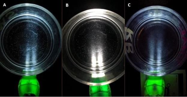
10273.3 Swimming Behavior
Qualitative observations of the swimming behavior of L. pictus embryos fertilized with untreated sperm, sperm
pretreated with DMSO, and sperm pretreated with thalidomide recorded at 72 hours after fertilization (pluteus stage)
showed differences between the treatment groups, pictured in Figure 3. The embryos from cultures fertilized by
untreated sperm and DMSO pretreated sperm ordinated towards the light source, within the beam of the light. The
embryos from cultures fertilized by sperm pretreated with thalidomide were not observed to orient to a particular area
of the culture dish, but instead were evenly distributed among the dish.
Figure 4. Swimming behavior observations of sea urchin embryos (L. pictus) 72 hours after fertilization using a light
test. 4A. The embryos fertilized with untreated sperm showed a response to the light and clustered within the beam of
light. 4B. The embryos fertilized with sperm pretreated with DMSO showed a response to the light and are clustered
within the beam of the light. 4C. The embryos fertilized with sperm pretreated with thalidomide showed no response
to the light stimulus and were scattered randomly throughout the culture dish.
4. Discussion
These results, as well as other studies in the literature, indicate that male exposure to thalidomide may pose a
teratogenic risk to a developing fetus. The results from the abnormality data analyses, the serotonergic staining, and
swimming behavior all support the hypothesis. While the exact mechanism by which sperm induces teratogenic effects
remains unknown, a study by Lutwak-Mann et al. observed that thalidomide penetrated into the spermatozoa and
could not be dislodged after washings.17 However, studies from Lutwak-Mann et al. and Teo et al. have found
thalidomide to be present in both semen and sperm after dosing and exposure to the drug.17,19
Previous research also supports the hypothesis that sperm exposure to thalidomide can exert a negative influence on
reproduction and embryonic development. The rabbit study by Lutwak-Mann and others showed that the number of
offspring could decrease when thalidomide is given to the paternal parent.17 The morphological results from the sea
urchin embryos further suggest that when sperm are exposed to thalidomide and used for fertilization, it can cause
teratogenic effects.
The results from the immunohistochemical staining suggest that embryos (S. Purpuratus) fertilized with sperm
exposed to thalidomide may negatively affect the development of serotonergic neurogenesis. These embryos not only
presented a higher average of malformations but were also observed to have a decrease in serotonergic neurons
compared to control treatments. Another study performed by Miyazaki and others on rat embryos exposed to
thalidomide during embryonic development showed a reduced number of serotonergic neurons compared to control
1028mice embryos.11 The results of immunohistochemical staining of the sea urchins suggest that embryos resulting from
sperm pretreated with thalidomide can exhibit altered serotonergic neural development. Previous research by Miyazaki
et al. revealed that thalidomide exposure to human embryos during the first trimester is linked to a higher occurrence
of autism.11 A reduced number of serotonergic neurons could lead to compromised neurochemical properties in the
brain.
The swimming behavior test showed that the embryos fertilized with sperm pretreated with thalidomide are
unresponsive to the light source, supporting the theory that peripheral neuron function was impacted. The embryos
fertilized with sperm pretreated with thalidomide are thought to have a decreased number of serotonergic neurons in
the apical organ, thus causing a decrease in calcium ion release. A decrease in calcium ions being released in turn
causes the cilia to not function properly and consequently, the embryos cannot respond to light. Evidence in the
literature from Yaguchi and Katow suggests that alteration of serotoninergic neurons can lead to abnormal swimming
behavior.15 The swimming behavior test suggests that thalidomide does affect the ciliary action by decreasing the
expression of serotonergic neurons.15
Thalidomide, a drug once withdrawn from many countries due to its teratogenic effects, is currently being used as a
therapeutic agent for various diseases. With an increased thalidomide use by males, it is essential that the
mechanism(s) of thalidomide induced teratogenesis becomes delineated. Understanding thalidomide’s mechanism(s)
of action would allow scientists to better study the teratogenic effects of sperm exposed to thalidomide which in turn
would allow for more educated warnings and distribution of thalidomide. Asatsuma-Okumura et al. investigated
thalidomide’s teratogenicity and found that cereblon (CRBN), a receptor of the E3 ubiquitin ligase, is a target of
thalidomide.20 Asatsuma-Okumura et al. also discovered that when thalidomide binds to the receptor, CRBN is
ubiquitinated and cellular proteins are degraded. Some of the cellular proteins degraded include p63 and SALL4. 20
Gao and others identify the p63 gene as an important transcription factor for the development of the cochlea, hearing,
and limbs of a developing embryo.21 These studies provide advances into the understanding of thalidomide’s
mechanism of action, but the mechanism remains widely unknown.
5. Conclusion
While thalidomide’s mechanism(s) of action is still unknown today, thalidomide is still provided as a useful treatment
for several diseases. Sea urchin embryos (L. pictus) fertilized with sperm pretreated with thalidomide presented a
higher average number of malformations than control embryos. The increased occurrence of malformations suggests
that paternal exposure to thalidomide may pose teratogenic effects to a developing embryo. A reduced number of
serotonergic neurons were observed in embryos fertilized with thalidomide pretreated sperm, indicating that
thalidomide may affect serotonergic neurogenesis. Embryos fertilized with thalidomide pretreated sperm displayed
unusual swimming behaviors and did not respond to a light stimulus. All these results indicate that paternal exposure
to thalidomide has the potential for teratogenicity and males should be warned of thalidomide’s effects when taking
the drug. Understanding the similarities and differences between maternal and paternal exposure can potentially help
further research to understand the mechanism of thalidomide.
6. Acknowledgments
The authors would like to express their appreciation to the Susquehanna University Biology Department for the
resources they provided to make this research possible. A special thanks to Dr. Reichard-Brown for her valuable and
constructive suggestions during the planning and development of our research.
7. References
1. Neil Vargesson, “Thalidomide-Induced Teratogenesis: History and Mechanisms,” Birth Defects Research Part
C: Embryo Today 105, no. 2 (2015), https://onlinelibrary.wiley.com/doi/10.1002/bdrc.21096.
2. Bara Fintel et al., “The Thalidomide Tragedy: Lessons for Drug Safety and Regulation,” Helix Magazine, Jul 28,
2009.
3. George M. Ing et al., “Drug-Induced (Thalidomide) Malformations,” Cancer Medical Association Journal 87,
no. 24 (1962), https://www.ncbi.nlm.nih.gov/pmc/articles/PMC1920822/.
10294. Anusha Sudulaguntla et al., “A Review: Uses of Thalidomide in Cancer Treatment,” Research & Reviews: A
Journal of Pharmacology 8, no. 1 (2018),
http://pharmajournals.stmjournals.in/index.php/RRJoP/article/view/55.
5. United States Food and Drug Administration, “Approved Risk Evaluation and Mitigation Strategies (REMS),”
U.S. FDA (2017), https://www.fda.gov/drugs/drug-safety-and-availability/risk-evaluation-and-mitigation-
strategies-rems.
6. Maria Di Bernardo and Marta Di Carlo, Sea Urchin - From Environment to Aquaculture and Biomedicine
(InTech, 2017), 119-132.
7. David R. McClay, “Evolutionary Crossroads in Developmental Biology: Sea Urchins,” Development 138, no. 13
(2011), https://dev.biologists.org/content/138/13/2639.
8. Charles A. Ettensohn, “Sea Urchins as a Model System for Studying Embryonic Development,” Reference
Module in Biomedical Sciences (2017).
9. Jan L. Reichard-Brown et al., “Sea Urchin Embryos Exposed to Thalidomide During Early Cleavage Exhibit
Abnormal Morphogenesis Later in Development” Birth Defects Research Part B: Developmental and
Reproductive Toxicology 86, no. 6 (2009), https://pubmed.ncbi.nlm.nih.gov/20025048/.
10. Scott F. Gilbert, “Developmental Biology: 6th ed.,” (Sinauer Associates, 2000).
11. Kaoru Miyazaki et al., “Maternal Administration of Thalidomide or Valproic Acid Causes Abnormal Serotonergic
Neurons in the Offspring: Implication for Pathogenesis of Autism,” International Journal of Developmental
Neuroscience 23, no. 2-3 (2005), https://pubmed.ncbi.nlm.nih.gov/15749253/.
12. Sarah Garner et al., “Neurogenesis in Sea Urchin Embryos and the Diversity of Deuterostome Neurogenic
Mechanisms,” Development 143, no. 2 (2016), https://dev.biologists.org/content/143/2/286.
13. S. Yaguchi et al., “Initial Analysis of Immunochemical Cell Surface Properties, Location and Formation of
Serotonergic Apical Ganglion in Sea Urchin Embryos,” Development, Growth & Differentiation 42, no. 5 (2000),
https://pubmed.ncbi.nlm.nih.gov/11041489/.
14. Zheng Wei et al., “Neurogenic Gene Regulatory Pathways in the Sea Urchin Embryo,” Development 143, no. 2
(2016), https://pubmed.ncbi.nlm.nih.gov/26657764/.
15. Shunsuke Yaguchi and Hideki Katow, “Expression of Tryptophan 5-hydroxylase Gene During Sea Urchin
Neurogenesis and Role of Serotonergic Nervous System in Larval Behavior,” Journal of Comparative Neurology
466, no. 2 (2003), https://onlinelibrary.wiley.com/doi/abs/10.1002/cne.10865.
16. Hideki Katow et al., “Serotonin Stimulates [Ca2+] Elevation in Ciliary Ectodermal Cells of Echinoplutei through
a Serotonin Receptor Cell Network in the Blastocoel,” Journal of Experimental Biology 210, part 3 (2007),
https://jeb.biologists.org/content/210/3/403.
17. C. Lutwak-Mann et al., “Thalidomide in Rabbit Semen,” Nature 214, no. 5092 (1967),
https://www.nature.com/articles/2141018a0.
18. Courteny Conrad and Lou Ann Tom in discussion with authors regarding unpublished data, Susquehanna
University, 2017.
19. Steve K. Teo et al., “Thalidomide Is Distributed into Human Semen After Oral Dosing,” Drug Metabolism and
Disposition 29, no. 10 (2001), https://pubmed.ncbi.nlm.nih.gov/11560881/.
20. Tomoko Asatsuma-Okumura et al., “p63 is a Cereblon Substrate Involved in Thalidomide Teratogenicity” Nature
Chemical Biology 15, no. 11 (2019), https://pubmed.ncbi.nlm.nih.gov/31591562/.
21. Shaobing Gao et al., “Recent advances in the molecular mechanism of thalidomide teratogenicity,” Biomedicine
and Pharmacotherapy 127 (2020), https://www.sciencedirect.com/science/article/pii/S0753332220303061.
1030You can also read