Elongation of the disc fovea distance and retinal vessel straightening in high myopia in a 10 year follow up of the Beijing eye study - Nature
←
→
Page content transcription
If your browser does not render page correctly, please read the page content below
www.nature.com/scientificreports
OPEN Elongation of the disc‑fovea
distance and retinal vessel
straightening in high myopia
in a 10‑year follow‑up of the Beijing
eye study
Rahul A. Jonas1,2, Yan Ni Yan3, Qi Zhang1,4, Ya Xing Wang1,7* & Jost B. Jonas1,5,6,7
To assess changes in the disc-fovea distance (DFD) in highly myopic eyes in a 10-year population-based
follow-up study. The case control study included all highly myopic eyes (myopic refractive error ≥ − 6.0
diopters or axial length ≥ 26.0 mm) and a randomized group of non-highly myopic eyes examined
in the population-based Beijing Eye Study 2001 and 2011. Using fundus photographs and optical
coherence tomographic images, we assessed changes in DFD, parapapillary gamma zone, angle
kappa (angle between the temporal arterial arcades), and course of papillo-macular retinal vessels.
The study included 89 highly myopic eyes and 86 non-highly myopic eyes. DFD elongation, gamma
zone widening, angle kappa decrease and straightening of papillo-macular retinal vessels were
detected more often (all P < 0.001) in the highly myopic group than in the non-highly myopic group
(63/89 versus 9/86;75/89 versus 18/86;61/89 versus 9/86; and 58/89 versus 7/86,respectively). Gamma
zone enlargement, angle kappa reduction and papillo-macular retinal vessel straightening were
significantly (all P < 0.001) associated with DFD elongation. The length of macular Bruch’s membrane
on the disc-fovea line and the vertical distance between the temporal arterial arcade did not change
during follow-up. DFD elongation (10-year incidence 70.8% in highly myopic eyes) was associated with
gamma zone enlargement, while macular Bruch’s membrane length remained unchanged. It supports
the notion of a temporal shift of an otherwise stable posterior Bruch’s membrane in axially elongated
eyes. Straightening of the papillo-macular vessels with increasing gamma zone width suggests a
coincident stretching of the papillo-macular retinal nerve fibers and inner limiting membrane.
Within the optic nerve head canal, three layers can be differentiated: Bruch’s membrane opening (BMO), the
opening in the choroidal layer delineated by the peripapillary border tissue of the choroid, and the opening in
the scleral flange, covered by the lamina cribrosa and delineated by the peripapillary border tissue of the scleral
flange1–3. Recent studies have suggested that the process of axial elongation in moderately myopic eyes is associ-
ated with a backward shift of the BMO, usually in direction towards the fovea4,5. Since the two deeper layers of
the optic nerve head canal, i.e. the choroidal opening and the peripapillary scleral flange opening, do not fully
follow this movement, the backward shift of the BMO leads to an overhanging of BM into the intrapapillary
compartment at the nasal disc border, and a lack of BM in the temporal parapapillary region6. A parapapillary
region without BM has been termed gamma zone5,7–12. Presence and size of gamma zone in the temporal region
is associated with a longer disc-fovea distance (DFD) while the length of the macular BM, measured as distance
1
Beijing Institute of Ophthalmology, Beijing Tongren Eye Center, Beijing Tongren Hospital, Capital Medical
University, Beijing Ophthalmology and Visual Science Key Laboratory, Beijing, China. 2Department of
Ophthalmology, University Hospital of Cologne, Cologne, Germany. 3Department of Ophthalmology, Beijing
Tongren Eye Center, Beijing Tongren Hospital, Capital Medical University, Beijing Ophthalmology and Visual
Science Key Laboratory, Beijing, China. 4Eye Center, The 2nd Affiliated Hospital, Medical College of Zhejiang
University, Hangzhou, China. 5Department of Ophthalmology, Medical Faculty Mannheim, Heidelberg University,
Mannheim, Germany. 6Institute of Molecular and Clinical Ophthalmology, Basel, Switzerland. 7These authors
jointly supervised this work: Ya Xing Wang and Jost B. Jonas. *email: yaxingw@gmail.com
Scientific Reports | (2021) 11:9006 | https://doi.org/10.1038/s41598-021-88579-9 1
Vol.:(0123456789)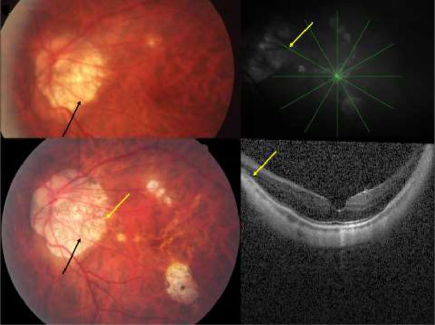
www.nature.com/scientificreports/
between the fovea and the temporal gamma zone border, is independent of axial l ength10. In a parallel manner, the
fovea is located more inferiorly in eyes with an inferior gamma zone than in eyes with a superior gamma z one13.
These observations made in cross-sectional studies led to the notion that BM with its associated retinal pig-
ment epithelium and photoreceptor layer may shift during the process of axial elongation into the posterior direc-
tion, potentially caused by a formation of new BM in the equatorial r egion14. To further assess that hypothesis,
we examined in the present follow-up study longitudinal associations between changes in the DFD and changes
in the parapapillary gamma zone. To avoid a potential bias by the referral of patients to a hospital, we choose a
population-based recruitment of the study population.
Methods
The Beijing Eye Study is a population-based longitudinal study performed in a rural region and in an urban area
of Greater B eijing15. The Medical Ethics Committee of Beijing Tongren Hospital approved the study protocol
according to the declaration of Helsinki and all study participants gave their written informed consent. The
ethics committee confirmed that all methods were performed in accordance with the relevant guidelines and
regulations. First conducted in 2001, the survey was repeated in 2011 inviting all participants from the survey of
2001. Included into the present study were all those individuals who had participated in the surveys in 2001 and
in 2011 and who were highly myopic, defined by an axial length of ≥ 26.0 mm or if axial length measurements
were not present, defined by a myopic refractive error of more than − 6 diopters in 2001 or 2011.
As also described in detail previously, all participants underwent a structured questionnaire, systemic exami-
nations, and a comprehensive ophthalmic examination16. The latter included measurements of visual acuity, slit
lamp examination of the anterior and posterior segment of the eye, and photography of the cornea, lens, optic
disc and macula (fundus camera Type CR6-45NM; Canon Inc., Tokyo, Japan). In 2011, the optic nerve head
including the peripapillary area and the macula were additionally imaged by spectral-domain optical coherence
tomography (OCT) (enhanced depth imaging mode) (Spectralis, Heidelberg Engineering Co, Heidelberg, Ger-
many). The protocol for imaging the optic disc and the macula included six radial scan lines with a scan length of
6 mm, centered on the optic disc and macula, respectively, and each comprising 100 A-scans. The parapapillary
region was examined with the intrinsic viewer (Heidelberg Eye Explorer software version 1.7.0.0; Heidelberg
Engineering), which automatically synchronized the vertical lines of each B-scan and the infrared image taken by
the OCT device. The parapapillary gamma zone was defined as the region between the end of BM and the border
isc16. The optic disc border was defined as the end of the lamina cribrosa or as the inner side of the
of the optic d
peripapillary border tissue of the peripapillary scleral flange. The OCT image of the macula served to determine
the localization of the foveola, and the optic nerve head imaging served for defining the border of gamma zone.
On the fundus photographs, we defined gamma zone as the parapapillary region, in which the sclera was
clearly visible and covered by superficial retinal tissue (i.e. retinal vessels and retinal nerve fibers) while the cho-
roid, except for few large choroidal vessels, was completely absent (Fig. 1)7,17. The delineation of gamma zone on
the fundus photographs was controlled by marking the border of gamma zone on the OCT images. The DFD
was assessed as the distance between the foveola and the optic disc center (Fig. 1)10. The position of the foveola
was manually identified on the fundus images with the help of OCT images. The angle kappa was defined as the
angle between the crossing point of the temporal superior arterial arcade with a vertical line drawn through the
fovea, the optic disc center, and the crossing point of the temporal inferior arterial arcade with a vertical fovea
line18. The length of the macular BM on the disc-fovea line was defined as the distance between the foveola and
the border of gamma zone. The vertical distance between the temporal superior and temporal inferior temporal
arterial arcade was the distance between both arterial arcades measured on a vertical line running through the
fovea. Presence and stage of myopic macular degeneration were specified according to the recommendations
made by the Pathologic Myopia Study G roup19. Lacquer cracks were considered as part of category 3 of myopic
macular degeneration. Aligning and flickering the photographs of the optic disc and macula, taken in 2001 and
2011, we assessed and noted any changes in the DFD, in the presence, size and location of parapapillary gamma
zone, in angle kappa, and in the course of the papillomacular retinal vessel. Retinal vessel straightening was
defined as a more straight, less contorted, course of a retinal vessel in the papillomacular region (Fig. 2).
Using a commercially available statistical software package (SPSS for Windows, version 25.0, IBM-SPSS,
Chicago, IL, USA), we first calculated the mean and standard deviations of the demographic parameters. Using
the chi-square test, we assessed associations between the occurrence of changes in the DFD and the occurrence
of changes in gamma zone, changes in angle kappa, and the course of the retinal vessels in the temporal region.
All P-values were two-sided and considered statistically significant when they were < 0.05.
Results
Out of 4439 individuals originally participating in the survey in 2001, 379 individuals had died and 1365 indi-
viduals had moved or did not want to be re-examined in 2011, so that the survey in 2011 included 2695 individu-
als who had also participated in the survey of 2001. They had a mean age of 64.7 ± 9.7 years (range 50–93 years)
in 2011 and a mean refractive error of − 0.20 ± 2.13 diopters (range − 22.0 to + 7.5 diopters). Out of 204 highly
myopic eyes with a myopic refractive error of ≥ − 6.0 diopters and/or an axial length of more than 26.0 mm in
the survey of 2001, 89 eyes (61 participants; 24 (39.3%) men) were re-examined in the survey of 2011 and had
assessable fundus photographs from both surveys. The age of these participants in 2011 (65.0 ± 9.8 years; median:
66 years; range 50–88 years) did not differ significantly (P = 0.17) from the age of the remaining participants in the
survey of 2011 (64.7 ± 19.7 years (median: 64 years; range 50–83 years). The mean axial length was 27.6 ± 1.3 mm
(range 26.06–30.88 mm). Cataract surgery had been performed in 11 out of the 89 eyes. For the remaining eyes
without cataract surgery, the mean refractive error was − 9.41 ± 3.52 diopters in 2001 and − 9.43 ± 3.26 diopters
in 2011 (Table 1).
Scientific Reports | (2021) 11:9006 | https://doi.org/10.1038/s41598-021-88579-9 2
Vol:.(1234567890)www.nature.com/scientificreports/
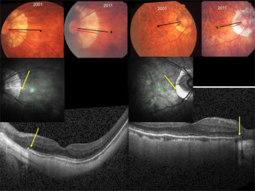
Figure 1. Upper part: Fundus photographs of two highly myopic eyes examined in 2001 and in 2011, showing
an elongation of the disc-fovea distance (black bar, as measured in 2001; green bar: elongation in 2011) and the
enlargement of gamma zone (red bar, as measured in 2001; green bar: amount of elongation of the disc-fovea
distance); black cross: foveola. Lower part: Optical coherence tomographic images showing the end of Bruch´s
membrane (yellow arrows) indicating the border of parapapillary gamma zone.
The present study additionally included a group of 86 randomly selected eyes (57 individuals) out of the
non-highly myopic group who had been examined in both surveys in 2001 and 2011. Mean refractive error in
2001 and in 2011 was − 0.97 ± 2.61 diopters and − 1.02 ± 2.45 diopters, respectively (Table 1). The mean axial
length was 23.6 ± 1.1 mm (range 21.64–25.90 mm). The highly myopic group and the non-highly myopic group
did not differ significantly in age (65.0 ± 9.8 years versus 66.7 ± 9.7 years (median: 68 years; range 50–84 years:
P = 0.25) and gender (P = 0.89).
Within the highly myopic group, 42 (48%) of the 89 highly myopic eyes showed a progression of myopic
maculopathy. Progression was observed in two (18.2%) out of 11 eyes with no fundus tessellation, 24 (41.4%) of
51 eyes with tessellated fundus (category 1) at the initial examination, in 6 (60%) of 10 eyes with diffuse chori-
oretinal atrophy (category 2), in 8 (100%) of the 8 eyes with patchy chorioretinal atrophy (category 3), and in two
(100%) out of two eyes with macular atrophy (category 4) in 2001. In 2011, 38 (42.7%) eyes were categorized as
“tessellated fundus” (category 1), 25 (28.1%) eyes as “diffuse chorioretinal atrophy” (category 2), 13 (14.6%) eyes
as “patchy chorioretinal atrophy” (category 3), and 4 (4.5%) eyes as “macular atrophy” (category 4).
The mean DFD was significantly longer in the highly myopic group than in the non-highly myopic group
(5.74 ± 0.50 mm versus 4.87 ± 0.48 mm; P < 0.001). In the 89 highly myopic group, an elongation of the DFD was
detected in 63 (70.8%) of the eyes, in 25 (28.1%) of the eyes the DFD remained constant, and in one (1.1%) eye,
the fovea moved inferiorly. In the 86 non-highly myopic group, the DFD elongated in 9 (10.5%) eyes and became
shorter in one eye (1.2%), while in 76 (88.4%) eyes, the DFD did not change. The difference in the prevalence of
an elongation of the DFD between both groups was significant (P < 0.001). Within the non-highly myopic group,
the subgroup of eyes with DFD elongation as compared to the eyes without DFD elongation was significantly
more myopic (− 3.97 ± 3.92 diopters versus − 0.67 ± 1.98 diopters; P < 0.001).
A widening of parapapillary gamma zone was detected significantly more often (P < 0.001) in the highly
myopic group (75/89 (84.3%) eyes) than in the non-highly myopic group (18/86 (20.9%) eyes). In the total study
group, and in the non-highly myopic group and in the highly myopic group taken separately, the enlargement of
gamma zone was significantly (all P < 0.001) associated with the elongation of the DFD. In the group of 93 eyes
Scientific Reports | (2021) 11:9006 | https://doi.org/10.1038/s41598-021-88579-9 3
Vol.:(0123456789)www.nature.com/scientificreports/
Figure 2. Left part: Fundus image of a highly myopic eye, taken in 2001 and in 2011 and showing the
straightening of the retinal vessel (black arrow) in association with a widening of parapapillary gamma zone
and an elongation of the disc-fovea distance in a highly myopic eye. Right part: Optical coherence tomographic
images showing the end of Bruch´s membrane (yellow arrows, also on the lower left image) indicating the
border of parapapillary gamma zone.
Highly myopic group Non-highly group P-value
n (eyes/participants) 89/61 86/57
Age (years) 65.0 ± 9.8 66.7 ± 9.7 0.25
Gender (women) 37 (60.7%) 36/57 0.89
Refractive error (spherical equivalent) in 2001 (diopters) − 9.41 ± 3.52 − 0.97 ± 2.61 < 0.001
Refractive error (spherical equivalent) in 2011 (diopters) − 9.43 ± 3.26 − 1.02 ± 2.45 < 0.001
Axial length (mm) in 2011 27.6 ± 1.3 23.6 ± 1.1 < 0.001
Myopic maculopathy grading in 2001 (Category 0/1/2/3/4) 11/58/10/8/2 73/13/0/0/0 < 0.001
Myopic maculopathy grading in 2011 (Category 0/1/2/3/4) 9/38/25/13/4 72/14/0/0/0 < 0.001
Disc-fovea distance elongation 63/89 (70.8%) 9/86 (10.5%) < 0.001
Gamma zone widening 75/89 /84.3%) 18/86 (20.9%) < 0.001
Angle kappa decrease 61/80 (68.5%) 9/86 (10.5%) < 0.001
Papillo-macular retinal vessel straightening 58/89 (65.2) 7/86 (8.1%) < 0.001
Table 1. Demographic and ocular characteristics and comparisons between the highly myopic group and the
control group.
with a widening of gamma zone, 72 (77.4%) eyes showed a DFD elongation, while in the group 82 eyes without
gamma zone widening, the DFD did not elongate in any eye.
In the highly myopic group, a decrease in angle kappa without evident cause was observed in 61 (68.5%) eyes,
and angle kappa became smaller in association with a rotation of the temporal arterial arcade downward in two
(2.2%) eyes. In the 86 non-highly myopic group, 9 (10.5%) eyes showed a decrease in angle kappa, one (1.2%)
eye showed a decrease in angle kappa in association with the development of an epiretinal membrane, and in 75
(88.4%) eyes angle kappa remained unchanged. In the total study group, and in the non-highly myopic group
Scientific Reports | (2021) 11:9006 | https://doi.org/10.1038/s41598-021-88579-9 4
Vol:.(1234567890)www.nature.com/scientificreports/
and in the highly myopic group taken separately, the reduction in angle kappa was associated (all P < 0.001) with
the DFD elongation.
The distance between the temporal superior arterial arcade and the temporal inferior arterial arcade assessed
on a vertical line through the fovea did not change in any of the 93 highly myopic eyes without macular BM
defects (i.e. category 2 or less of myopic maculopathy), except for one eye with a decrease in angle kappa in asso-
ciation with the development of an epiretinal membrane, and it did not change in any non-highly myopic eye.
A straightening of retinal vessels in the temporal sector was observed in 58 (65.2%) highly myopic eyes, while
in 31 (34.8%) of the highly myopic eyes, the vessel course remained unchanged. In the non-highly myopic group,
the vessel course remained unchanged in 77 (89.5%), while the vessels appeared to be straightened in 7 (8.1%)
eyes and to be less straight in 2 (2.3%) eyes. The difference in the incidence of vessel straightening between both
groups was significant (P < 0.001). Temporal vessel straightening was present (P < 0.001) in 64 of the (88.9%) 72
eyes with an DFD elongation and one (1.0%) of the 103 eyes without DFD elongation. In the total study group,
and in the non-highly myopic group and in the highly myopic group taken separately, the retinal vessel straight-
ening was associated (all P < 0.001) with the DFD elongation.
In multivariable analysis, the frequency of a DFD elongation was associated with a higher prevalence of an
enlargement of gamma zone (P < 0.001), a more myopic refractive error (P = 0.03), and a higher prevalence of
progression of myopic maculopathy (P < 0.001).
Discussion
In this population-based sample of highly myopic eyes, the prevalence of a DFD elongation during a follow-up
of 10 years was relatively high (70.8%), and it was associated with a widening of gamma zone, a decrease in angle
kappa, and a straightening of the papillo-macular retinal vessels. In a multivariable analysis, a higher prevalence
of a DFD elongation additionally depended on a more myopic refractive error and a higher prevalence of pro-
gression of myopic maculopathy. In all eyes without macular BM defects and without epiretinal membranes, the
length of the macular BM on the disc-fovea line and the vertical distance between the temporal arterial arcades
did not change during the study period.
The observations made in this longitudinal study agree with findings obtained in a recent cross-sectional
investigation on a larger group of study participants in which a longer DFD was correlated with a wider gamma
zone and a longer axial length10. In that study, the longer DVD in axially elongated eyes could fully be explained
by the wider gamma zone, while the length of the macular BM on the disc-fovea line, measured as distance
between the foveola and the border of gamma zone, was independent of axial l ength10. In a similar manner, the
vertical distance between the temporal arterial arcades was not related with axial length if eyes with macular
BM defects were e xcluded18. Similar findings were made in the present longitudinal investigation. It suggests
that the widening of gamma zone is the cause of the DFD elongation, while BM in the macular region remained
unaffected by axial elongation, unless macular BM defects developed. It leads to the notion that the macular
region, outside of the parapapillary area with gamma zone and delta zones, remains primarily untouched by the
process of axial elongation, except for an axial elongation-associated thinning of the choroid and a potential, axial
elongation-associated stretching of the retinal nerve fibers in the papillo-macular bundle region (see below). The
finding of a constancy in the macular BM dimensions is paralleled by observations made in histologic and clini-
cal studies, which showed that the thickness of the retina, the density of the retinal pigment epithelium and the
density of the choriocapillaris in the macular region were not related to axial l ength20–23. Correspondingly, best
corrected visual acuity was not dependent on axial length if eyes with a myopic maculopathy were e xcluded24. It
agrees with the notion that the distance of the macular photoreceptors was not enlarged in axially elongated eyes,
in agreement with the histologic findings of an independence of the density of the retinal pigment epithelium
and the choriocapillaris thickness of axial length. The decrease in angle kappa in eyes with a DFD elongation and
a constancy of the vertical distance between the arterial arcades can geometrically be explained. The observa-
tions support the notion of a shifting, and otherwise stable, BM at the posterior pole in axially elongating e yes18.
Another finding of the present study was the straightening of the papillo-macular retinal vessels in eyes with
an enlarging DFD and widening of gamma zone. The retinal nerve fibers and the inner limiting membrane are
the only anatomical structures which connect the other retinal layers, including the retinal ganglion cell layer,
with the optic disc. Both, the inner limiting membrane and the retinal nerve fibers are firmly connected to the
optic nerve head. The lengthening of the distance between the macular ganglion cells and the optic disc by the
enlarging gamma zone may thus lead to a straightening of the retinal nerve fibers, parallel to the straightening
of the papillo-macular retinal vessels. If the retinal nerve fibers had a curved course to the optic disc before the
DFD elongation started, the elongation of the distance to the optic disc could be compensated by a straightening
of the course of the nerve fibers. If however the retinal nerve fibers were located in the center of the papillo-
macular bundle and had a straight course to the optic disc already before an enlargement of gamma zone started,
the fibers could not compensate the elongated DFD by straightening their course. One may assume that they get
stretched in that situation. Such a stretching of papillo-macular nerve fibers might be associated with a dam-
age to the nerve fibers and may lead to the development of deep central scotomas in highly myopic eyes with
a large gamma zone and without myopic macular changes explaining such perimetric d efects25–27. Fitting with
that notion, a higher prevalence of myopic maculopathy and longer axial length was associated with a thinner
peripapillary retinal nerve fiber layer in the recent Ural Eye and Medical Study, after adjusting for the prevalence
and amount of glaucomatous optic neuropathy or after excluding eyes with glaucomatous optic neuropathy from
the statistical a nalysis28. In a parallel manner, Omoto and colleagues reported that in 138 healthy eyes without
any known eye disease the macular ganglion cell-inner plexiform layer thickness decreased significantly with
narrowing of the peripapillary retinal artery angle (or angle kappa)29.
Scientific Reports | (2021) 11:9006 | https://doi.org/10.1038/s41598-021-88579-9 5
Vol.:(0123456789)www.nature.com/scientificreports/
Narrowing of the angle kappa was strongly correlated with an elongation of the DFD. One may discuss that
a posterior shift of the fovea in association with the elongation of the disc-fovea distance at the nasal side of
the fovea would imply the development of a secondary divergent pseudo strabismus in myopic eyes, unless the
posterior pole with the fovea is shifted backwards evenly in all meridians. Since the majority of myopic eyes does
not develop a pseudo-strabismus, one may infer that indeed the foveal backward shift occurs in all meridians. It
would fit with the notion of a circular enlargement of BM in the equatorial region of the g lobe14.
The inner limiting membrane is, besides the retinal nerve fibers and the large retinal vessels, the only other
structure connecting the retina with the optic disc. As the retinal nerve fibers, also the inner limiting membrane
in the papillo-macular region may get stretched in eyes with an elongated DFD and enlarged gamma zone. The
inner limiting membrane as the basal membrane of the Müller-cells is assumed to have a relatively low elasticity,
as may also be the case with the lens capsule as basal membrane of the lens epithelium or Descemet’s membrane
as the basal membrane of the corneal endothelium cells. If the DFD elongates more than the low elasticity of
the inner limiting membrane allows, the inner limiting membrane may get elevated to shorten the elongated
distance to the optic disc. It may hold true in particular in eyes with a scleral staphyloma at the posterior pole.
The phenomenon may be similar to the effect of a strained washing line. One may discuss whether such a pulling
force exerted on the inner limiting membrane in highly myopic eyes with a large gamma zone may be forwarded
to the deeper retinal layers, leading to an increase in the inner-retinal strain with the sequel of a macular schisis30.
Peeling and removal of the inner limiting membrane in such a situation may then release the strain from the
deeper retinal layers and might lead to a decrease in the height of the macular schisis. It may however increase
the strain on the retinal nerve fibers which then are the only remaining structure (except for the large retinal
vessels) connecting the macular retina with the optic disc.
Interestingly, 9 (10.5%) out of the 86 non-highly eyes showed a DFD elongation. A similar result was reported
in a previous study by Yanagisawa and colleagues who observed 125 eyes of 72 patients (mean refractive error:
− 3.5 ± 3.1 diopters (− 13.0 to + 3.25 diopters) with primary open-angle glaucoma for a mean follow-up time of
about 5 years and who found that an increase in axial length (mean: 0.035 ± 0.10 mm) was significantly related
with a lower axial length at baseline31. Therefore, the DFD elongation was observed not only in eyes with longer
axial length but also in eyes with shorter axial length. A longer axial length is associated with a longer DFD.
The reasons for the elongation of the DFD in our non-highly myopic study population may have been that the
subgroup of non-highly myopic eyes with DFD elongation as compared to the non-highly myopic eyes without
DFD elongation was significantly (P < 0.001) more myopic, and a partially arbitrary cut-off value of − 6 diopters
for the definition of high myopia. It may indicate that some eyes of adults with a myopic refractive error of less
than − 6 diopters may also undergo further myopic changes at their posterior pole.
The observations made in our study may have some clinical meaning. The potential stretching of the retinal
nerve fibers in the papillo-macular bundle area, parallel to straightening of the retinal vessels may indicate the
presence of paracentral scotomas in highly myopic eyes without that myopic macular changes such as macular
BM defect might explain such scotomas. The elongation of the DFD in myopic eyes may have the psychophysi-
cal equivalent of an increased distance between the visual center and blind spot in perimetry. It may influence
the morphologic-functional relationship in optic neuropathies such as the glaucomas. The enlarged gamma
zone in myopic eyes is the equivalent of an enlargement of the blind spot in myopic eyes32,33. The decrease in
angle kappa with longer axial length may lead to an accompanying change in the position in the peaks of the
peripapillary retinal nerve fiber layer profile, so that in an OCT-based assessment of the optic nerve the peaks,
despite a normal volume of the retinal nerve fiber layer, come to lie outside of the normal position and may be
marked as abnormal or pathological.
When discussing the findings of our study, its limitations should be taken into account. First, only 89 (43.6%)
out of 204 highly myopic eyes which were examined in the survey in 2001 were included into the present study.
This figure of re-participation of 44% in a 10-year follow-up study roughly compares with the 5-year follow par-
ticipation rate of 68.7% in the Blue Mountains Eye Study on the re-examination of eyes with myopic r etinopathy34.
As in any population-based study, however, the non-participation might have influenced the assessment of the
prevalence of a DFD elongation within the study population. It may however not have had an effect on the main
result, that the DFD elongation is mostly due to an enlargement of gamma zone and that it is associated with
a decrease in angle kappa and a straightening of the papillomacular retinal vessels. Second, OCT images were
not obtained at the baseline examination in 2001. The assessment of the OCT images taken in 2011 allowed
however the identification of gamma zone and of the location of the fovea. Third, we measured the DFD on
two-dimensional fundus images of the curved posterior ocular segment. It led to an underestimation of the real
distance between the optic disc and the fovea. Although this principal measurement error was present in all
eyes, it was more marked in eyes with a more profoundly curved posterior segment, in particular in eyes with a
posterior staphyloma. Fourth, as determined by Littmann and others, the magnification of images of elements of
the posterior fundus depends on the axial length35. Any comparison in the DFD or gamma zone width between
eyes and between individuals would have necessitated the inclusion of the axial length into the assessment and
comparison of the parameters. In our study however, we performed an intra-eye comparison of the DFD and
other morphologic variables between images taken in 2001 and obtained in 2011. Although the axial length
will have increased in a substantial number of eyes of the highly myopic group, this increase, according to data
from previous studies, may have been relatively low, so that the change in axial length between 2001 and 2011,
in agreement with the calculations by Littmann, may not have markedly influenced the image magnification by
the optical system of the e ye35–37. Fifth, the study participants were born in 1961 or earlier, so that the type of
high myopia examined in our study may differ from the type of “school children myopia” common in the young
generation today. It has therefore remained elusive whether the findings obtained in this study can be transferred
onto the young highly myopic individuals of today. Sixth, future studies may address whether the findings made
in our Chinese population can be transferred on population of different ethnicity.
Scientific Reports | (2021) 11:9006 | https://doi.org/10.1038/s41598-021-88579-9 6
Vol:.(1234567890)www.nature.com/scientificreports/
In conclusion, the prevalence of a DFD elongation in the highly myopic eyes of our population-based sam-
ple was relatively high with a value of 64.8% during a 10-year follow-up. The DFD elongation was due to an
enlargement of gamma zone, while the length of BM measured on the disc-fovea line and as distance between
the temporal arterial arcades remained constant. These findings support the notion of a shifting, but otherwise
stable BM at the posterior pole in axially elongated and elongating eyes. The straightening of the papillo-macular
vessels with increasing gamma zone may suggest a stretching of the papillo-macular retinal nerve fibers in highly
myopic eyes, potentially leading to a non-glaucomatous optic nerve damage. It may also indicate a stretching of
the inner limiting membrane in the papillo-macular region.
Received: 4 February 2021; Accepted: 6 April 2021
References
1. Jonas, J. B., Ohno-Matsui, K. & Panda-Jonas, S. Myopia: Anatomic changes and consequences for its etiology. Asia Pac. J. Ophthal-
mol. (Phila). 8(5), 355–359 (2019).
2. Jonas, R. A. & Holbach, L. Peripapillary border tissue of the choroid and peripapillary scleral flange in human eyes. Acta Ophthal-
mol. 98(1), e43–e49 (2020).
3. Hong, S. et al. OCT-detected optic nerve head neural canal direction, obliqueness, and minimum cross-sectional area in healthy
eyes. Am. J. Ophthalmol. 208, 185–205 (2019).
4. Zhang, Q., Xu, L., Wei, W. B., Wang, Y. X. & Jonas, J. B. Size and shape of Bruch´s membrane opening in relationship to axial length,
gamma zone and macular Bruch´s membrane defects. Invest. Ophthalmol. Vis. Sci. 60(7), 2591–2598 (2019).
5. Wang, Y. X., Panda-Jonas, S. & Jonas, J. B. Optic nerve head anatomy in myopia and glaucoma, including parapapillary zones alpha,
beta, gamma and delta: histology and clinical features. Prog. Ret. Eye Res. 1, 1 (2021).
6. Reis, A. S. et al. Optic disc margin anatomy in patients with glaucoma and normal controls with spectral domain optical coherence
tomography. Ophthalmology 119(4), 738–747 (2012).
7. Jonas, J. B. et al. Parapapillary atrophy: Histological gamma zone and delta zone. PLoS ONE 7(10), e47237 (2012).
8. Kim, T. W. et al. Optic disc change with incipient myopia of childhood. Ophthalmology 119(1), 21 (2012).
9. Jonas, J. B., Ohno-Matsui, K., Spaide, R. F., Holbach, L. & Panda-Jonas, S. Macular Bruch’s membrane defects and axial length:
association with gamma zone and delta zone in peripapillary region. Invest. Ophthalmol. Vis. Sci. 54(2), 1295–1302 (2013).
10. Jonas, R. A. et al. Optic disc-fovea distance, axial length and parapapillary zones: The Beijing Eye Study 2011. PLoS ONE 10(9),
e0138701 (2015).
11. Yamada, H. et al. Microstructure of peripapillary atrophy and subsequent visual field progression in treated primary open-angle
glaucoma. Ophthalmology 123(3), 542–551 (2016).
12. Kim, M., Choung, H. K., Lee, K. M., Oh, S. & Kim, S. H. Longitudinal changes of optic nerve head and peripapillary structure dur-
ing childhood myopia progression on OCT: Boramae Myopia Cohort Study Report 1. Ophthalmology 125(8), 1215–1223 (2018).
13. Jonas, R. A., Brandt, C. F., Zhang, Q., Wang, Y. X. & Jonas, J. B. Location of parapapillary gamma zone and vertical fovea location:
The Beijing Eye Study 2011. Invest. Ophthalmol. Vis. Sci. 62(1), 18 (2021).
14. Jonas, J. B., Ohno-Matsui, K., Jiang, W. J. & Panda-Jonas, S. Bruch membrane and the mechanism of myopization. A new theory.
Retina 37(8), 1428–1440 (2017).
15. Yan, Y. N. et al. Ten-year progression of myopic maculopathy: The Beijing Eye Study 2001–2011. Ophthalmology 125(8), 1253–1263
(2018).
16. Zhang, Q., Wang, Y. X., Wei, W. B., Xu, L. & Jonas, J. B. Parapapillary beta zone and gamma zone in a normal population: The
Beijing Eye Study 2011. Invest. Ophthalmol. Vis. Sci. 59(8), 3320–3329 (2018).
17. Jonas, J. B., Fang, Y., Weber, P. & Ohno-Matsui, K. Parapapillary gamma zone and delta zone in high myopia. Retina 38(5), 931–938
(2018).
18. Jonas, J. B., Weber, P., Nagaoka, N. & Ohno-Matsui, K. Temporal vascular arcade width and angle in high axial myopia. Retina
38(9), 1839–1847 (2018).
19. Ohno-Matsui, K. et al. International photographic classification and grading system for myopic maculopathy. Am. J. Ophthalmol.
159(5), 877-883.e7 (2015).
20. Jonas, J. B. et al. Retinal thickness and axial length. Invest. Ophthalmol. Vis. Sci. 57(4), 1791–1797 (2016).
21. Jonas, J. B., Ohno-Matsui, K., Holbach, L. & Panda-Jonas, S. Retinal pigment epithelium cell density in relationship to axial length
in human eyes. Acta Ophthalmol. 95(1), e22–e28 (2017).
22. Jonas, J. B., Holbach, L. & Panda-Jonas, S. Bruch’s membrane thickness in high myopia. Acta Ophthalmol. 92(6), e470-474 (2014).
23. Panda-Jonas, S., Holbach, L., & Jonas, J.B. Choriocapillaris thickness and density in axially elongated eyes. Acta Ophthalmol. https://
doi.org/10.1111/aos.14486 (2020).
24. Shao, L. et al. Visual acuity and subfoveal choroidal thickness: The Beijing Eye Study. Am. J. Ophthalmol. 158(4), 702-709.e1 (2014).
25. Sawada, Y., Araie, M., Ishikawa, M. & Yoshitomi, T. Multiple temporal lamina cribrosa defects in myopic eyes with glaucoma and
their association with visual field defects. Ophthalmology 124(11), 1600–1611 (2017).
26. Park, H. Y. L., Jeon, S. J. & Park, C. K. Features of the choroidal microvasculature in peripapillary atrophy are associated with visual
field damage in myopic patients. Am. J. Ophthalmol. 192, 206–216 (2018).
27. Jeon, S. J., Park, H. Y. L., Kim, Y. C., Kim, E. K. & Park, C. K. Association of scleral deformation around the optic nerve head with
central visual function in normal-tension glaucoma and myopia. Am. J. Ophthalmol. 217, 287–296 (2020).
28. Bikbov, M. M. et al. Prevalence of myopic maculopathy among adults in a Russian population. JAMA Netw. Open. 3(3), e200567
(2020).
29. Omoto, T. et al. Relationship between macular ganglion cell thickness and ocular elongation as measured by axial length and
retinal artery position. Invest. Ophthalmol. Vis. Sci. 61(11), 16 (2020).
30. Shinohara, K. et al. Ultrawide-field OCT to investigate relationships between myopic macular retinoschisis and posterior staphy-
loma. Ophthalmology 125(10), 1575–1586 (2018).
31. Yanagisawa, M. et al. Changes in axial length and progression of visual field damage in glaucoma. Invest. Ophthalmol. Vis. Sci.
59(1), 407–417 (2018).
32. Jonas, J. B., Gusek, G. C. & Fernández, M. C. Correlation of the blind spot size to the area of the optic disc and parapapillary
atrophy. Am. J. Ophthalmol. 111(5), 559–565 (1991).
33. Rensch, F. & Jonas, J. B. Direct microperimetry of alpha zone and beta zone of parapapillary atrophy. Br. J. Ophthalmol. 92(12),
1617–1619 (2008).
34. Vongphanit, J., Mitchell, P. & Wang, J. J. Prevalence and progression of myopic retinopathy in an older population. Ophthalmology
109(4), 704–711 (2002).
35. Littmann, H. Bestimmung der wahren Größe eines Objektes auf dem Hintergrund des lebenden Auges. Klin Monbl Augenheilkd.
192(1), 66–67 (1988).
Scientific Reports | (2021) 11:9006 | https://doi.org/10.1038/s41598-021-88579-9 7
Vol.:(0123456789)www.nature.com/scientificreports/
36. Saka, N. et al. Long-term changes in axial length in adult eyes with pathologic myopia. Am. J. Ophthalmol. 150, 562–568 (2010).
37. Fang, Y. et al. Progression of myopic maculopathy during 18-year follow-up. Ophthalmology 125, 863–877 (2018).
Author contributions
Design: R.A.J., Y.X.W., J.B.J.; collection of data: R.A.J., Y.N.Y., Q.Z., Y.X.W., J.B.J.; statistical analysis: R.A.J., J.B.J.;
drafting of the manuscript: R.A.J., Y.N.Y., J.B.J.; revising and final approval of the manuscript: R.A.J., Y.N.Y.,
Q.Z., Y.X.W., J.B.J.
Funding
National Natural Science Foundation of China (#81570835).
Competing interests
Jost B. Jonas and Rahul A. Jonas: European patent application 16 720 043.5 and US patent application US 2019
0085065 A1: „Agents for use in the therapeutic or prophylactic treatment of myopia or hyperopia). Jost B. Jonas:
Advisory Board Novartis; Patent holder with Biocompatibles UK Ltd. (Farnham, Surrey, UK) (Title: Treatment
of eye diseases using encapsulated cells encoding and secreting neuroprotective factor and/or anti-angiogenic
factor; Patent number: 20120263794), Patent application: Agents for the use in the therapeutic or prophylactic
treatment of retinal pigment epithelium associated diseases. All other authors declare no competing interests.
Additional information
Correspondence and requests for materials should be addressed to Y.X.W.
Reprints and permissions information is available at www.nature.com/reprints.
Publisher’s note Springer Nature remains neutral with regard to jurisdictional claims in published maps and
institutional affiliations.
Open Access This article is licensed under a Creative Commons Attribution 4.0 International
License, which permits use, sharing, adaptation, distribution and reproduction in any medium or
format, as long as you give appropriate credit to the original author(s) and the source, provide a link to the
Creative Commons licence, and indicate if changes were made. The images or other third party material in this
article are included in the article’s Creative Commons licence, unless indicated otherwise in a credit line to the
material. If material is not included in the article’s Creative Commons licence and your intended use is not
permitted by statutory regulation or exceeds the permitted use, you will need to obtain permission directly from
the copyright holder. To view a copy of this licence, visit http://creativecommons.org/licenses/by/4.0/.
© The Author(s) 2021
Scientific Reports | (2021) 11:9006 | https://doi.org/10.1038/s41598-021-88579-9 8
Vol:.(1234567890)You can also read