Epileptic seizures in patients with COVID-19: A systematic review of early evidences - Nepal Journals ...
←
→
Page content transcription
If your browser does not render page correctly, please read the page content below
Open Access                                                                             Journal of Biomedical Sciences
SYSTEMATIC REVIEW                                                          DOI: https://doi.org/10.3126/jbs.v8i1.38459

Epileptic seizures in patients with COVID-19: A systematic
review of early evidences
Roy B1*, Banerjee I2

*Corresponding author:
Dr Bedanta Roy, Ph.D.
                                                          ABSTRACT
Senior Lecturer, Department of Physiology, Faculty of      Background
Medicine, Quest International University, Ipoh, Perak,     Global emergence of SARS-CoV-2 surfaced neurological
Malaysia                                                   complications amongst the patients. COVID-19 resembles
Email: bedanta.roy@gmail.com ORCID                         with other coronavirus strains follows a trend of
                                                           neurological complication, damage and encephalopathy,
                                                           which entails considerable risks, requires attention for the
Information about the article:                             neurologists. This is, to our knowledge, the first systematic
                                                           review of the literature to investigate solely to elucidate the
Received: Oct. 05, 2020                                    seizure spectrum by unfolding epileptogenicity of the SARS
Accepted: April 04, 2021                                   CoV-2 and potential pathways of neuroinvasion.
Published online: July 11, 2021

Cite this article:                                         Methods
                                                           A systematic literature search was performed in PubMed
Roy B, Banerjee I. Epileptic seizures in patients          and Embase database following standard guidelines, using
with COVID-19: A systematic review of early                specific keywords based on epileptic seizure onset described
evidences. Journal of Biomedical Sciences. 2021;           from December 01, 2019, to July 17, 2020
8(1):33-44
                                                           Results
Publisher                                                  A total of 17 studies were included ranging from case
Nepal Health Research Society, Bahundhara           -6,    reports, series of cases, multicentre cross-sectional study
Gokarnesowor Municipality, Kathmandu, Nepal                with the first-time onset of seizure associated with an
eISSN 2382-5545, ISSN 2676-1343 (Print)                    epileptic origin. We excavated causes of complex COVID-
© The Author(s). 2021                                      19 related neurological manifestations, e.g., cerebrovascular
Content licensing: CC BY 4.0                               diseases, encephalitis, demyelinating lesions, cytokine storm
                                                           and proposed routes of SARS-CoV-2 entry into the nervous
                                                           system to understand the mechanism of an epileptic seizure.

                                                           Conclusion
                                                           COVID-19 is a potent neuropathogen which causes the new
                                                           onset of epileptic seizures should get diagnostic recognition
                                                           to evade possible deterioration of neurological conditions.
                                                           However, more shreds of evidence from the future will
                                                           further elucidate the epileptogenic potential of the pandemic.

                                                           Keywords
                                                           Brain diseases, Coronavirus infections, Epilepsia Partialis
                                                           Continua, Epilepsy, neurologic manifestations, SARS-CoV-
                                                           2, Seizures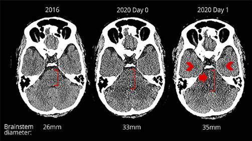
JBS 2021;8(1):33-44
Journal of Biomedical Sciences Epileptogenesis in COVID-19
Background references in the included studies to confirm the literature
Severe acute respiratory syndrome coronavirus-2 (SARS- saturation. All authors scanned the titles and full-text
CoV-2) is recently emerged human pandemic stormed the reports independently against the standard search criteria
whole world. The virus was identified as a beta coronavirus for the systematic review to identify the eligibility and
is dissimilar with severe acute respiratory syndrome inclusion. Disagreement pertaining for the inclusion of
coronavirus (SARS-CoV) and Middle East respiratory articles were resolved through the discussions. The authors
syndrome coronavirus (MERS-CoV), hence got a distinct extracted the following data from all included studies:
identity. The clinical presentation of SARS-CoV-2 is acute Author/year, age, gender, design, the interval of COVID-19
respiratory distress syndrome (ARDS) and viral pneumonia symptom onset and first seizure, clinical presentation,
[1,2]. The disease was first surfaced in Wuhan, Hubei neurological manifestations, diagnostic findings,
Province, since December 12, 2019, in China, conceivably interventions, and limitations. The level of evidence and
linked to the Huanan Seafood Wholesale Market located in quality of the research was also carefully observed.
Jianghan District [3]. The virus was named as Coronavirus Reference management was done by EndNote X5 software
disease 2019 "COVID-19" by the World Health (Clarivate Analytics, Boston, MA, USA). This systematic
Organization (WHO), affected 216 countries with review protocol was not registered earlier. We mostly
2,102,6758 confirmed cases and 75,5786 deaths as of followed the WHO recommended gold standard guidelines
August 16, 2020 [4]. The spread of this virus and ongoing (epidemiological history, clinical symptoms, and laboratory
devastation around the world shows no evidence of ending or radiological findings) to consider the articles in our
of this global pandemic and it impacted deadly on study. All cases included in this review were confirmed
economic, financial, social, and mental wellbeing on the cases of SARS-CoV-2, which was diagnosed by SARS-
humanity. CoV-2 PCR testing using a nasopharynx swab. We
Shortness of breath, fever, and cough reported since the reviewed the clinical researches, including original articles,
beginning of the pandemic [1, 2], but clinical manifestations case series, and case reports, for neurological involvement
of SARS-CoV-2 are not limited to the respiratory system; it by COVID-19 on the incidence of epilepsy and organized
infects the nervous system too. Mao et al. published the first them into tables.
hospital-based report on the SARS-CoV-2 infected patients
revealed that 36.4% of the patients had neurological Results
complications: CNS (53 [24.8%]), PNS (19 [8.9%]). Through our search strategy, we have identified a total of
Dizziness, headache, taste and smell impairment was the 160 abstracts. After exclusion and eligibility of full text, 17
most common symptoms reported [5]. Growing shreds of articles were selected for systematic review, involved
evidence of nausea, vomiting, myalgia, asthenia, dizziness, seizures, or epilepsy as a new-onset due to SARS-CoV-2
and reduced consciousness imply the viral neuroinvasive infection. Among these articles, 12 were case reports; four
potential which increases with the severity of infection [5, were case series, and one study was a multicentre cross-
6]. SARS-CoV-1 outbreak in 2002-2003 documented sectional study. Table-1 and Table-2 shows the summary of
numerous neurological manifestations ranging from the included studies [5, 10-25].
moderate complications like seizures, status epilepticus, All studies were critically analyzed based on the standard
myopathy, to severe consequences stroke and diagnosis for epilepsy (e.g., EEG, head CT /MRI, CSF
polyneuropathy [7, 8]. analysis). We also included CSF-PCR to obtain information
This systematic review aims to outline a spectrum of the for neuroinvasion of SARS-CoV-2 and the intervention
seizures and epileptic symptoms of SARS-CoV-2 infected strategy towards the administration of Anti-epileptic drugs.
patients and enlighten the viral invasion to CNS and Besides, we rigorously reviewed a few relevant literature
mechanism of epileptogenicity. (26-30) to understand the mechanism of entry of SARS-
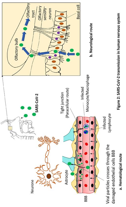
CoV-2 in CNS summarized in Figure 2.
Material and methods Figure 2: Explains the possible routes of SARS-CoV-2
A systematic literature search was conducted from entry in the nervous system.
December 01, 2019, to July 17, 2020, in PubMed and 2a. shows the hematologic route where viral particles cross
Embase database. We followed the recommendations of the the endothelial cells of the blood-brain barrier (BBB) either
Preferred Reporting Items for Systematic Review and Meta- directly or enter by using the infected cells of the
Analysis (PRISMA) protocol [9]. The search strategy was reticuloendothelial system (RE) or lymphocyte as a vehicle
developed by an expert panel of neurologists and through the paracellular route.
neurophysiologists. Search terms included “COVID-19” 2b. shows the neurologic route where virus enters in the
OR “SARS-CoV-2” OR “2019-nCoV” OR “novel olfactory epithelium, olfactory bulb and later enters in the
coronavirus” in conjunction with “epilepsy” OR “Seizure.” olfactory tract. Retrograde axonal transport and CoVs
(Figure 1). clathrin-dependent endocytotic/exocytotic pathway may
Articles written in the English language were included for help in this process.
this review. The authors carefully examined the list of
Page | 34JBS 2021;8(1):33-44
Journal of Biomedical Sciences Epileptogenesis in COVID-19
Figure 1: Inclusion of articles by Preferred Reported
Items for Systematic reviews
Page | 35JBS 2021;8(1):33-44
Journal of Biomedical Sciences Epileptogenesis in COVID-19
Page | 36JBS 2021;8(1):33-44
Journal of Biomedical Sciences Epileptogenesis in COVID-19
Table 1: Summary of the studies, infection interval and clinical presentation
Author, Age(years)/mean/ Design No (% in Interval of Clinical presentation towards epileptic
year mean [SD], total COVID manifestations
Gender participants) symptom onset /Seizure description/
and first characteristics
seizure activity
(days)
Lyons et al. 20, M Case report 1 +3 Light-headedness with blurred and double
2020 vision, lower limb weakness, generalised tonic-
clonic seizure
Moriguchi et 24, M Case report 1 +9 consciousness disturbance, transient
al. 2020 generalized
seizures for about a minute
Atere et al. 46, M Case report 1 +3 Episode of seizures, syncope; conscious after
2020 30 seconds; an involuntary loss of feces
Somani et al. 49, F Case report 1 -3 Case 1: altered mental status, seizure
2020
73, F Case report 1 +1 Case 2: Persistent face and arm myoclonus
with worsening altered mental status (status
epilepticus)
Fasano et al. 54, M Case report 1 +7 Single seizure characterized by clonic
2020 movements in the right arm and loss of
consciousness.
Zanin et al. 54, F Case report 1 Not clear Unconscious, later unrest
2020
Stefano et al. 56, F Case report 1 +18 Agitation
2020
Dixon et al. 59, M Case report 1 +10 Vacant staring, speech arrest, flexion of both
2020 shoulders and a brief witnessed generalized
tonic-clonic seizure (GTCS), followed by
postictal state
Sohal et al. 72, M Case report 1 +2 Multiple episodes of persistent tonic colonic
2020 movements of upper and lower extremities
Elgamasy et 73, F Case report 1 +2 Painful muscle stiffening and twitching in the
al. 2020 left arm and leg
Hepburn et 76, M Case report 1 +2 Multiple episodes of left upper extremity clonic
al. 2020 activity and worsening encephalopathy
82, M Case report 1 +15 Right eyelid and facial twitching
Farhadian et 78, F Case report 1 +2 Sudden-onset uncontrolled limb movements
al. 2020 with ocular deviation followed by several
minutes of unresponsiveness.
Galanopoulou Age: 63.23 ± 11.9 Case series 11(64.7%) NA Motor seizure like events
et al. 2020 (30-83) (retrospective)
Morassi et al. 76, F Case series 1 +10 Transient loss of consciousness, followed by
2020 confusion
Garazzino et 5, not mentioned Multicentre cross- 2(1.2) 1.6 Febrile seizure
al. 2020 sectional (prospective)
Mao et al. 58.2±15.0 Case series 1(0.5) NA Sudden onset of limb twitching, foaming in the
2020 (retrospective mouth, and loss of consciousness, lasted for 3
observational) minutes.
Pinna et al. 59.6 Case series 13(26) NA Not available
2020 (retrospective
observational)
Page | 37JBS 2021;8(1):33-44
Journal of Biomedical Sciences Epileptogenesis in COVID-19
Table 2: Diagnostic findings, interventions, and limitations
Author, year EEG findings Head CT /MRI CSF analysis/ Intervention Main findings Potential limitations Outcome
findings CSF PCR for (Anti
SARS-CoV2 epileptic
drugs)
Lyons et al., Normal CT: Normal; MRI: Mild Lymphocytic Levetiracetam Meningoencephalitis No known limitations Survived
2020 mucosal thickening in pleocystosis (21
the sphenoid sinus cells/mm3, 99 %
mononuclear, 1%
polymorphs);
PCR Negative
Moriguchi et Not performed CT: Normal; MRI: Normal; PCR Levetiraceta Right lateral EEG not performed, Alive
al., 2020 Hyperintensity along the positive ventriculitis and no follow up during
wall of inferior horn of encephalitis mainly on information reporting
right lateral ventricle. right mesial lobe and of the
Fluid-attenuated hippocampus case
inversion
recovery (FLAIR)
images showed
hyperintense signal
changes in
the right mesial
temporal lobe and
hippocampus with slight
hippocampal atrophy.
Atere et al., Not performed Normal Not performed Not SARS-CoV-2 associated No EEG, SARS-CoV- Survived
2020 administered neurological symptoms 2 PCR in CSF; CSF
PCR for other viruses
was done to rule out as
DD
Somani et al., Frequent (4-6/hour) Normal Not performed Lorazepam, De novo status No CSF Survived
2020 cyclical seizures Levetiracetam epilepticus investigations(analysis,
emanating from left PCR), CSF PCR was
fronto-central regions performed to rule out
other viral infections
as DD
Frequent (5/hour) CT: Normal, MRI: Not Not performed Lorazepam, New-onset refractory No CSF Passed
cyclical seizures performed levetiracetam, status epilepticus investigations(analysis, away
emanating from left lacosamide, (NORSE) PCR), CSF PCR was
and right fronto- phenytoin, performed to rule out
central regions midazolam other viral infections
as DD
Fasano et al., Normal CT: Normal; MRI: Not Not performed Not First focal motor seizure No CSF Survived
2020 performed administered investigations(analysis,
PCR); CSF PCR for
other viruses was not
performed to rule out
as DD, Anti epileptic
drug was not
administered
Zanin et al., Two seizures in CT: Normal, MRI: Normal; PCR Lacosamide, CNS involvement and No known limitations Survived
2020 frontotemporal region Alterations of the negative levetiracetam, demyelinating lesions
and diffusing in periventricular white phenytoin associated with SARS-
homologous matter. Lesions present CoV-2
contralateral in bulbo-medullary
hemisphere junction (cervical and
dorsal spinal cord)
Stefano et al., Intermittent onset of 4 CT: Not performed; Increased protein Sedatives (not Focal injury in the CT not performed; Survived
2020 Hz rhythms over the MRI: numerous level (1.31 g/l) mentioned) absence of standard CSF virology
bilateral parasagittal punctiform signal voids and encephalopathy, critical PCR was not done for
regions, lasting from in bilateral juxtacortical Immunoglobulins; illness–associated other viruses to rule
5 s to maximum 25 s. white matter, corpus PCR negative cerebral microbleeds out as DD
callosum, and internal (related to cytokine
capsule, compatible with release syndrome)
cerebral microbleeds,
without any ischemic or
necrotizing lesion.
Page | 38JBS 2021;8(1):33-44
Journal of Biomedical Sciences Epileptogenesis in COVID-19
Dixon et al., Not performed CT: Brain stem Increased protein Levetiracetam Rapidly evolving Patient passed away, Passed
2020 swelling, subtle intrinsic concentration (2.3 encephalopathy involving no follow up away
pontine hemorrhage, g/L); PCR brain stem; hemorrhagic
symmetrical negative ANE
hypodensities in the
deep gray matter and
amygdalae; MRI:
Extensive, relatively
symmetrical changes
throughout the
supratentorial and
infratentorial
compartments.
Sohal et al., Six left temporal CT: Chronic Not performed Levetiracetam, Cytokine storm, No SARS-CoV-2 PCR Passed
2020 seizures and left microvascular ischemic valproate encephalitis in CSF; standard CSF away
temporal sharp waves changes; MRI: Not virology PCR was not
which were performed done for other viruses
epileptogenic. to rule out as DD
patient passed away,
no follow up
Elgamasy et Normal CT: Mild dilatation of Normal, slightly magnesium, Focal epilepsy, chronic No SARS-CoV-2 PCR Survived
al., 2020 the lateral ventricles elevated levetiracetam small vessel ischemia in CSF; standard CSF
with prominent fissures leukocytes (0.5 lacosamide, virology PCR was not
and sulci. Scattered per cubic clobazam done for other viruses
deep white matter millimeter); not to rule out as DD
hypodensities, MRI: performed
dilated ventricular
system with a patent and
prominent aqueduct of
Sylvius
Hepburn et al., Three focal seizures Normal Not performed Levetiracetam Focal seizure, co- No SARS-CoV-2 PCR Passed
2020 lasting approximately morbidity in CSF; CSF PCR for away
30 s each arising from other viruses was not
the right done to rule out as DD
centroparietal region
EEG seizures mainly CT: Hypodensities Not performed Levetiracetam Status epilepticus, brain No SARS-CoV-2 PCR Passed
in left frontal– within the supratentorial damage in CSF; CSF PCR for away
temporal regions white matter, consistent other viruses was not
eventually progressed with mild microvascular done to rule out as DD
to focal status disease but without
epilepticus acute intracranial lesion;
MRI: Not performed
Farhadian et Mild generalized No CT, MRI: atrophy CSF Not available Sequelae of small vessel No treatment data was Survived
al.,2020 slowing in ECG and patchy inflammation; ischemic disease; available towards
periventricular and inflammatory encephalopathy seizure management
subcortical white matter cytokines present,
hyperintensities 350 red cells/uL,
protein 43 mg/dL;
PCR negative
Galanopoulou Sporadic epileptiform Not performed Not performed Sedatives, Myoclonic seizures, Scanty Not
et al., 2020 discharges present antiseizure abnormal tremulous neurological/diagnostic applicable
7(41.18%) medications movements concerning work-up
(ASMs) for seizure, motor
seizures, abnormal
movements or shaking
movements, concerning
for seizures.
Morassi et al., Recurrent sharp slow CT: hypodense area Normal; PCR not Levetiracetam encephalopathy, No SARS-CoV-2 PCR Survived
2020 waves over the left (5 mm) in the head of performed characterized by focal in CSF; CSF PCR for
temporal region, with the right caudate seizures other viruses was not
occasional nucleus referable to a done to rule out as DD
observation on the lacunar infarction. MRI:
right homologous a small rounded area of
regions diffusion restriction on
the left pre-rolandic
gyrus
Page | 39JBS 2021;8(1):33-44
Journal of Biomedical Sciences Epileptogenesis in COVID-19
Garazzino Not performed Not performed Not performed Not Non-encephalopathic Scanty Not
et al., 2020 mentioned seizure neurological/diagnostic applicable
work-up
Mao et al., Not performed Not performed Not performed Not clear SARS-CoV-2 induced Scanty Not
2020(a) seizure neurological/diagnostic applicable
work-up
Pinna et al., Not available Not available Not available Not Seizure was more Retrospective approach Not
2020 applicable prominent in patients and patient selection applicable
with COVID-19 bias, a full neurological
symptoms as first onset, evaluation was not
other than neurological done, no long-term
symptoms follow-up and outcome
data were unavailable
NA-not applicable clinical presentations towards seizure amongst COVID-19
SE-status epilepticus victims were status epilepticus, tonic colonic movements,
ANE-acute necrotizing encephalopathy and loss of consciousness. According to a series of COVID-
NORSE-new-onset refractory status epilepticus 19 patients from the USA, seizure events occurred in 11
ASMs-antiseizure medications (64.7%) of the patients. Authors reported gaze deviation,
DD-differential diagnosis motor seizure-like events such as myoclonic seizures,
abnormal tremulous movements concerning seizure, motor
Discussion seizures, abnormal movements, and associated these events
We are now passing through a tough time due to the SARS- with new onset of encephalopathy. These symptoms may
CoV-2 pandemic affected almost all the countries [4]. arise due to COVID-19 infection as a consequence of
Previous outbreaks of Coronaviruses showed potential for damage to the nervous system, alteration in metabolic
CNS invasion, neuronal infection, and cytokines' entry activities, hypoxia, and organ failure. The presence of
along with immune cells in brain tissue. Human coronavirus frontal sharp waves as epileptic discharge indicated frontal
OC43, a single-stranded RNA virus, can affect neurons and epileptogenic anomaly, which authors connected with the
cause pervasive destruction [31, 32]. In a study of 70 nasopharyngeal mucosal entry of SARS-CoV-2 or via the
patients infected with MERS-CoV, showed epileptic olfactory nerve [22] . Mao et al reported 14.8% patients
seizures and altered mental state(33). SARS-CoV-2 possess with severe COVID-19 disease, displayed encephalopathy
high homology with other coronavirus strains, so in this [5].
current pandemic neurologists and medical practitioners Lorazepam, Levetiracetam are the commonest seizure
face challenges to confront the central and peripheral management drugs used [10, 11, 13, 15, 17-19, 23]. Other
nervous systems manifestations. An updated systematic drugs namely lacosamide, phenytoin, midazolam [13, 15],
review may enlighten the spectrum of epileptic seizure for valproate [18], magnesium, lacosamide, clobazam [19] also
the diagnosis of SARS-CoV-2 infection, helping clinicians used.
understand the underlying mechanism to start intervention
earlier. Probable mechanism of seizure in SARS-CoV-2 patients
Infection in CNS is the origin of unprovoked seizure and
Prevalence and management of seizures associated epilepsy. Viral encephalitis is associated with the
clinical manifestations development of seizures may go up to 22% [34].
In this study, we observed that evidence of seizures and Encephalitis is observed in SARS-CoV-2 infection is
epilepsy is closely associated. However, articles are scarce. associated with seizures in its acute phase [10, 11, 16, 18,
Moriguchi et al. documented the first reported case of 21, 23]. The toxins generated by SARS-CoV-2 and
seizures in COVID-19 patients [11]. Earlier instances of inflammatory cytokines by the brain [35] ignite a vicious
coronavirus infections showed the potential for seizure. A cycle of inflammation resulting in a hyperexcitable state for
study by Saad M showed that six patients 8.6% have a neurons. This leads to the activation of the glutaminergic
seizure onset in the Middle East respiratory syndrome receptor via neurotransmitter glutamate, which heavily
(MERS)-CoV infection [33]. On average, the interval of the implicates anomalous signalling leading to acute epileptic
infection and onset of symptoms is 3-5 days, but it may go seizures [35, 36]. Stephano et al reported focal injury in
up to 10 days (Table-1). absence of encephalopathy and cerebral microbleeds – a
Seizure was less commonly observed in the study by Ling result of cytokine release [16], whereas others reported
Mao, 2020, where 1(0.5%) case was reported. The authors cytokine storm [21] with encephalopathy [18]. Past
acknowledged that clinical outcomes were unavailable researchers connected status epilepticus (SE) induced
during the time of analysis because of the patients' glutamate release and excessive stimulation of
hospitalization [5]. Whereas others documented more glutaminergic receptors and N-methyl-D-aspartate receptors
number of cases [22, 25]. We observed that most common (NMDARS) [37, 38], linked with seizure and neurological
morbidity in SARS-CoV-2 outbreak [12, 13, 20].
Page | 40JBS 2021;8(1):33-44
Journal of Biomedical Sciences Epileptogenesis in COVID-19
Electrolyte imbalance was the less likely cause of new onset Tick-borne encephalitis virus (TBEV) and Zika virus
of seizures, observed in two of the reports, but a clear (ZIKV) is well documented [28,29]. TBEV enters via
indication towards encephalitis and normocapnic hypoxia as clathrin-dependent endocytosis, which resembles with entry
a cause [12, 15]. Dixon et al reported acute necrotizing pattern for other family members like West Nile virus,
encephalopathy with rapid progression of seizures and Dengue virus, Hepatitis C virus, and Bovine Viral
reduced consciousness [17]. Diarrhoea virus [29] and another coronavirus, porcine
haemagglutinating encephalomyelitis (HEV67) [30].
In the neurologic route, retrograde axonal transport through
the specific peripheral nerves is the key route of SARS-
CoV-2 transmission to the CNS. Olfactory epithelium and
nerve fibers play a vital role in this viral transmission [46].
The trans-synaptic viral transfer is reported for other CoVs
clathrin-dependent endocytotic/exocytotic pathway.
Although SARS-CoV-2 enters through BBB in Nervous
system, interestingly except one case study [11] all tested
SARS-CoV-2 PCR in CSF was negative in our review [5,
10, 12-25].
Diminished viral RNA in the brain in autopsy studies (7 out
of 22) of SARS-CoV-2 related fatality is suggestive of it
[47]. There is a possibility that early stages of infections
Figure 3: Head CT findings from a SARS-CoV-2 may not allow viral entry in CNS but causes
infected patient with acute necrotizing encephalopathy neuroinflammation. So COVID-19 treatment should be
[17]. oriented towards host-inflammation. Farhadian et al.
reported increased Monocyte Chemoattractant Protein-
Axial CT head images on different dates (left to right) 2016, 1(MCP-1), a key chemokine in CSF – a clear indication of
2020 day 0, day 1 follow up. Early admission CT deployment of inflammatory infiltrate into the nervous
demonstrates subtle new swelling of the brain stem which tissue [21].
progressed on follow-up. Fig 3c shows swelling with new
central hemorrhagic foci (closed arrow) and symmetrical Conclusion
hypodensities in both amygdalae (chevrons) [17].
COVID-19 infects nervous system which causes the new
There may be a possible role of cytotoxic granules of CD8+
onset of epileptic seizures is an important aspect of
T cells that overexcites N-methyl-D-aspartate receptor
diagnosis. Cerebrovascular diseases, encephalitis,
(NMDA) in the process of immune reactions, substantially
demyelinating lesions, cytokine storm are possible
contributing to neuronal degenerations [39]. Most of the
underlying pathology for epileptogenicity. Seizure amongst
viral infections increase T cell populations in brains [40,
hospitalized patients varied from mild to life-threatening
41]. Lyons reported Lymphocytic pleocystosis (21
complications, such as hemorrhagic acute necrotizing
cells/mm3, 99 % mononuclear, 1% polymorphs) is
encephalopathy (ANE). The neuroinflammatory potential
indicative of this [10]. Interleukin-12 (IL-12), secreted by
unveils the viral entry through hematologic and neurogenic
macrophages and microglia [42, 43] involved in potential
route and underpin existing knowledge. More evidence
CNS damage in Kainic acid (KA) induced seizures [44].
from cohort studies with complete diagnostic findings and
differential diagnosis will strengthen the association of
SARS-Cov-2 and neuroinvasion
COVID-19 virus with an epileptic seizure.
The underlying mechanisms of viral transmissions to CNS
We believe our work's novelty lies in the breadth of
involves BBB and modulates functional ramifications.
coverage, guiding neurologists by cumulative evidence of
There are two possible routes – hematological and
seizure associated with neurological damage from a
neurological are available for the SARS-CoV-2 entry to the
practical point of view. However, we humbly acknowledge
CNS. In the hematologic route, the virus crosses the
the shortfalls. First, we used mostly single case reports and
specialized brain microvascular endothelial cells (BMECs)
relatively small case series which restrict to generalize our
of the BBB either directly or by using the infected cells of
findings. Secondly, due to the patient's critical condition or
the reticuloendothelial system as a vehicle through the
morbidity, it was not possible to follow-up even to perform
paracellular route(26). Circulating lymphocytes may be
CT/MRI scan and CSF analysis in some studies we listed.
another possibility for the hematological invasion [11].
Although number of studies are relatively less and
COVID virus is capable of direct BBB penetration and
predominance of suboptimal level of evidence, still sporadic
meningeal inflammation reported by Tohidpour et al., 2017
epileptic seizure episodes indicates encephalopathy, brain
[27].
damage in SARS-CoV-2 infection.
Structurally endfeet of the astrocytes covers intracranial
blood vessels [45]. Astrocyte mediated endocytic route of
Page | 41JBS 2021;8(1):33-44
Journal of Biomedical Sciences Epileptogenesis in COVID-19
Abbreviations References
acute necrotizing encephalopathy (ANE), acute respiratory 1. Perlman S. Another Decade, Another
distress syndrome (ARDS), blood-brain barrier (BBB), Coronavirus. N Engl J Med. 2020 Feb
brain microvascular endothelial cells (BMECs), CD8 20;382(8):760-762.
(cluster of differentiation 8), Central nervous system (CNS) https://doi.org/10.1056/NEJMe2001126
Cerebrospinal fluid (CSF), Computed Tomography (CT) 2. Wang C, Horby PW, Hayden FG, Gao GF. A
Coronavirus disease 2019 (COVID-19), novel coronavirus outbreak of global health
electroencephalogram (EEG), Interleukin (IL), Kainic acid concern. The Lancet. 2020;395(10223):470-3.
(KA), Magnetic Resonance Imaging (MRI), Middle East https://doi.org/10.1016/S0140-6736(20)30185-9
respiratory syndrome coronavirus (MERS-CoV), monocyte 3. Huang C, Wang Y, Li X, Ren L, Zhao J, Hu Y, et
Chemoattractant Protein-1(MCP-1), N-methyl-D-aspartate al. Clinical features of patients infected with
receptor (NMDA), novel coronavirus (2019-nCoV), 2019 novel coronavirus in Wuhan, China. The
polymerise chain reaction (PCR), porcine lancet. 2020;395(10223):497-506.
haemagglutinating encephalomyelitis (HEV67), Preferred https://doi.org/10.1016/S0140-6736(20)30183-5
Reporting Items for Systematic Review and Meta-Analysis 4. Organization WH. Novel Coronavirus (2019-
(PRISMA), reticuloendothelial system (RE), Ribonucleic nCoV): situation report, 3. 2020. [online 2021]
Acid (RNA), Severe acute respiratory syndrome [cited 12 Jan 2021].
coronavirus-2(SARS-CoV-2), status epilepticus (SE), Tick- Available from: URL:
borne encephalitis virus (TBEV), Zika virus (ZIKV) https://apps.who.int/iris/handle/10665/330762
5. Mao L, Jin H, Wang M, Hu Y, Chen S, He Q, et
Authors’ contribution al. Neurologic manifestations of hospitalized
a. Study planning: BR patients with coronavirus disease 2019 in Wuhan,
b. Literature search: BR, IB China. JAMA neurology. 2020;77(6):683-90.
c. Manuscript writing: BR, IB https://doi.org/10.1001/jamaneurol.2020.1127
d. Manuscript revision: BR, IB 6. Wang D, Hu B, Hu C, Zhu F, Liu X, Zhang J, et
e. Final approval: BR, IB al. Clinical characteristics of 138 hospitalized
f. Agreement to be accountable for all aspects of the work: patients with 2019 novel coronavirus-infected
BR, IB pneumonia in Wuhan, China. Jama.
2020;323(11):1061-9.
Funding https://doi.org/10.1001/jama.2020.1585
No funding has been obtained to conduct the study. 7. Tsai L, Hsieh S, Chang Y. Neurological
manifestations in severe acute respiratory
syndrome. Acta neurologica Taiwanica.
Availability of data and materials 2005;14(3):113.
All data underlying the results are available as part of the 8. Lau K-K, Yu W-C, Chu C-M, Lau S-T, Sheng B,
article. Yuen K-Y. Possible central nervous system
infection by SARS coronavirus. Emerging
Competing interests infectious diseases. 2004;10(2):342.
None declared. https://doi.org/10.3201/eid1002.030638
9. Moher D, Liberati A, Tetzlaff J, Altman DG,
Publisher’s Note Group P. Preferred reporting items for systematic
NHRS remains neutral with regard to jurisdictional reviews and meta-analyses: the PRISMA
claims in published maps and institutional affiliations. statement. PLoS med. 2009;6(7):e1000097.
The publisher shall not be legally responsible for any https://doi.org/10.1371/journal.pmed.1000097
types of loss, actions, claims, proceedings, demand or 10. Lyons S, O'Kelly B, Woods S, Rowan C, Brady
costs or damages whatsoever or howsoever caused D, Sheehan G, et al. Seizure with CSF
arising directly or indirectly in connection with or lymphocytosis as a presenting feature of COVID-
arising out of the use of this material. 19 in an otherwise healthy young man. Seizure-
European Journal of Epilepsy. 2020;80:113-4.
https://doi.org/10.1016/j.seizure.2020.06.010
Author information
1
11. Moriguchi T, Harii N, Goto J, Harada D,
Dr Bedanta Roy, Senior Lecturer, Department of Sugawara H, Takamino J, et al. A first case of
Physiology, Faculty of Medicine, Quest International meningitis/encephalitis associated with SARS-
University, Ipoh, Perak, Malaysia. ORCID Coronavirus-2. Int J Infect Dis. 2020 May;94:55-
2
Dr Indrajit Banerjee, Associate Professor, Department of 58.
Pharmacology, Seewoosagur Ramgoolam Medical College, https://doi.org/10.1016/j.ijid.2020.03.062
Belle Rive, Mauritius. ORCID
Page | 42JBS 2021;8(1):33-44
Journal of Biomedical Sciences Epileptogenesis in COVID-19
12. Atere M, Singh S, Arora K, Khan Z, Muzangwa findings in acutely ill patients investigated for
L, Bhavsar U, et al. COVID-19: The Case of SARS-CoV-2/COVID-19: A small case series
Three Patients with the Same Diagnosis but preliminary report. Epilepsia Open. 2020 May
Different Clinical and Laboratory Features. Case 17;5(2):314-324.
Rep Med. 2020 May 24;2020:9185041 https://doi.org/10.1002/epi4.12399
https://doi.org/10.1155/2020/9185041 23. Morassi M, Bagatto D, Cobelli M, D'Agostini S,
13. Somani S, Pati S, Gaston T, Chitlangia A, Gigli GL, Bnà C, Vogrig A. Stroke in patients
Agnihotri S. De Novo Status Epilepticus in with SARS-CoV-2 infection: case series. J
patients with COVID-19. Ann Clin Transl Neurol. 2020 Aug;267(8):2185-2192.
Neurol. 2020 Jul;7(7):1240-1244. https://doi.org/10.1007/s00415-020-09885-2
https://doi.org/10.1002/acn3.51071 24. Garazzino S, Montagnani C, Donà D, Meini A,
14. Fasano A, Cavallieri F, Canali E, Valzania F. Felici E, Vergine G, et al. Multicentre Italian
First motor seizure as presenting symptom of study of SARS-CoV-2 infection in children and
SARS-CoV-2 infection. Neurol Sci. 2020 adolescents, preliminary data as at 10 April 2020.
Jul;41(7):1651-1653. Eurosurveillance. 2020;25(18):2000600.
https://doi.org/10.1007/s10072-020-04460-z https://doi.org/10.2807/1560-
15. Zanin L, Saraceno G, Panciani PP, Renisi G, 7917.ES.2020.25.18.2000600
Signorini L, Migliorati K, et al. SARS-CoV-2 25. Pinna P, Grewal P, Hall JP, Tavarez T, Dafer
can induce brain and spine demyelinating lesions. RM, Garg R, et al. Neurological manifestations
Acta Neurochirurgica. 2020:1-4. and COVID-19: Experiences from a tertiary care
https://doi.org/10.1007/s00701-020-04374-x center at the Frontline. J Neurol Sci. 2020 Aug
16. De Stefano P, Nencha U, De Stefano L, 15;415:116969.
Mégevand P, Seeck M. Focal EEG changes https://doi.org/10.1016/j.jns.2020.116969
indicating critical illness associated cerebral 26. Bohmwald K, Galvez N, Ríos M, Kalergis AM.
microbleeds in a Covid-19 patient. Clin Neurologic alterations due to respiratory virus
Neurophysiol Pract. 2020 Jun 10;5:125-129. infections. Frontiers in cellular neuroscience.
https://doi.org/10.1016/j.cnp.2020.05.004 2018;12:386.
17. Dixon L, Varley J, Gontsarova A, Mallon D, https://doi.org/10.3389/fncel.2018.00386
Tona F, Muir D, et al. COVID-19-related acute 27. Tohidpour A, Morgun AV, Boitsova EB,
necrotizing encephalopathy with brain stem Malinovskaya NA, Martynova GP, Khilazheva
involvement in a patient with aplastic anemia. ED, et al. Neuroinflammation and infection:
Neurol Neuroimmunol Neuroinflamm. 2020 May molecular mechanisms associated with
26;7(5):e789. dysfunction of neurovascular unit. Frontiers in
https://doi.org/10.1212/NXI.0000000000000789 cellular and infection microbiology. 2017;7:276.
18. Sohal S, Mansur M. COVID-19 Presenting with https://doi.org/10.3389/fcimb.2017.00276
Seizures. IDCases. 2020 May 1;20:e00782. 28. Jorgačevski J, Korva M, Potokar M, Lisjak M,
https://doi.org/10.1016/j.idcr.2020.e00782 Avšič-Županc T, Zorec R. ZIKV strains
19. Elgamasy S, Kamel MG, Ghozy S, Khalil A, differentially affect survival of human fetal
Morra ME, Islam SMS. First case of focal astrocytes versus neurons and traffic of ZIKV-
epilepsy associated with SARS-coronavirus-2. J laden endocytotic compartments. Scientific
Med Virol. 2020 Oct;92(10):2238-2242. reports. 2019;9(1):1-14.
https://doi.org/10.1002/jmv.26113 https://doi.org/10.1038/s41598-019-44559-8
20. Hepburn M, Mullaguri N, George P, Hantus S, 29. Zorec R, Županc TA, Verkhratsky A.
Punia V, Bhimraj A, et al. Acute Symptomatic Astrogliopathology in the infectious insults of the
Seizures in Critically Ill Patients with COVID- brain. Neuroscience Letters. 2019;689:56-62.
19: Is There an Association? Neurocritical Care. https://doi.org/10.1016/j.neulet.2018.08.003
2020:1. 30. Li YC, Bai WZ, Hirano N, Hayashida T,
https://doi.org/10.1007/s12028-020-01006-1 Taniguchi T, Sugita Y, et al. Neurotropic virus
21. Farhadian S, Glick LR, Vogels CBF, Thomas J, tracing suggests a membranous‐coating‐mediated
Chiarella J, Casanovas-Massana A, et al. Acute mechanism for transsynaptic communication.
encephalopathy with elevated CSF inflammatory Journal of Comparative Neurology.
markers as the initial presentation of COVID-19. 2013;521(1):203-12.
BMC Neurol. 2020 Jun 18;20(1):248. https://doi.org/10.1002/cne.23171
https://doi.org/10.1186/s12883-020-01812-2 31. Arbour N, Côté G, Lachance C, Tardieu M,
22. Galanopoulou AS, Ferastraoaru V, Correa DJ, Cashman NR, Talbot PJ. Acute and persistent
Cherian K, Duberstein S, Gursky J, et al. EEG infection of human neural cell lines by human
Page | 43JBS 2021;8(1):33-44
Journal of Biomedical Sciences Epileptogenesis in COVID-19
coronavirus OC43. Journal of virology. https://doi.org/10.1128/JVI.00008-07
1999;73(4):3338-50. 41. Nair A, Hunzeker J, Bonneau RH. Modulation of
https://doi.org/10.1128/JVI.73.4.3326-3337.1999 microglia and CD8+ T cell activation during the
32. Jacomy H, Talbot PJ. Vacuolating encephalitis in development of stress-induced herpes simplex
mice infected by human coronavirus OC43. virus type-1 encephalitis. Brain, behavior, and
Virology. 2003;315(1):20-33. immunity. 2007;21(6):791-806.
https://doi.org/10.1016/S0042-6822(03)00323-4 https://doi.org/10.1016/j.bbi.2007.01.005
33. Saad M, Omrani AS, Baig K, Bahloul A, Elzein 42. Aloisi F, Penna G, Cerase J, Iglesias BM,
F, Matin MA, et al. Clinical aspects and Adorini L. IL-12 production by central nervous
outcomes of 70 patients with Middle East system microglia is inhibited by astrocytes. The
respiratory syndrome coronavirus infection: a Journal of Immunology. 1997;159(4):1604-12.
single-center experience in Saudi Arabia. 43. Olson JK, Girvin AM, Miller SD. Direct
International Journal of Infectious Diseases. activation of innate and antigen-presenting
2014;29:301-6. functions of microglia following infection with
https://doi.org/10.1016/j.ijid.2014.09.003 Theiler's virus. Journal of virology.
34. Schmutzhard E. Viral infections of the CNS with 2001;75(20):9780-9.
special emphasis on herpes simplex infections. https://doi.org/10.1128/JVI.75.20.9780-
Journal of neurology. 2001;248(6):469-77. 9789.2001
https://doi.org/10.1007/s004150170155 44. Chen Z, Duan R-S, Concha QH, Wu Q, Mix E,
35. Libbey JE, Kirkman NJ, Smith MC, Tanaka T, Winblad B, et al. IL-12p35 deficiency alleviates
Wilcox KS, White HS, et al. Seizures following kainic acid-induced hippocampal
picornavirus infection. Epilepsia. neurodegeneration in C57BL/6 mice.
2008;49(6):1066-74. Neurobiology of disease. 2004;17(2):171-8.
https://doi.org/10.1111/j.1528- https://doi.org/10.1016/j.nbd.2004.07.018
1167.2008.01535.x 45. Verkhratsky A, Nedergaard M. Physiology of
36. Singhi P. Infectious causes of seizures and astroglia. Physiological reviews. 2018;98(1):239-
epilepsy in the developing world. Developmental 389.
Medicine & Child Neurology. 2011;53(7):600-9. https://doi.org/10.1152/physrev.00042.2016
https://doi.org/10.1111/j.1469- 46. Swanson II PA, McGavern DB. Viral diseases of
8749.2011.03928.x the central nervous system. Current opinion in
37. McDonough JH Jr, Shih TM. virology. 2015;11:44-54.
Neuropharmacological mechanisms of nerve https://doi.org/10.1016/j.coviro.2014.12.009
agent-induced seizure and neuropathology. 47. Puelles VG, Lütgehetmann M, Lindenmeyer MT,
Neurosci Biobehav Rev. 1997 Sep;21(5):559-79. Sperhake JP, Wong MN, Allweiss L, et al
https://doi.org/10.1016/s0149-7634(96)00050-4 .Multiorgan and Renal Tropism of SARS-CoV-2.
38. Dorandeu F, Barbier L, Dhote F, Testylier G, N Engl J Med. 2020 Aug 6;383(6):590-592.
Carpentier P. Ketamine combinations for the https://doi.org/10.1056/NEJMc2011400
field treatment of soman-induced self-sustaining
status epilepticus. Review of current data and
perspectives. Chemico-biological interactions.
2013;203(1):154-9.
https://doi.org/10.1016/j.cbi.2012.09.013
39. Malipiero U, Heuss C, Schlapbach R, Tschopp J,
Gerber U, Fontana A. Involvement of the
N‐methyl‐D‐aspartate receptor in neuronal cell
death induced by cytotoxic T cell‐derived
secretory granules. European journal of
immunology. 1999;29(10):3053-62.
https://doi.org/10.1002/(SICI)1521-
4141(199910)29:103.0.CO;2-I
40. Getts MT, Kim BS, Miller SD. Differential
outcome of tolerance induction in naive versus
activated Theiler's virus epitope-specific CD8+
cytotoxic T cells. Journal of virology.
2007;81(12):6584-93.
Page | 44You can also read