Evaluation of toxicity of copper oxide nano particles on human blood - Academic Journals
←
→
Page content transcription
If your browser does not render page correctly, please read the page content below
Vol. 9(1), pp. 10-19, January-June 2021
  DOI: 10.5897/JBSB2020.0076
  Article Number: 28C529D66179
  ISSN 2141-2200
  Copyright © 2021
  Author(s) retain the copyright of this article             Journal of Biophysics and Structural Biology
  http://www.academicjournals.org/JBSB

Full Length Research Paper

Evaluation of toxicity of copper oxide nano particles on
                      human blood
                 R. S. Shams Eldine1*, A. M. El kader2, T. I. Shalaby1 and O. A. Balbaa3
          1
           Department of Medical Biophysics, Medical Research Institute, Alexandria University, Sharqi, Egypt.
            2
             Basic Science Department, Faculty of Physical Therapy, Pharos University in Alexandria, Egypt.
            3
              Department of Hematology, Medical Research Institute, Pharos University in Alexandria, Egypt.
                                          Received 27 October 2020; Accepted 11 January, 2021

    Copper oxide (CuO) nanoparticles are of incredible interest because of its efficacious applications
    including electronic devices, optoelectronic devices, such as microelectromechanical frameworks, field
    effect transistors, electrochemical cells, gas sensors, magnetic storage media, sun-powered cells, field
    emitters and nanodevices (for catalysis and medical applications). Examination by Transmission
    Electron Microscope (TEM) revealed that CuO nanoparticles depicted as wire-like nature with an
    average size of 27 nm. The results of the particle size analyzer showed the hydrodynamic diameter of
    chemically syntheized CuO nanoparticles was 82.24 nm with polydisperisty index (PDI) of 0.426. The
    zeta potential of prepared CuO nanoparticles was -4.69 mV. After 24 h of incubation, CuO nanoparticles
    produced deformation in the erythrocytes, the deformation enhanced at the most elevated concentration
    of CuO nanoparticles (400 ppm) suspended phosphate buffer saline (PBS)/citrate; erythrocytes
    influence was time and dose-dependent. The results of the toxicity study shows that the red blood cell
    count is substantially reduced after 24 h of incubation and progressively transparnent with a
    concentration of 400 ppm CuO nanoparticles, when compared with negative control and positive control
    samples. The prothrombin time (PT) and partial thromboplastin time (PTT) test cannot be detected,
    which means that CuO nanoparticles appear as potent inhibitors anti-partial thromboplastin time (APTT)
    agents by retarding clotting time in PT and PTT test. After 24-h incubation with CuO nanoparticles, there
    was a substantial decrease in the platelets (PLT) count in blood sample relative to negative control and
    positive control samples. The exposure to CuO nanoparticles should be minimized.

    Key words: CuO nanoparticles, toxicity, human blood, prothrombin time.

INTRODUCTION

The study of “Nanotechnology” includes the tailoring of               possible technological applications in zones, for example,
materials at an atomic level to accomplish unique                     device technology and drug delivery, but are also of vital
properties that can be appropriately influenced for the               interest in that the properties of a material can alter in this
desired applications (Jadhav et al., 2011; Gleiter, 2000).            transitional regime between the bulk and molecular
Materials with nanoscopic measurements not only contain               scales (Martin, 1994). Metals of silver, zinc, copper and

*Corresponding author. E-mail: rasha_shams17@yahoo.com.

Author(s) agree that this article remain permanently open access under the terms of the Creative Commons Attribution
License 4.0 International License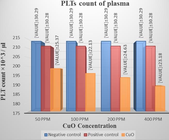
Shams Eldine et al.            11

iron nanoparticles were eco-friendly synthesized using                dissolving of the copper sulfate.
the method reported by Pattanayak and Nayak (2013)                    (iv) After 15 min, sodium hydroxide solution (2.0 M) was added,
                                                                      black precipitate was formed Cu(OH) 2.
with a slight modifcation. Nanoparticles clinical                     (v) This precipitate was filtered out and washed with distilled water
applications is one of the most attractive areas of science           and ethanol several times alternatively to remove the impurities.
nowadays, nanoparticles are used for bioremediation of                (vi) After washing the precipitate was kept in an oven at 200°C for 3
diverse contaminants, water treatment, antimicrobial                  h. During this process all copper hydroxide was converted to
applications and drug delivery. Due to their essential role           copper oxide.
in the blood and systemic homeostasis, red blood cells
can be considered as a landmark in any health evaluation              Characterization of CuO nanoparticles
of all NP compounds (Iancu et al., 2009; Mocan et al.,
2011; Ilie et al., 2013).                                             The prepared CuO nanoparticles were characterized using
   Unlike metal particles, oxide nanoparticles exhibit                Transmission Electron Microscope (TEM) (JEOL-100 CX), X-ray
expansion in their lattice parameters relative to their bulk          Diffraction (XRD) (Shimadzu, XRD-7000, Maxima, Japan), Fourier
                                                                      Transform Infrared spectroscopy (FTIR) (Bruker, Tensor 37 FT-IR),
parameter (Rao et al., 2004). Often, because of their
                                                                      and Zeta size (Malvern, UK).
small size and high density of corner or edge surface
sites, they may exhibit unusual physical and chemical
properties (Rodríguez et al., 2007; Ayyub et al., 1995).              The synthesis of CuO nanoparticles suspensions
Both the oxidation processes and size reduction form the
essential structures that transcribe a nanostructured                 Stock suspensions of CuO nanoparticles were dispersed in a
                                                                      mixture of dimethyl sulphoxide (DMSO) and PBS/Citrate (10 ml
oxide’s behavior (Sun, 2003). Copper oxide (CuO) is a                 DMSO+15 ml PBS/Citrate). The mixture was sonicated using bath
semi-conducting, monoclinically formed compound. CuO                  sonicator (Ultrasonic cleaner, Sonica, Soltec, Italy) until the
is the simplest member of the copper family and has a                 particles were completely dispersed in the solution. Different
variety of potentially useful physical properties like high           concentrations of CuO nanoparticles (50, 100, 200, and 400 ppm)
temperature superconductivity, electron correlation                   by adding 8 mg of CuO nanoparticles to 100 ml of the stock
effects, and spin dynamics (Ren et al., 2009).                        solution, and then diluted this solution to the other concentration, by
                                                                      the addition of distilled water. The procedure was carried out at The
   The high toxicity of CuO particles resulting in nearly             research laboratory, Pharos University, Alexandria.
100% cell death after 18 h may be a response to DNA
damage as a means of preventing mutagenic results
(Gurr et al., 2005). Another possibility is that the CuO              Study of the effect of CuO nanoparticle on red blood cells
                               2+                                     (RBCs)
nanoparticles release Cu ions. An eco-toxicological
analysis revealed that CuO nanoparticles were mostly
                         2+                                           Separate erythrocytes at 37°C were washed with PBS/Citrate by
toxic by soluble Cu          ions (Heinlaan et al., 2008),            centrifugation at 150 g and 37°C for 10 min. Washed erythrocytes
whereas another study concluded that the dissolution of               were aliquotted into equal parts of 50 μl. CuO nanoparticles with
    2+
Cu nanoparticles was insufficient to clarify the toxicity             different concentrations (50, 100, 200, and 400 ppm) suspended in
(Griffitt et al., 2007). While limited surveys have focused           PBS/citrate with DMSO (or PBS-citrate with DMSO alone for the
on the effects of copper oxide nanoparticles on the blood             positive control) were added to aliquot in a volume/volume (v/v)
                                                                      ratio of 2:1 for erythrocyte. Samples were incubated at 37°C for
parameters of different species of fish, this resulted to             different periods (1/2, 3, and 24 h). Samples were tested for RBCs
changes in hematological and biochemical parameters                   count after incubation periods and examined using Scanning
(Jahanbakhshi et al., 2015). The need of this work is to              Electron Microscopy (SEM), Phase Contrast Microscopy (PCM),
study the toxicity of CuO nanoparticle on human blood.                and Osmotic Fragility Measurements (OFM).
   The objective of this study was to investigate the
effects of different dosage of copper oxide nanoparticles
                                                                      Determination of RBCs count
on some hematological parameters of human blood.
                                                                      The RBCs samples were analyzed after incubation of blood
                                                                      samples in an incubator (Incubator, BTC, Egypt) at 37°C for (1/2, 3,
MATERIALS AND METHODS                                                 and 24 h) using automated hematology analyzer (Sysmex, XT-
                                                                      1800i, Japan) at The Hematology Laboratory at Hematology
The present study was carried out in the following sequence:          Department, Medical Research Institute, Alexandria University.

(1) The synthesis of copper oxide nanoparticles by chemical
precipitation method according to                                     Examination of CuO nanoparticles using Scanning Electron
Suleiman et al. (2015).                                               Microscope (SEM)
(2) Copper sulfate 5-hydrate (CuSO4.5H2O) (Sigma Aldrich).
(3) Sodium hydroxide (1 NaOH) (Sigma Aldrich).                        (1) After the incubation period (1/2, 3, and 24 h), RBCs samples
(i) A 0.4 M aqueous solution of copper sulfate (1.00 g copper         were fixed in 1% glutaraldehyde.
sulfate in 10ml deionized water)                                      (2) Fixed samples were washed by exchanging supernatant with
(ii) 2.0 M aqueous solutions of sodium hydroxide (NaOH) were          citrated Phosphate Buffer Saline (PBS) and incubated for 20 min at
prepared in distilled water (1.25 g NaOH in 10 ml distilled water).   room temperature.
(iii) Then, the solution of copper sulfate was heated to 85°C and     (3) This procedure was repeated four times, while the last
kept under constant stirring using magnetic stirrer till complete     incubation was performed overnight at 4°C.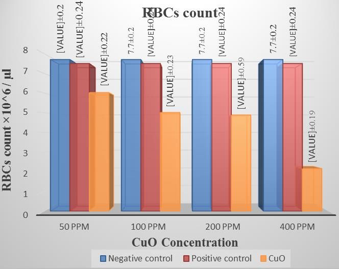
12 J. Biophys. Struct. Biol.
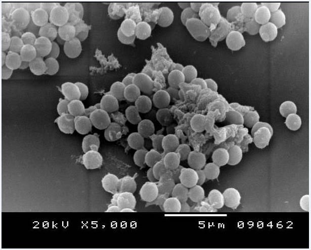
Figure 1. Scanning electron micrograph for copper oxide nanoparticles.
(4) Samples of RBCs were then post-fixed for 60 min at 4°C in 1% Scanning Electron Micrograph of CuO nanoparticles
OsO4 dissolved in PBS.
(5) Dehydrated in a graded series of alcohol/water (10%-100% v/v),
critical-point dried, gold-sputtered, and examined using a scanning
The shape and the size of CuO nanoparticles were
electron microscope (JEOL. JSM 5300 LA, Japan) at Electronic determined by SEM image. The SEM micrograph of CuO
Microscope Unit, Faculty of Science, Alexandria University. nanoparticles appeared as wire-like images with an
Osmotic fragility measurements were done according to Hobbie average size of 27 nm and are shown in Figure 1.
and Roth (2006).
Study of the effect of CuO nanoparticles on platelet-rich
Particle size analysis and zeta potential
plasma measurements
Washed platelet-rich plasma was aliquotted into equal parts of 200 Particle size: The average hydrodynamic diameter and
μl each. CuO nanoparticles with different concentrations (50, 100, poly-dispersity index (PDI) of CuO nanoparticles were
200, 400 ppm) suspended in PBS/citrate with DMSO (or PBS-
determined by Dynamic Light Scattering (DLS) using
citrate with DMSO alone for the positive control) and added to
aliquot in a v/v ratio of 3:1 using Sysmex, XT-1800i, Japan at The Malvern zeta sizer. The hydrodynamic diameter of
Hematology Laboratory, Hematology Department, Medical Research chemically synthesized CuO nanoparticles was 82.24
Institute, Alexandria University. nm, and the PDI was 0.426 as shown in Figure 2.
Zeta potential measurement: The potential of the
RESULTS synthesized CuO nanoparticles was performed using
Zeta Sizer (Malvern, UK). The zeta potential of
Characterization of copper oxide nanoparticles synthesized CuO nanoparticles was -4.69 mV as shown
in Figure 3.
The synthesized CuO NPs were characterized using
Transmission Electron Microscope (TEM), X-ray X-ray diffractometer: The crystalline nature of the
Diffraction (XRD), Fourier Transform Infrared synthesized CuO nanoparticles was identified from their
spectroscopy (FTIR), and Zeta potential (ζ). The results corresponding powder XRD patterns as shown in Figure
obtained are further explained. 4. The diffraction peaks were well matched with the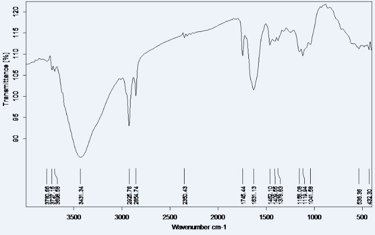
Shams Eldine et al.   13

      Figure 2. Particle size distribution of synthesized copper oxide nanoparticles.

       Figure 3. The zeta potential of synthesized copper oxide nanoparticles.

Figure 4. X-Ray diffraction pattern of copper oxide nanoparticles.
14 J. Biophys. Struct. Biol.
Figure 5. Fourier transform infrared spectrum of chemically synthezised copper oxide
nanoparticles.
surface to volume ratio, they can absorb moisture. The
-1
peaks at 1631 cm depict the Cu-O symmetrical
stretching. The high-frequency mode at 536.36 and
-1
432.30 cm can be assigned to the Cu-O stretching
vibration. Moreover, no other IR active mode was
-1
observed in the range of 605 to 660 cm , which totally
rules out the existence of another phase, that is, Cu2O.
Thus, the pure phase CuO nanoparticles with monoclinic
structure is also confirmed from the FTIR analysis.
Copper ions are redox-active, which means that the
high intracellular concentration gained after the dissolution
of CuO nanoparticles within the cell likely results in
massive oxidative stress. Various signs of oxidative
stress and genotoxicity have been observed upon cellular
exposure to CuO nanoparticles.
Effect and evaluation of copper oxide nanoparticle
Figure 6. Red blood cells count after 24 h incubation at different toxicity on human RBCs
concentrations of CuO nanoparticles.
From data obtained from the research study, there is no
notable effect of CuO nanoparticles on RBC counts after
½ h incubation as compared with controls. After 3 h
monoclinic phase of CuO (standard JCPDS File No: 048- incubation, there is no significant reduction in the RBC
1548). Diffraction peaks with 2Ө values of 33.6°, 35.45°, count as compared with negative control and positive
38.73°, 48.92°, 61.99°, and 66.49°, respectively were control samples. On the other hand, there is a significant
indexed to (110), (002), (111), (020), (022) and (113) reduction in the RBC count after 24 h incubation because
planes. of hemolysis as shown in Figure 6 and Table 1.
Fourier-transform infrared spectroscopy: The Fourier
Transform Infrared spectra of CuO nanoparticles as Scanning electron micrograph of RBCs examination
shown in Figure 5 depicts that broad absorption peak at
3431 cm-1 was caused by adsorbed water molecules. The examination of the RBCs using SEM were done by
Since the nanocrystalline materials possess a high fixation in 01% glutaraldehyde and incubated for anotherShams Eldine et al. 15
Table 1. Red blood cell count after 24 h of incubation with different concentrations of CuO
nanoparticles.
CuO concentration (ppm)
Group
Negative control Positive control 50 100 200 400
6
Mean×10 /µl 7.77 7.53 6.02 5.01 4.88 2.16
6
SD×10 0.20 0.24 0.22 0.23 0.59 0.19
F 397
P 0.0
6
LSD (5%) 0.24×10
Different superscripts are statically significant. F: F test (ANOVA) by Minitab program. Least significant
difference at 5%.
Figure 7. Scanning Electron Micrograph (SEM) of normal Red Blood Cells, incubated with
Copper Oxide Nanoparticles of (400 ppm) for 24 h. The Red Blood Cells appeared
swelled, aggregated, and degenerated.
hour at room temperature and centrifuged at 1550 g and Fragility (MCF); that for the control group at 35, group
37°C for 10 min. The supernatant was exchanged with (100 ppm) at 40, group (200 ppm) at 45, and group (400
PBS-citrate, and the samples were vortexed, centrifuged ppm) at 50 indicating a decrease in the RBCs resistivity
at 1550 g and 37°C for 10 min and fixed in 2% to hemolysis. These results appear more evident in the
glutaraldehyde for an hour. differentiation curves as shown in Figure 9 for blood
samples. The formed peaks indicate the increase in the
hemolysis of RBCs. From the differential curves, we
Osmotic fragility of RBCs determined the maximum hemolysis rate (Cmax), the
half-maximum width of the peak (Whmax), which
Figures 8 and 9 show the variation of hemolysis represents the elastic range of the cell membrane and
percentage as a function of NaCl concentration percent. H50, which represent the concentration percentage of
The figures indicate a shift in the Median Corpuscular NaCl that leads to 50% hemolysis.16 J. Biophys. Struct. Biol.
Figure 8. The osmotic fragility curve for red blood cells incubated with copper oxide
suspension (400 ppm) for 24 h in comparison with control.
Figure 9. The differential curve for red blood cells incubated with copper oxide
suspension (400 ppm) for 24 h in comparison with control.
The effect of copper oxide nanoparticles on active significant oxidative stress is possibly due to the high
platelets intracellular concentration obtained after the dissolution
of CuO nanoparticles within the cell. At cellular exposure
To study the effect of CuO nanoparticles on the number to CuO nanoparticles, various signs of oxidative stress
of active platelets, different concentrations of CuO and genotoxicity were observed (Ahamed et al., 2006;
nanoparticles were incubated with platelets for 24 h. Hanagata et al., 2015). The CuO nanoparticles were
Table 2 and Figure 10 show the change in the number of successfully synthesized using a chemical process. The
active platelets after 24 h incubation. nanoparticles were characterized in detail using Zeta
sizer, Transmission Electron Microscopy (TEM), X-Ray
Diffraction (XRD) analysis, and Fourier Transform
DISSCUSION Infrared (FTIR). The TEM analysis of CuO nanoparticles
revealed their irregular wire-like nature with an average
Copper ions are redox-active, which means that size of 27 nm. Zeta potential (ζ) measured by Zeta sizerShams Eldine et al. 17
Table 2. Platelets count in plasma after 24 h incubation at different concentrations of copper oxide
nanoparticles.
CuO concentration (ppm)
Group
Negative control Positive control 50 100 200 400
3
Mean×10 /µl 214.06 211.33 198.85 196.12 193.8 188.97
3
SD×10 30.29 30.28 25.37 22.13 24.63 23.18
F 1.46
P 0.22
3
LSD (5%) 73.87×10
Different superscripts are statically significant. F: F test (ANOVA) by Minitab program. Least significant difference at
5%. *Statistically significant at p ≤ 0.05.
Figure 10. Platelets count in plasma after 24 h incubation at different
concentrations of copper oxide.
revealed that the particles carry a small negative charge nanoparticles. The FTIR spectra of CuO nanoparticles
-1
of -4.69 mV. The XRD pattern showed that all the showed the presence of peaks at 3431 cm , which
diffractions peaks were well matched with the monoclinic indicates that the nanoparticles are surrounded by water
phase of CuO nanoparticles with 2Ө values of 33.6°, molecules which contained the hydroxyl group. The
35.45°, 38.73°, 48.92°, 61.99°, and 66.49°, respectively peaks at 1631 may be for the Cu-O symmetrical
were indexed to (110), (002), (111), (020), (022) and stretching. The high-frequency mode at 536.36 and
-1
(113) planes. XRD and TEM analysis confirmed the high 432.30 cm can be assigned to the Cu-O stretching
crystallinity and uniform non-agglomeration of vibration. Moreover, no other infra-red active mode was
-1
synthesized CuO nanoparticles. observed in the range of 605 to 660 cm , which totally
FTIR analysis was performed in order to understand rules out the existence of another phase, that is, Cu 2O.
the chemical and structural character of the synthesized Thus, the pure phase CuO with monoclinic structure is
nanomaterials and the influence of the chemicals used in confirmed from the FTIR analysis. The data obtained for
the synthesis. Also, infrared studies were carried out in characterizing chemically synthesized Copper Oxide
order to ascertain the purity and nature of the metal Nanoparticles was verified by those obtained by El-Trass18 J. Biophys. Struct. Biol.
et al. (2012). In agreement with our results, several other properties. Journal of Physical Review B 51(9): 6135-6138.
research groups have also documented dose-dependent El-Trass A, ElShamy H, El-Mehasseb I, El-Kemary M (2012). CuO
nanoparticles: synthesis, characterization, optical properties and
cytotoxicity of nanoparticles exposed cells (Fernández- interaction with amino acids. Journal of Applied Surface Science
Alberti and Fink, 2000). CuO nanoparticles showed toxicity 258(7):2997-3001.
in mammalian cells, leading to the generation of reactive Fernández-Alberti A, Fink NE (2000). Red blood cell osmotic
oxygen species (ROS), oxidant injury, excitation of fragility confidence intervals: a definition by application of a
inflammation, and cell death (Oyawale et al., 1997). The mathematical model. Journal of Clinical Chemical Laboratory
nanoparticles are capable of penetrating, translocating Medicine 38(5):433-436.
Gleiter H (2000). Nanostructured materials basics concepts and
within, and destroying living cells. This ability results microstructure. Journal of Acta Materialia 48:1-29.
primarily from their small size, which allows them to Griffitt RJ, Weil R, Hyndman KA, Denslow ND, Powers K, Taylor D,
penetrate or attach to the cell membrane causing et al (2007). Exposure to Copper Nanoparticles Causes Gill
deformation and subsequent cell death. Injury and Acute Lethality in Zebrafish (Danio rerio). Journal
The outcomes of the osmotic fragility curves provide of Environmental Science and Technology 41(23):8178-8186.
Gurr JR, Wang AS, Chen CH, Jan KY (2005). Ultrafine titanium
information on the changes in the elasticity and ionic
dioxide particles in the absence of photoactivation can induce
permeability of the red blood cells membrane, which oxidative damage to human bronchial epithelial cells. Journal
plays a significant role in the RBCs metabolic activities. of Toxicology 213(1-2):66-73.
Since the diameters of the RBCs are in the range of 12 to Hanagata N, Zhuang F, Connolly S, Li J, Ogawa N, Xu M (2015).
16 μm, each RBCs has to flow through narrow blood Molecular responses of human lung epithelial cells to the toxicity
capillaries of smaller diameters (~8 μm) to reach the of copper oxide nanoparticles inferred from whole genome
expression analysis. Journal of American Chemical Society Nano
target cells in the body to carry-out the metabolic 5(12):9326-9338.
reaction, the RBCs have to be folded several times to Heinlaan M, Ivask A, Blinova I, Dubourguier HC, Kahru A (2008).
decrease their diameters in order to pass through blood Toxicity of nanosized and bulk ZnO, CuO and TiO2 to bacteria
capillaries under high pressures according to Bernoulli's Vibrio fischeri and crustaceans Daphnia magna and
equations. The elasticity of the RBCs membrane will play Thamnocephalus platyurus. Journal of Chemosphere 71(7):1308-
a significant role in the folding process, any decrease in 1316.
Hobbie RK, Roth BJ (2006). Curves differentiation. In: intermediate
its elasticity will lead to the loss of the folding physics for medicine and biology. 4th ed New York NY: Springer.
mechanisms that will permit the RBCs to carry-out its Iancu C, Ilie IR, Georgescu CE, Ilie R, Biris AR, Mocan T, Mocan
function, which will cause anemic diseases. The change LC, Zaharie F, Todea-Iancu D, Susman S, Ciuca DR, Biris AS
in cell membrane elasticity can be distinguished from the (2009). Applications of nanomaterials in cell stem therapies and
Whmax values in the differential curves of osmotic the onset of nanomedicine. Journal of Particulate Science and
Technology 27:562-574.
fragility, that is, the decrease in the Whmax clearly shows
Ilie I, Ilie R, Mocan T, Iancu C, Mocan L (2013). Nicotinamide-
the decrease in the elastic range of the RBCs membrane. functionalized multiwalled carbon nanotubes increase insulin
Furthermore, platelet counts decrease as the production in pancreatic beta cells via MIF pathway. Interntional
incubation period with CuO nanoparticles increased, and Journal of Nanomedicine 8:3345-3353.
its dosage increased. The significant decrease in the Jadhav S, Gaikwad S, Nimse M, Rajbhoj A (2011). Copper Oxide
count of platelet indicated competent and supportive Nanoparticles: Synthesis, Characterization and Their
Antibacterial Activity. Journal of Clust Science 22:121-129.
immune responses which could have negative effect of Jahanbakhshi A, Hedayati A, Pirbeigi A (2015). Determination of
Cu-NPs or CuO to promote blood thickening, which in acute toxicity and the effects of sub-acute concentrations of CuO
return caused platelet damage. The same results nanoparticles on blood parameters in Rutilus rutilus. Journal of
obtained for the whole blood PLT count showed a Nanomedicine 2(3):195-202.
substantial decrease in the PLT after 24 h of incubation Martin CR (1994). Nanomaterials: A Membrane-Based Synthetic
with CuO nanoparticles and decreased the count as the Approach. Journal of Science 266(5193):1961-1966.
Mocan T, Clichici S, Biris AR, Simon S, Catoi C, Tabaran F, Filip A,
concentration of CuO nanoparticles increased (Noureen, Daicoviciu D, Decea N, Moldovan R, Mocan L, Muresan A
2018). (2011). Dynamic effects over plasma redox ballance following
subcutaneous injection of single walled carbon nanotubes
functionalized with single strand DNA. Digest Journal of
CONFLICT OF INTERESTS Nanomaterials and Biostructures 6(3):1207-1214.
Noureen A, Jabeen F, Tabish TA, Yaqub S, Ali M, Chaudhry AS
(2018). Assessment of copper nanoparticles (Cu-NPs) and
The authors have not declared any conflict of interests. copper (II) oxide (CuO) induced hemato-and hepatotoxicity in
Cyprinus carpio. Journal of Nanotechnology 29(14):144003.
Oyawale JO, Okewumi TO, Olayemi FO (1997). Haematological
REFERENCES Changes in West African Dwarf Goats Following Haemorrhage.
Journal of Veterinary Medicine A 44: 619-624.
Ahamed M, Siddiqui MA, Akhtar MJ, Ahmad I, Pant AB, Alhadlaq Pattanayak M, Nayak PL (2013). Ecofriendly green synthesis of iron
HA (2006). Genotoxic potential of copper oxide nanoparticles in nanoparticles from various plants and spices extract. The
human lung epithelial cells. Journal of Biochemical Biophysical International Journal of Plant, Animal and Environmental
Research Communications 396(2):578-583. Sciences 3(1):68-78.
Ayyub P, Palkar VR, Chattopadhyay S, Multani M (1995). Effect of Rao CN, Müller A, Cheetham Ak (eds) (2004). The Chemistry of
crystal size reduction on lattice symmetry and cooperative Nanomaterials: Synthesis, Properties and Applications. Weinheim:Shams Eldine et al. 19 WILEY-VCH Verlag GmbH & Co. KGaA pp. 1-11. Suleiman M, Mousa M, Hussein A (2015). Wastewater disinfection Ren G, Hu D, Cheng EW, Vargas-Reus MA, Reip P, Allaker RP by synthesized copper oxide nanoparticles stabilized with (2009). Characterisation of copper oxide nanoparticles for surfactant. (Doctoral dissertation). antimicrobial applications. International Journal of Antimicrobial Sun CQ (2003) Oxidation electronics: bond–band–barrier Agents 33(6):587-590. correlation and its applications. Journal of Progress in Materials Rodr guez , Fern ndez- arc a (eds) (2007). Synthesis, Science 48:521-685. Properties, and Applications of Oxide Nanomaterials. Hoboken N.J: Wiley-Interscience.
You can also read