Experimental model in hamster - (Mesocricetus auratus) to study heterologous graft of scars and cutaneous diseases in plastic
←
→
Page content transcription
If your browser does not render page correctly, please read the page content below
Experimental model in hamster
                    (Mesocricetus auratus) to study heterologous
                   graft of scars and cutaneous diseases in plastic
                                                  surgery1

                                                                                Bernardo Hochman2
                                                                             Lydia Masako Ferreira3
                                                                          Flaviane Cássia Vilas Bôas4
                                                                                    Mario Mariano5

____________________________________________________________________________________
Hochman B, Ferreira LM, Vilas Bôas FC, Mariano M. Experimental model in hamster (Mesocricetus
auratus) to study heterologous graft of scars and cutaneous diseases in plastic surgery. Acta Cir Bras
[serial online] 2004 Vol 19 Special Edition. Available on URL: http://www.scielo.br/acb.

ABSTRACT: Syrian golden hamster (Mesocricetus auratus) has in its cheek pouches sub-epithelium an
"Immunologically Privileged Site" which allows the integration of homo- and heterologous graft. This
paper describes some anatomical and histological characteristics of that site, as well as analyzes aspects
related with its immune properties. It also focuses the advantages of this experimental model over other
models which are natural or induced carriers of immunodeficiency. Based on both these advantages and
literature, this study aims to establish this model, through the performance of heterologous graft, as
another option for the investigation of scar disturbances, as keloids and other diseases which may
interest Plastic Surgery, as benign cutaneous lesions, and malignant neoplasias such as skin carcinomas
and melanomas. The work also addresses perspectives for using this model, which still is a source
scarcely known by Brazilian medical class.

KEY WORDS: Mesocricetus. Transplantation, heterologous. Skin transplantation. Keloid.
____________________________________________________________________________________

Introduction

         Technological advances in the area of genetic Bioengineering allowed the creation of
experimental models, mutant and isogenic mice and rats which are deficient in T and B cells, K
(Killer) cells, and NK (Natural Killer) cells, or even with congenital absence of thymus.
____________________

1. This paper is a part of a Thesis presented to Federal University of São Paulo–Paulista School of Medicine in
   2002 by the Post-Graduation Program of Recovering Plastic Surgery (UNIFESP–EPM), to reach the Master
   Degree. The experimental work was performed in the Discipline of Immunology of the Parasitology,
   Microbiology, and Immunology Department of UNIFESP-EPM.
2. Master Degree in Recovering Plastic Surgery and Post-Graduation at Doctor level by the Post-Graduation
   Program of Recovering Plastic Surgery of UNIFESP-EPM.
3. Titular Professor and Head of the Discipline Plastic Surgery of the UNIFESP-EPM Surgery Department, and
   Coordinator of the Post-Graduation Program for Recovering Plastic Surgery of UNIFESP-EPM.
4. Medical Doctor of UNIFESP-EPM.
5. Titular Professor of the Biomedical Sciences Institute of São Paulo University Immunology Department, Titular
   Professor of the Pathology Department of Veterinary Medical and Zootechnical School of USP, and Visitor
   Professor of the Discipline Immunology in the Parasitology, Microbiology, and Immunology of UNIFESP-
   EPM.

69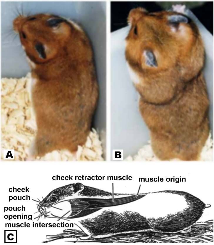
These models reached the preference of the global medical literature as experimental animals, to
study heterologous grafts1 instead of animals undergone to neonatal thymectomy or chemical
immunosupression in spite of their weaknesses.
However, due to their partial or total immunodeficiency, these models demand, for maintaining
them, a laboratory infrastructure of sound environmental asepsis, as well as alimentary and operational
special care.2 Nevertheless, these experimental models present high rates of morbidity and mortality. Said
demands may also impair the models for some types of studies, or even, be very expensive or not viable
for many investigators.3
The hamster (Mesocricetus auratus) is an experimental animal with a natural Immunologically
Privileged Site placed in the sub-epithelium of a pouch exisiting at both sides of the oral cavity.
Conceptually, an immunologically privileged site would result from the absence or deficiency of a
suitable afferent anatomical pathway to drain antigenetic material to a regional lymphatic station, whose
presence would recognize the antigen and would start the immune response.4
The species Mesocricetus auratus is also known as golden Syrian hamster or Lebanese squirrel
because its origin is a region in the North of Syria, near the Euphrates River, named Aleppo. The Syrian
golden hamster was described in 1839 and by unknown reason, in the beginning of the 20th Century, it
was considered as being extinct. In 1930, Professor Aharoni of Jerusalem Hebraic University collected in
Aleppo one female and its 12 broods. After one year of breeding, some from this strain specimens were
sent to the United Kingdom in 1931 and to the United States in 1938. The species adpated easily to
Western countries and became the most common amongst all the hamsters. Nowadays, all the exisiting
animals of the species are descendants from that strain breeded in Jerusalem.5 The Chinese hamster
(Cricetulus griseus) was already used in China as common experimental model since 1919. The Syrian
golden hamster, however, was only explored as a model carrier of an immunologic privilege site from the
middle of the 40s.
The decades 1960, 1970 up to approximately 1985 represented the apex period of researches in
medical literature, using the hamster cheek pouch, a trend which was afterwards substituted for the use of
animals coming from Bioegineering.
Proposition
Hamster’s cheek pouch sub-epithelium may be indicated for studying grafts of benign cutaneous
diseases, such as: nevus, warts, seborrheic keratosis, sebum hyperplasia, xanthomas, angiomas,
dermatofibromas and malignant neoplasias as basocellular and spinocellular carcinoma6, and cutaneous
melanoma.(7) In the same way, the hamster cheek pouch may also be indicated for chemotherapeutic7
drugs action and qualification tests, besides identification of carcinogenic substances.8
Due to the transparency of the epithelium and to its rich vascularization, hamster cheek pouch
may be used as a model for performing angiogenesis studies with cutaneous tissues,6,9 to biochemical
studies in both inflammatory and allergic reactions,10 as well as for in vivo evaluation of dynamics of the
microcirculation.11 The cheek pouch may also be useful for researches on infectious and parasitary
cutaneous diseases.12
This study purpose is both standardize and analyze hamster cheek pouch as an integration
experimental model for human skin heterologous grafts.
Model description
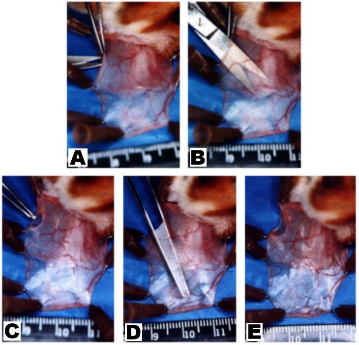
Hamster cheek or jugal pouches are highly distensible diverticles or bilateral sack invaginations
of the jugal mucosas. They are extended under the skin from the posterior border of the oral cavity to
about the scapulas of the animal. The main function of these pouches is storing and transporting food. In
females, specifically, they also serve to transport broots in case of perceived danger (“hamstern”, in
German, means “to catch”). The way for emptying the content of the pouch is the contraction of the
pouch retractor muscle (FIGURE 1).
70
FIGURE 1 – Syrian golden hamster (Mesocricetus auratus).
                          A - Animal with empty cheek pouches.
                          B – Cheek pouches with food to be carried out.
                          C – Scheme of the anatomical disposition of cheek pouch and
                          pouch retractor muscle in hamster.

         Each pouch, after being everted, presents two walls or epithelium layers. Each layer is formed by
epithelial cells strata, which are sustained on areolar conjunctive tissue, without lymphatic vases, 13
excepting amongst the pouch retractor muscle fibers of insertion.14 Therefore, this virtual space between
both epithelial walls constitutes the cheek pouch sub-epithelial space which is naturally immunodeficient.
Regional drainage lymphatic station of each pouch sub-epithelium woyld be, theoretically, the surface
cervical regional lymphonode.12,15 Pouch wall epithelium ultra-structure is similar to the human skin, and
has been named “skin without follicles and glands”16 and "richely vascularized pouch skin".4 It presents
spiny, granular and corneous basal layer with keratinocytes, similar to the epidermis and gums epithelium
or hard palate of humans.17,18 Besides the absence of annexial structures, the hamster cheek pouch is also
without pigmentation. The vascular supply of hamster cheek pouches is performed by superior, mean and
inferior sac arteries, which are branches of inferior labial artery. All this vascular complex is linked by
plenty of arterial and venous anastomosis, which stil links, under the mandible, both pouches. Thus, this
model of blood supply brings an anatomic subsidy which assures, from a vascular point of view, the
integration and the growth of grafted tissues on any part of the pouch (FIGURE 2).

71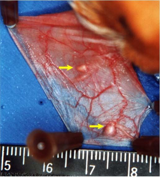
FIGURE 2 – Layers of hamster cheek pouch: macro- and microscopic aspects.
              A – Demonstrative dissection of everted cheek pouches layers.
              B – Microscopy of hamster cheek pouch layer in a sagital section (H.E. x 200).
              C – Vascular aspect of hamster cheek pouch after contrast injection, evidencing a
                 rich network of arterial and venous anastomosis [Figure obtained in the article:
                 Poor E, Lutz BR. Anat Rec 1958;132(2):121-6].
              [E = Pouch epithelium; SE = Subepihelium; TC = Conjunctive tissue; M = Pouch
                 retractor muscle; A = Artery; V = Vein; aa = arterial anastomosis; av = venous
                 anastomosis].

        Operation technique

         Cutaneous tissues to be grafted are obtained from exceeding operatory pieces which are
posteriorly undergone to defatting procedure. They have to be transported from the surgical suite to the
experimental laboratory immerse into a flask with physiologic solution at 0.9% into a covered styrofoam
container with ice. Tissue fragments are obtained from a 2 mm circular punch and should remain until the
moment of the graft insertion also immerged into chilled physiological solution.
         Animals are preferably anesthetized via intraperitoneal. The combination of hydrochloride of 2-
(2,6-xylidine)-5,6-dihydro-4H-1,3-tiazine (Rompun®) (hypnotic) at a dose of 0.1 mL/kg and ketamine
hydrochloride (Francotar®) (myorelaxing) at a dose of 0.075 mL/kg is recommended. With the animal
under anesthetic induction, in order to remove residuals from the cheek pouches, they are washed through
gentle water jets injected from a syringe by the oral cavity, with the head of the animal positioned below
the rest of its body to avoid accidental water inhalation.
         Then, each pouch is everted with the help of two Adson-Brown forceps on styropor foam and it is
fixed on it with 13 x 4.5 needles. Traction with the same forceps the proximal area of pouch epithelium
by an avascular site, and with straight iris scissors a 5 mm incision is made. Through this incision, with
the scissors, a rhombic divulsion of sub-epithelial tissue, in tunnel, to the most distal extremity of the
pouch. With a narrow and long pincer the cutaneous tissue to be grafted is placed at the distal extremity
of the disected tunnel. The pincer is removed while the fragment is grasped with a finger. Incision borders
are approximated only by manoeuvre of bi-digital coaptation without the need of sutures. Finally, the
needles are removed and the pouch is reintroduced into the oral cavity (FIGURE 3). Procedures may be
performed without the conventional rigors of sepsis and antisepsis.

72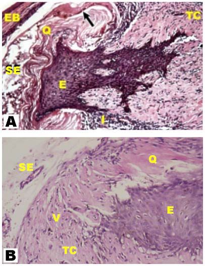
FIGURE 3 – Technique of grafting on hamster cheek pouch.
            A – Everted and fixed on styropor cheek pouch, tracioning avascular area of epithelium,
               proximal to the animal’s mouth where the incision will be performed.
            B – Rhombic divulsion, in tunnel, from sub-epithelium to the distal site of the pouch.
            C - Indroduction, by the incision, of the fragment to be grafted.
            D – Positioning of the fragment to be grafted in the sub-epithelium of distal site of the
               pouch.
            E - Final aspect of the hamster pouch with grafted fragment of human cutaneous tissue.

        At the end of the pre-established time of grafting, animals are anesthetized again, pouches are
resected and then animals are undergone euthanasia. These pouches are distended with needles on a
styropor piece, and the grafted fragment site is marked with needle, in justadistal position, before the
piece is immerged in 12% formaldehyde for fixing. This tactic aims to make easy its location within the
pouch to histopathologic test, since after formaldehyde immersion the fragment may have difficult
macroscopical enhancement. It is recommended that this marker needle is put at the longest ax
(logitudinal) of the grafted fragment, with the aim of directing the laboratory technician and making easy
the epithelium and conjunctive tissue inclusion on the glass plate to be examined (FIGURE 4).

73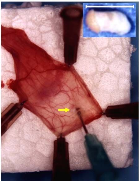
Figure 4 – Preparation of the pouch for histopathological
                           examination.
                           After distending the pouch with needles on the styropor
                           piece, the position of the grafted fragment is marked with a
                           needle different of others (arrow), at distal position, before
                           the graft is undergone formaldehyde. In the detail above,
                           with a white line, the longest ax of the fragment is
                           represented before it is grafted, enfolding epithelium and
                           conjunctive tissue of cutaneous tissue (in this case, keloid).

        Advantages of the model

         This is a larger animal than mice, it is easily adapted to captivity, it is more gentle and capable of
domestication, with easier operational handling. Unlike immunodepressed animals, hamsters can be fed
with common water and food without needing laboratories and bioteries with special infrastrucutre. Only
needed basic conditions of environmental basic hygiene with a cheap operational management are
needed, which is accessible to researchers, in general. 3 Morbidity and post-surgical complications rate in
grafting is lower than 5%, including conditions where asepsis rules are not followed.
         At operatory level, the model presents easy access to pouch sub-epithelium by simple eversion
manoeveur; disection is easy due to the pouch translucence, which means a low surgical trauma, hides
grafted fragments and protect them from trauma, exempts from protective bands with no need of
insulating each animal.
         In skin fragments grafts, scars and cutaneous diseases in the cheek pouch of hamster, there is no
need of de-epitelizing the fragment. In other experimental models, as athymic mice, this procedure is
generally needed, with the purpose of avoiding inclusion cysts, since the transplant is performed on
subcutaneous cellular tissue of the animal. Epithelium preservation, besides making easy the operatory
act, enhances researches results (FIGURE 5).

74
FIGURE 5 – Microscopic aspect of human skin fragment at 12 days and keloid at 21
days in hamster cheek pouch.
A – Integration and viability of grafted fragment is noted by the active keratin production
by the epithelium, with 12 days after grafting, in the pouch sub-epithelium (arrow).
(Color HE, x 200).
B – Grafted and integrated keloid fragment at 21 days (Color HE, x 200).
[EB = Pouch epithelium; SE = Pouch sub-epithelium; E = Epithelium of grafted
fragment; Q = Keratin; V = Blood vase; I = Inflammatory infiltrated; TC = Graft
conjunctive tissue].
Hamster cheek pouch epithelium have a similar ultra-structure than human skin and, more
specifically, similar to the epidermis or epithelium of human gum or hard palate,17,18 and so it is also
named "skin without follicles and glands"16 and "richly pouch vascularized skin".4 It has basal, spiny,
granulous, and corneal layers with keratinocytes, while the mouse skin only has 2 or 3 layers. Thus,
mouse granulous layer is different in human being because granules are not intra-cellular. Cheek pouches
also present a rich perivascular network of mastocytes and, similarly the human being, without serotonin
secretion, while mice mastocytes secrete significantly serotonin.10
Since the hamster has thymus and normal immunity, the risk of mice or transplanted
immunodepressed mutant rats is avoided in more prolonged studies of developing immunity,
mechanisms of rejection by producing T lymphocytes by extra-thymic vias, when they become
older.Thus, the hamsters, because they have intact immune system, do not have naturally hormonal
dysfunctions.
Therefore, all morphological and physiological similarities with the human being, above
described, would reinforce that the hamster cheek pouch is an experimental model more suitable than
dorsal subcutaneous cellular tissue of mice and rats, from the phylogenetic point of view, to perform
human skin related researches.
75Perspectives
Hamster, as an experimental model, has peculiar characteristics that may represent a wide range
of perspectives. As, for instance, the anatomical location and the immunological privilege of the animal
cheek pouches maintain independence between each other, this is, the nature of the graft in a pouch does
not have influence on the other side one, each pouch may be addressed and and approached as if it was a
sub-unit of this experimental model. Therefore, if it is possible grafting tissues of different origins
(organs) and donor source in each pouch (auto, homo, and heterologous grafts), it is possible indicating
combinations in the same animal, be them auto-homo, auto-hetero, and hetero-heterologous grafts, as
examples.
Because the hamster is an animal with qualified immune system, with sites of immunological
privilege only in its cheek pouches, in the rest of its body it has entire afferent lymphatic vias,
lymphonodes, and efferent vias. Therefore, vases are able to transport immune effectors from the
synthesis sites to the target antigen. So, in case of previous antigenetic sensitivation by cutaneous
heterologous grafts in any part of the body, excepting the cheek pouches, their sub-epithelium would lose
the status of being an Immunologically Privileged Site for this determined heterologous graft.8
This characteristic would allow to indicate cross tests study with human skin antigens and keloid
tissue, for example, with previously sensitized animals at one of these tissues.
Results could reveal either differences or analogies of antigenetic characteristics of each tissue
obtained from the same individual. So, it could be possible to open a large screen of possibilities of
immunological studies by following the current trend of researches.
It is possible grafting more than one fragment of the same tissue in each pouch, since the rich
vascular network assures the survival of all the grafts in the pouch. This way, it is possible to multiply 2
ou 3-folds the amount of fragments obtained from each pouch, reaching a total of 4 or 6 fragments grafted
at the same time in a same animal, with the suitable margin of separation between them. This situation
could be demanded, for instance, where there is a need of a great amount of grafts, for certain studies
(FIGURE 6).
FIGURE 6 – Grafting of 2 fragments in the same cheek pouch. Macroscopic
aspect at 21 days of two fragments of keloid in the same pouch (arrows).
76Immunobiology is still searching after an “ideal” experimental animal model to receive human
skin heterologous grafts and related disturbs. Initially, this “ideal” model is described as an animal,
which would have a small immunogenic potential but which would have excellent metabolic and
physiologic compatibility with transplants coming from human beings. By all the above mentioned,
characteristics, although researchers are still after this “ideal” animal, the authors may consider that the
hamster would represent, a priori, a highly faithful and precise experimental model, over all other
experimental small sized models, to study human cutaneous grafts18.
References
1.Thomas F, Araneda D, Henretta J, Pittman K, Marchman W - Extended and expanded studies of
xenograft rejection in immunodeficient animals. Transplant Proc. 1994;26(3):1224-7.
2.Ferreira LM, Borsanyi JP, Benhaim P, Anthony JP, Mathes SJ, Andrews JM, Laredo Filho J - The
incidence of lethal graft-versus-host disease in rat limb allotransplantations. Rev Hosp Sao Paulo-Esc
Paul Med. 995;6(1/2):7-10.
3. Hochman B, Ferreira LM, Bôas FCV, Mariano M - Investigação do transplante heterólogo de quelóide
na bolsa jugal do hamster (Mesocricetus auratus). Acta Cir Bras. 2003;18(4):266-71.
4.Head JR, Billingham RE - Immunologically privileged sites in transplantation immunology and
oncology. Perspec Biol Med. 1985;29(1):115-31.
5.Adler S - Origin of the golden hamster Cricetus auratus as a laboratory animal. Nature. 1948;162:256-
7.
6.Wolf JE, Hubler WR - Tumor angiogenic factor and human skin tumors. Arch Dermatol. 1975;111:321
7.Dufour FD, Okada GT, Mah SG, Morton DL - Chemotherapy against human melanoma in the hamster
cheek pouch. J Surg Oncol. 1980;15:355-61.
8.Ziegler MM, Lopez V, Barker CF - Carcinogenesis in an immunologically privileged site. J Surg Res.
1975;18:201-7.
9.Wolf J, Harrison RG - The angiogenic stimulus of epidermis and epidermal components on the
microvasculature of the hamster cheek pouch. Bibl Anat. 1973;12:490-6.
10.Raud J, Dahlen SE, Smedegard G, Hedqvist P - An intravital microscopic model for mast cell-
dependent inflammation in the hamster cheek pouch. Acta Physiol Scand. 1989;135:95-105.
11.Svensjo E, Bouskela E - Microcirculação na bolsa da bochecha do hamster. In: Castro e Silva Jr. O,
Zucoloto S, Beer Jr. A - Modelos experimentais de pesquisa em cirurgia. Sao Paulo: Robe, 1998.
p.795-02.
12.Arruda MSP, Montenegro MR - The hamster cheek pouch: an immunologically privileged site suitable
to the study of granulomatous infections. Rev Inst Med Trop Sao Paulo. 1995;7(4):303-9.
13.Barker CF, Billinghan FRS - The lymphatic status of hamster cheek pouch tissue in relation to its
properties as a graft and as a graft site. J Exp Med. 1971;133:620-39.
14.Goldenberg DM, Steinborn W - Reduced lymphatic drainage from hamster cheek pouch. Proc Soc Exp
Bio Med. 1970;135:724-6.
15.Sinhorini IL, Merusse JLB, Mariano M - The role of lymphatic drainage on the development of BCG-
induced granulomas in the hamster. Int Arch Allergy Immunol. 1994;103:166-74.
16.Duling BR - The preparation and use of the hamster cheek pouch for studies of the microcirculation.
Microvasc Res. 1973;5:423-9.
17.White FH, Gohari K - Cellular and nuclear volumetric alterations during differentiation of normal
hamster cheek pouch epithelium. Arch Dermatol Res. 1982;273:307-18.
18.Hochman B,Ferreira LM, Bôas FCV, Mariano M - Integração do enxerto heterólogo de pele humana
no subepitélio da bolsa jugal do hamster (Mesocricetus auratus). Acta Cir Bras. 2003;18(5):415-30
77____________________________________________________________________________
Hochman B, Ferreira LM, Vilas Bôas FC, Mariano M. Modelo experimental no hamster (Mesocricetus
auratus) para estudo de transplantes heterólogos de cicatrizes e doenças cutâneas em cirurgia plástica.
Acta Cir Bras [serial online] 2004 Vol 19 Edição Especial. Disponível em URL: http://www.scielo.br/acb.
RESUMO: O hamster sírio dourado (Mesocricetus auratus) possui no subepitélio de suas bolsas jugais
um "Local de Privilégio Imunológico", permitindo a integração de homo e heteroenxertos. Este artigo
descreve as características anatômicas e histológicas desse local, assim como os aspectos relativos as suas
propriedades imunológicas. Cita, também, as vantagens desse modelo experimental sobre outros modelos
portadores, naturais ou induzidos, de imunodeficiência. Com base nessas vantagens, e também baseado na
literatura, tem o intuito de estabelecer esse modelo, mediante a realização de heteroenxertos, como uma
outra opção para investigar distúrbios cicatriciais, como o quelóide, e doenças de interesse em Cirurgia
Plástica, como lesões cutâneas benignas e neoplasias malignas como carcinomas e melanoma cutâneo.
Tece, ainda, considerações sobre perspectivas de aplicações desse modelo, ainda bastante desconhecido
entre a classe médica brasileira.
DESCRITORES: Mesocricetus. Transplante heterólogo. Transplante de pele. Quelóide.
_____________________________________________________________________________________
Conflito de interesse: nenhum
Fonte de financiamento: CAPES
Correspondence:
Dr. Bernardo Hochman
UNIFESP-EPM, Plastic Surgery Division, Surgery Division
Rua Napoleão de Barros, 715, 4° andar.
CEP 04024-900 São Paulo
Tel: (11)55760418 FAX: (11) 55716579
sandra.dcir@epm.br
lydia.dcir@epm.br
78You can also read