Extracorporeal shock wave treatment modulates skin fibroblast recruitment and leukocyte infiltration for enhancing extended skin-flap survival
←
→
Page content transcription
If your browser does not render page correctly, please read the page content below
Wound Repair and Regeneration

Extracorporeal shock wave treatment modulates skin
fibroblast recruitment and leukocyte infiltration for
enhancing extended skin-flap survival
Yur-Ren Kuo, MD, PhD1,2; Chun-Ting Wang, BS1,2; Feng-Sheng Wang, PhD2; Kuender D. Yang, MD,PhD2;
Yuan-Cheng Chiang, MD1; Ching-Jen Wang, MD3

1. Department of Plastic and Reconstructive Surgery,
2. Department of Medical Research, and
3. Department of Orthopedics, Chang Gung Memorial Hospital, Kaohsiung Medical Center, Chang Gung University College of Medicine,
Kaohsiung, Taiwan

Reprint requests:                             ABSTRACT
Yur-Ren Kuo, MD, PhD, Department of
Plastic and Reconstructive Surgery, Chang     Extracorporeal shock wave (ESW) treatment has a positive effect of rescuing is-
Gung Memorial Hospital, Kaohsiung             chemic skin flaps. This study assessed whether ESW treatment rescues the com-
Medical Center, Chang Gung University,        promised flap tissue by suppressing the apoptosis of ischemic tissue and
123 Ta-Pei Road, Niao-Sung Hsiang,            recruiting tissue remodeling. We used a random-pattern extended dorsal–
Kaohsiung 83305, Taiwan.                      skin-flap (103 cm) rodent model. Thirty-six male Sprague–Dawley rats were
Tel: 1886-7-731 7123, ext. 8002;
                                              divided into three groups. Group I, the control group, received no treatment.
Fax: 1886-7-731 1696;
Email: t1207816@ms22.hinet.net
                                              Group II received one session of ESW treatment (500 impulses at 0.15 mJ/mm2)
                                              immediately after surgery. Group III received two sessions of ESW treatment,
                                              immediately and the day after the surgery. Results indicated that the necrotic
This paper was presented at the 53rd
Annual Meeting of The Plastic Surgery
                                              area in the flaps in group II was significantly smaller than that of the flaps in
Research Council on May 28–31, 2008,          group I (p < 0.01). Transferase dUTP-nick end labeling (TUNEL) analysis re-
Springfield, IL, USA.                         vealed a significant decrease in the number of apoptotic cells in group II.
                                              Hydrogen peroxide (H2O2) expression in circulation blood was significantly de-
Manuscript received: May 8, 2008              creased in group II on the day after ESW treatment. Immunohistochemical
Accepted in final form: August 29, 2008       staining indicated that compared with no treatment, ESW treatment could sub-
                                              stantially increase proliferating cell nuclear antigen (PCNA), endothelial nitric
DOI:10.1111/j.1524-475X.2008.00444.x          oxide synthase, and prolyl 4-hydroxylase (rPH) expression, reduce CD45 expres-
                                              sion, and suppress 8-hydroxyguanosine (8-OG) expression in the ischemic zone
                                              of the flap tissue. In conclusion, ESW treatment administered at an optimal dos-
                                              age exerts a positive effect of rescuing ischemic extended skin flaps. The mecha-
                                              nisms of action of ESWs involve modulation of oxygen radicals, attenuation of
                                              leukocyte infiltration, decrease in tissue apoptosis, and recruitment of skin fibro-
                                              blasts, which results in increased flap tissue survival.

Random-pattern skin flaps are still widely used in recon-                studies have shown that ESWs could induce bony union,
structive plastic surgery. However, ischemic necrosis of the            cell differentiation, and neovascularization.15–20 However,
distal skin flap remains a serious problem, with a high                  these studies examined ESW treatment for only certain
morbidity in reconstructive surgical procedures. The                    musculoskeletal disorders. The cascade of biological ef-
pathogenesis of skin-flap ischemic necrosis remains                      fects associated with ESWs directly correlates with en-
unclear. The consensus is that cellular activation of pro-              hanced blood supply and tissue regeneration. Recent
inflammatory mediators, insufficient vascularity, and                     studies performed using a rat model have documented that
thrombosis are the principal factors in the pathogenesis                ESWs enhance the survival of the distal portion of the ex-
of flap ischemic necrosis.1–5 Clinical treatment of skin-flap             tended island skin flap.21 However, the biomechanisms of
ischemic necrosis remains controversial. Numerous ap-                   ESWs involved in rescuing ischemic skin flaps remain un-
proaches, such as hyperbaric oxygen, ischemic precondi-                 clear. In our previous study, ESW treatment was investi-
tioning, pharmacological agents, and growth factor                      gated using a random-pattern extended dorsal-skin-flap
delivery to ischemic tissues, have been applied to reduce is-           rodent model.22 We demonstrated that ESW treatment at
chemic necrosis in cases of impending skin-flap failure.6–13             an optimal dosage exerts a positive effect of rescuing is-
However, no effective clinical treatment exists for rescuing            chemic skin flaps by increasing tissue perfusion and vascu-
ischemic tissue necrosis.                                               lar endothelial growth factor (VEGF) expression and
   Shock waves are high-energy acoustic waves generated                 inducing neovascularization.22 In this study, we attempted
with high-voltage explosion and vaporization under wa-                  to determine whether ESW treatment rescues the compro-
ter.14 Recently, extracorporeal shock wave (ESW) treat-                 mised flap tissue by suppressing the apoptosis of ischemic
ment has been applied and adapted to different clinical                 tissue cells and leukocyte infiltration and recruiting fibro-
fields.14 The results of animal experiments and clinical                 blasts for tissue regeneration.

80                                                                          Wound Rep Reg (2009) 17 80–87
                                                                                                          c 2009 by the Wound Healing Society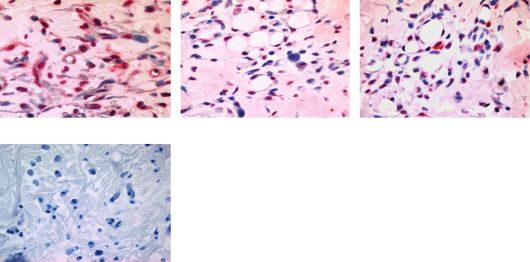
Kuo et al.                                                                Extracorporeal shock waves and ischemic skin-flap survival

MATERIALS AND METHODS                                               skin flap was well demarcated, and it could be easily iden-
                                                                    tified by gross observation on day 7 postoperatively. Oxy-
All animals were treated humanely according to the guide-           gen radicals in circulating blood were detected by flow
lines in the Guide for the Care and Use of Laboratory               cytometry on day 1 (immediately after ESW therapy ) and
Animals published by the National Institutes of Health              day 2 postoperatively (the day after ESW) and on the same
(USA). All animals were housed under conventional con-              days in group I. The expressions of anti-8-hydroxyguano-
ditions. The division of laboratory animal resources at             sine (8-OG; Serotecs, Oxford, UK) and endothelial nitric
Chang Gung memorial hospital (CGMH), Kaohsiung                      oxide synthase (eNOS; BD Pharmingens, SanDiego, CA)
medical center, provided veterinary care to the rodents.            were examined. Cellular proliferation was assessed by
All experiments were approved by the Institutional animal           examining the expression of proliferating cell nuclear anti-
care and use committee (IACUC) at CGMH.                             gen (PCNA; Upstate Biotechnologys, Lake Placid, NY);
                                                                    leukocyte infiltration, by detecting the expression of CD45
Random-pattern skin-flap model                                      (BD Pharmingens), and tissue remodeling and collagen
                                                                    processing was examined by the expression of prolyl 4-hy-
A modified McFarlane skin-flap model was used in this                 droxylase (rPH; Chemicons, Temecula, CA), a marker
study.22,23 The procedures followed in the study have been          within fibroblasts that are actively producing procollagen.
described previously.22 Rat dorsum was shaved, and a                The expressions of CD45, 8-OG, eNOS, rPH, and PCNA
caudal 103 cm random-pattern extended dorsal flap was               were examined by immunohistochemical (IHC) staining on
obtained. The palpable hip joints were used as the ana-             day 7 postoperatively. The terminal deoxynucleotidyl
tomical landmarks for defining the flap base. Under sterile           transferase digoxigenin-labeled uridine triphosphate
conditions, incisions were made and the entire flap was              (dUTP)-nick end labeling (TUNEL; Roches, Mannheim,
undermined below the level of the dorsal fascia. To harvest         Germany) assay was performed to detect apoptotic cells.
the flap, it was elevated without perforating the cutaneous          The surviving flap area was also determined on day 7 post-
blood vessels, which supply the base. The skin flap was              operatively. The animals were sacrificed by an intraperito-
sutured back to its native position using 4-0 silk sutures.         neal injection of 100 mg/kg ketamine on postoperative
Following the surgery, the rats were returned to their cages        day 7.
in the animal holding room after they recovered from
anesthesia.
                                                                    Estimation of flap necrosis area
Experimental design                                                 Nonviable and viable skin areas in the flap were assessed
                                                                    on day 7 postoperatively using the template technique de-
Thirty-six male Sprague–Dawley rats weighing 250–300 g
                                                                    scribed previously.11,22 Gross observation identified a clear
were divided equally into three groups (n512 in each
                                                                    line demarcating the living tissue from the necrotic tissue.
group). They were anesthetized with an intraperitoneal
                                                                    The weight of the entire flap, including the necrotic and
injection of 6% chloro-hydrate (5 mL/kg; Riedel-de
                                                                    viable areas, was measured by tracing the flap onto a
Haëns, Schnelldorf, Germany).22 To reduce saliva secre-
                                                                    transparent graph paper; the traced portion was cut and
tion, atropine (0.1 mg/kg) was administered intramuscu-
                                                                    its weight on the rat was estimated. The weight of the ne-
larly peri- and postoperatively.
                                                                    crotic tissue was calculated as A7/A0100, where A0 is the
   In all the three groups, skin flaps were elevated and su-
                                                                    weight of the original flap area and A7 is the weight of the
tured in their native position, as described above. Group I
                                                                    necrotic area on day 7 postoperatively.
(control group) flaps did not receive ESW treatment post-
operatively. Group II flaps were treated with one session of
ESW treatment (Ossatrons, HMT High Medical Technol-                 Immunohistochemical staining
ogies GmbH, Kreuzlingen, Switzerland) at a dose of 500
impulses at 14 kV (equivalent to 0.15 mJ/mm2 energy flux             Punch biopsy samples in the transitional zone at the mid-
density) immediately after the surgery. Group III flaps re-          third of the dorsal flap were obtained after various treat-
ceived two sessions of ESW treatment at the above-                  ments at 1 week. These transitional zones represented the
mentioned dose: one immediately after flap suturing and              various ischemic flap portions that typically undergo
the other on the following day. The time required to admin-         necrosis. IHC was performed using a horseradish per-
ister 500 impulses of ESW is 10 minutes. The dosages and            oxidase-diaminobenzidine (HRP-DAB) system staining
timing of ESW treatment were those used in our previous             kit (R&D Inc., Minneapolis, MN), as described previ-
studies.22 Ultrasound transmission gel (Pharmaceutical In-          ously.11,12 The transitional necrotic zone of the flap tissue
novations Inc., Newark, NJ) was applied as the contact me-          was examined. Polyclonal anti-8-OG, anti-eNOS, anti-
dium between the ESW apparatus and the skin. ESWs were              rPH, anti-CD45, and anti-PCNA antibodies at 1 : 100 di-
applied to five areas from the mid-part of the dorsal flap to         lutions in phosphate-buffered saline (PBS) were used as
the distal corner.22 These areas represented the various is-        the primary antibodies for 1 hour. Tissue sections were
chemic flap portions that typically undergo necrosis.                then incubated with biotinylated goat anti-rabbit antibod-
   All rats were observed daily and follow-up examina-              ies for 30 minutes. Specific binding to primary antibodies
tions were performed on postoperative day 7. Although               was visualized by enzymatic conversion of the chromo-
the edge effect of the flap wound margin might be a factor           genic substrate DAB into a brown precipitate by HRP.
affecting the flap survival, however, the edge effect was            The slides were mounted, cleared, and cover-slipped and
overlooked because the same procedure was used for all              subsequently examined under a light microscope (Carl
the animals. The necrotic area in the distal portion of the         Zeiss, Gottingen, Germany).

Wound Rep Reg (2009) 17 80–87
                              c 2009 by the Wound Healing Society                                                               81
Extracorporeal shock waves and ischemic skin-flap survival Kuo et al.
TUNEL assay the differences in the various expressions among the three
groups in a normal distribution. Post hoc comparisons
Biopsy samples were obtained after various treatments at 1 were analyzed by the Tukey method. A value of p < 0.05
week. To identify apoptotic cells by means of direct immu- was considered statistically significant.
noperoxidase detection of digoxigenin-labeled genomic
DNA, an in situ cell death detection kit (Roches) was used
according to the manufacturer’s instructions. Formalin- RESULTS
fixed and paraffin-embedded tissue blocks were sectioned at
6 mm thickness and placed on coated slides. The paraffin Optimal ESW treatment rescued ischemic skin flap
sections were deparaffinized in xylene and rehydrated in
tissue
graded ethanol series and PBS. The specimens were perme-
ated with 0.1% Triton X-100 in 0.1% sodium citrate. TdT The distal necrotic area in the random-pattern extended
and dUTP were added, and the sections were incubated for 1 skin flaps in group II (flaps treated with one session of
hour at 37 1C. The sections were washed with PBS, and the ESW treatment) was significantly smaller than that in
digoxigenin-dUTP incorporated was detected by incubation group I (control group) (13 2.6% vs. 42 5.7%;
with anti-digoxigenin Fab fragments conjugated with alka- p50.003). On the other hand, the necrotic area in group
line phosphate (AP) at 37 1C for 30 minutes. The reaction III (flaps treated with two sessions of ESW treatment) was
product was visualized using a 5-bromo-4-chloro-3-indolyl insignificantly smaller than that in group I (29 6.2% vs.
phosphate/nitro blue tetrazolium (BCIP/NBT)-buffered 42 5.7%; p50.09). These results indicate that ESW
substrate in AP buffer (dilution, 1 : 50) at room temperature treatment at an optimal dosage has a positive effect of
for 10 minutes. The TdT enzyme was replaced with distilled promoting flap tissue survival.
water in the negative control. Finally, each specimen was
mounted under a glass coverslip and analyzed for the num-
ESW suppressed leukocyte infiltration in the ischemic
ber of apoptotic cells under a light microscope.
zone of flap tissue
Histomorphometrical examination The transitional ischemic portion of the flap tissue was
subjected to a histological examination. Hematoxylin and
Punch biopsy samples were obtained (six rats’ samples/per eosin (H&E) staining indicated that a single ESW session
group) after various treatments at 1 week. For immuno- markedly reduced leukocyte infiltration as compared with
staining quantification, sections were analyzed using a no ESW treatment. Leukocyte infiltration was assessed by
Zeiss Axioskop 2 plus microscope (Carl Zeiss). Four ran- observing the CD45 expression in the ischemic zone of the
dom images from each selected area were then taken under flap tissue by HRP-DAB IHC staining. The results indi-
400 magnifications. All images of each specimen were cated that compared with no ESW treatment, both single
captured using a cool CCD camera (SNAP-Pro c.f. Digital and two ESW sessions markedly reduced CD45 expression
kit; Media Cybernetics, Silver Spring, MD). Images were in the region extending from the dermis to the subcutaneous
analyzed using IMAGE-PROr Plus image-analysis soft- muscular layers in the ischemic zone of the flap (Figure 1).
ware (Media Cybernetics) as described previously.15,16,24 On the other hand, group II flaps showed a significantly
The number of positive immunolabeled cells and total cells lower CD45 expression level than the group III flaps.
in each area were counted, and the percentages of positive-
labeled cells were calculated. One pathologist blinded to
the treatment regimen performed measurements on all ESW decreased apoptosis in the ischemic zone of flap
sections under 400 magnifications. tissue
The presence of apoptotic cells in the ischemic zone of the
Detection of oxygen radicals (O
2 and hydrogen
flap tissue after ESW treatment was investigated by the
peroxide [H2O2]) by flow cytometry TUNEL assay. The results revealed apoptotic cells with a
relatively greater DNA damage and fragmentation in
A 50 mL whole-blood sample was lysed using an ammo- group I flaps. Compared with group I flaps, groups II and
nium chloride potassium (ACK) buffer and washed until it III flaps exhibited a marked reduction in the number of
was almost clear. The sample was then centrifuged at apoptotic cells from the dermis to the subcutaneous mus-
1,450 g for 5 minutes. To all the samples, 50 mM of 2 0 ,7 0 - cular layers of the ischemic zone. This result indicates that
dichlorofluorescein diacetate (DCFH; Eastman Kodak, ESW, when applied at an optimal dosage, could decrease
Rochester, NY) was added to detect H2O2, and 10 mM of cell apoptosis in the ischemic skin-flap tissue (Figure 2).
histofluorescence (HE) was added to detect O 2 at 37 1C in
the dark for 15 minutes. Subsequently, the samples were
ESW recruited skin fibroblasts and tissue remodeling
subjected to flow cytometric analysis for the detection of
oxygen radicals. The results were analyzed using a soft- Cellular proliferation was analyzed by observing the
ware program LYSIS II (Becton–Dickinson, Palo Alto, PCNA expression in the ischemic zone of the flap tissue.
CA). PCNA expression was higher, especially in the basal layers
of the dermis and subcutaneous layers in groups II and III
Data management and statistical analysis than in group I (Figure 3A). However, PCNA expression
was significantly increased in group II compared with that
Experimental results are presented as means SE. One- in group III. In contrast, IHC staining revealed that the
way analysis of variance (ANOVA) was used to determine rPH expression in the ischemic zone of the flap tissue was
82 Wound Rep Reg (2009) 17 80–87
c 2009 by the Wound Healing SocietyKuo et al. Extracorporeal shock waves and ischemic skin-flap survival
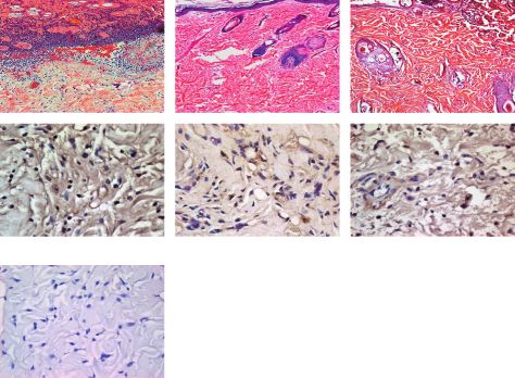
A Ctrl ESW-1 ESW-2
Figure 1. ESW treatment suppresses in-
flammatory response in the flap tissue.
(A) The transitional zone of the flap tis-
H&E sues was examined histologically. Hem-
atoxylin and eosin (H&E) staining revealed
that leukocyte infiltration in the flaps that
received single ESW treatment immedi-
ately after surgery was markedly lower
than that in the control flaps that did not
receive ESWs and the flaps that received
ESW twice. The scale bar520 mm. (B)
Leukocyte infiltration was analyzed by ex-
amining CD45 expression using HRP-
CD45+ DAB IHC staining. The results indicated
Negative control that compared with no treatment, a single
B 100
ESW session significantly reduced the
80 CD45 expression in the ischemic zone of
#
CD45 (%)
the flap tissues. The scale bar55 mm.
60 * * n
p < 0.001 vs. controls; #p50.006 signifi-
40 cant differences in ESW-1 vs. ESW-2
groups. ESW-1, ESW treatment once;
20 ESW-2, ESW twice; HRP-DAB IHC,
0 horseradish peroxidase-diaminobenzidine
Ctrl ESW-1 ESW-2 immunohistochemical.
significantly higher on day 7 in group II than in group I flap survival by suppressing oxygen radical burst and pro-
(Figure 3B). Meanwhile, the rPH expression in the is- moting eNOS expression.
chemic zone of the flap tissue was significantly higher in
group II than in group III. These results indicate that ESW
treatment at an optimal dosage significantly promotes DISCUSSION
fibroblast proliferation and tissue remodeling in the
ischemic skin flap. Random-pattern skin flaps are still widely used in recon-
structive surgery. However, necrosis of skin flaps remains
a serious complication in reconstructive surgical proce-
ESW down-regulated oxygen radical burst and dures.1,3 The distal part of a random-pattern flap is more
promoted eNOS expression prone to ischemia and consequent necrosis. Although dis-
tal–skin-flap ischemic necrosis is a common complication
Oxygen radical expressions in the circulating leukocytes after skin-flap surgery, the underlying pathogenic mecha-
were detected by flow cytometry. No significant differences nism remains unclear. Several approaches have been de-
existed in the expressions of H2O2–DCFH or O 2 -HE in veloped to reduce ischemic necrosis in unsuccessful skin
the circulating leukocytes between the control and the flaps.7–12 Although several methods exist for augmenting
ESW-treated groups at day 1 postoperatively (after ESW tissue perfusion in flap ischemia, suppression of leukocyte
therapy immediately). The O 2 -HE expression revealed a inflammation and induction of tissue regeneration are con-
mild decrease but no significant differences between the sidered as the primary factors involved in flap-tissue sur-
control and the ESW-treated groups at day 2 postopera- vival.2,25
tively (the day after ESW therapy). However, compared Several studies have proposed the beneficial effects of
with group I, groups II and III exhibited an apparent de- ESW treatment in bone fracture and tendon healing.19,26
crease in the H2O2–DCFH expression the day after ESW Recently, Meirer et al.21 demonstrated the rescue effect of
treatment (Figure 4). Further, we also examined the ex- ESW treatment on extended epigastric artery skin island
pression of 8-OG, a byproduct of ischemia, in the transi- flaps in a rodent model. In our previous study, we have in-
tional ischemic zone of the flap tissue by IHC. The 8-OG vestigated the efficacy of ESW treatment in random-pat-
expression in the ischemic zone was significantly lower on tern dorsal skin flaps in a rodent model.22 Experimental
day 7 postoperatively in group II than in group I (Figure results indicate that ESW treatment at an optimal dosage
5A). Compared with group I, group III exhibited a mini- rescues the compromised distal flap tissue by increasing tis-
mal decrease in 8-OG expression. In contrast, IHC re- sue perfusion and inducing neovascularization.22 Never-
vealed that the eNOS expression in the ischemic zone of theless, the biological mechanism by which ESWs enhance
the flap tissue, especially in the basal layers of the dermis ischemic flap-tissue healing remains to be determined.
and subcutaneous layers, was higher in groups II and III The literature has reported that leukocyte inflammation
than in group I (Figure 5B). These results indicate that is an important factor predisposing a flap to ischemic ne-
ESW treatment at an optimal dosage promotes ischemic crosis.1,5,27,28 In the present study, histological analysis of
Wound Rep Reg (2009) 17 80–87
c 2009 by the Wound Healing Society 83Extracorporeal shock waves and ischemic skin-flap survival Kuo et al.
A Ctrl ESW-1 ESW-2 Figure 2. ESW decreased apoptosis in
the transitional ischemic zone of the flap
tissue. The presence of apoptotic cells in
the ischemic zone of the flap tissue was
investigated in all the three groups using
the TUNEL assay. Experiment results re-
vealed apoptotic cells with relatively
greater DNA damage in the ischemic zone
of skin flap tissue in the control group.
Negative Control Compared with no treatment, the applica-
B 100 tion of ESW once or twice markedly re-
duced apoptotic cell expression in the
cell apoptosis (%)
80 *
* region extending from the dermis to the
subcutaneous muscular layers of the is-
60
chemic zone of the flap tissue. The scale
40 bar55 mm. np < 0.001 vs. controls;
#
p < 0.001 significant differences in
20 ESW-1 vs. ESW-2 groups. ESW-1, ESW
treatment once; ESW-2, ESW treatment
0 twice; TUNEL, transferase dUTP-nick end
Ctrl ESW-1 ESW-2 labeling.
the ischemic zone of the flap tissue shows that inflamma- In this study, we investigated whether ESW treatment
tory cell infiltration was attenuated by the one ESW rescues the compromised skin-flap tissue by suppressing
treatment as compared with no treatment. IHC revealed the apoptosis of ischemic tissue cells. The presence of ap-
that leukocyte infiltration, which was assessed by optotic cells was analyzed by the TUNEL assay. Experi-
detecting CD45 expression, in the ischemic zone of mental data revealed that compared with no treatment,
the flap tissue was markedly reduced by the one-session ESW treatment markedly reduced the number of apopto-
ESW treatment as compared with no treatment. tic cells in the ischemic zone of the flap tissue. This indi-
These data show that inflammatory cell infiltration was cated that ESW treatment at an optimal dosage could
attenuated by the immediate postoperative ESW decrease cell apoptosis in the ischemic zone of the flap,
treatment. Shock wave-enhanced flap survival is associ- thereby promoting flap-tissue survival.
ated with the suppression of a pro-inflammatory In contrast, we investigated the role of ESWs in recruit-
response. ing cellular proliferation and tissue remodeling. We
A Negative Control Ctrl ESW-1 ESW-2
Figure 3. ESW treatment up-regulated
the PCNA and rPH expressions in the
transitional ischemia zone of the flap tis-
PCNA sue, as revealed by HRP-DAB IHC stain-
ing. Cellular proliferation was assessed by
examining the PCNA expression in the is-
chemic zone of the flap tissue. IHC re-
sults indicated that PCNA expression was
increased in the flaps treated with one
rPH
ESW treatment as compared with that in
the control flaps. There was a significant
difference in PCNA expression between
the group that was treated with ESW
# # twice and the control group. The rPH ex-
B 100 * C 100 *
pression in the ischemic zone of the flap
80 80 * tissue was significantly increased on day
PCNA (%)
*
rPH (%)
60 60 7 in group II, which was treated with one
40 40 ESW treatment as compared with the
20 control group that received no treatment.
20
There was also significant increase in the
0 0 rPH expression in the ischemic zone
Ctrl ESW-1 ESW-2 Ctrl ESW-1 ESW-2
between the group treated with ESW
treatment twice and the controls. np < 0.001 vs. controls; #p < 0.001 significant differences in ESW-1 vs. ESW-2 groups. ESW-1,
ESW treatment once; ESW-2, ESW treatment twice; HRP-DAB IHC, horseradish peroxidase-diaminobenzidine immunohistochem-
ical; PCNA, proliferating cell nuclear antigen; rPH, prolyl 4-hydroxylase. Scale bar55 mm.
84 Wound Rep Reg (2009) 17 80–87
c 2009 by the Wound Healing SocietyKuo et al. Extracorporeal shock waves and ischemic skin-flap survival
A B Figure 4. ESW treatment down-regu-
lated hydrogen peroxide (H2O2) expres-
300 ESW-2 30 sions in the circulating leukocytes. H2O2–
ESW-1 25 DCFH expressions in the circulating leu-
Mean fluorescence
Control kocytes were detected by flow cytome-
intensity (MFI)
Control 20 try. Compared with the controls, there
15 was a significant decrease in the H2O2–
* * DCFH expression in the flaps treated with
10 ESW treatment on the day after ESW
5 application. np < 0.01 vs. controls. ESW-
1, ESW treatment once; ESW-2, ESW
0 treatment twice. MFI, mean fluorescence
100 101 102 103 Ctrl ESW-1 ESW-2
intensity.
FL1-DCFH
examined the ischemic zone 1 week after the ESW treat- modulates oxygen radical production, regulates osteopro-
ments. Experimental data showed markedly elevated genitor cell growth, and promotes bony module formation
PCNA expression, particularly in fibroblasts, in group II. in vitro.19 In this study, reactive oxygen species in the cir-
Tissue remodeling represented as rPH expression, an en- culating leukocytes were detected by flow cytometry. The
zyme that modified proline residues in procollagen to al- results revealed that ESW application for one day caused
low stable assembly of mature type I collagen, was an apparent decrease in the H2O2–DCFH expression of
obviously higher in group II than in group I. These find- the circulating leukocytes as compared with no treatment.
ings indicate that topical ESW application reduced tissue However, the O 2 -HE expression was not associated with
necrosis by increasing cellular proliferation, especially by significant decrease one day after ESW treatment. The
recruiting fibroblast proliferation and actively producing O2 -HE expression might have remained unchanged in
procollagen, thereby attenuating flap-tissue ischemic in- circulating leukocytes because the superoxide anion radi-
jury and increasing tissue repair. cal transformed largely via a reaction catalyzed by the en-
The contribution of free radicals to ischemic tissue zyme superoxide dismutase to H2O2 substances.29,30 In
damage has been investigated. Oxidative stress has been contrast, the results of IHC indicated that the expression
implicated as an early mediator of tissue damage in level of 8-OG, a byproduct of ischemia, was significantly
postischemic tissue injury in a variety of models.4,12,29 In lower on day 7 in group II than in group I. Further, the
contrast, oxygen radicals are also known to play an eNOS expression level in the ischemic zone of the flap
important role in regulating cell proliferation and meta- tissue after application of ESWs was significantly greater
bolism. Studies have indicated that ESW treatment than that after no treatment. Taken together, these results
A Figure 5. ESW treatment down-regu-
Negative control Ctrl ESW-1 ESW-2
lated 8-OG expression and promoted
eNOS expression in the transitional is-
chemia zone of the flap tissue, as as-
8-OG sessed by HRP-DAB IHC staining.
Experimental results indicated that 8-
OG expression in the ischemic zone of
the flap tissue was significantly de-
creased on day 7 in group II, which was
treated with one session of ESW as
eNOS compared with that in the control group,
which received no treatment. Compared
with the control group, group III, which
# was treated with ESW treatment twice,
B 100 C 100 # showed a minimal decrease in 8-OG ex-
* * *
80 80 pression. On the other hand, IHC results
eNOS(%)
8-OG(%)
60 revealed that eNOS expression in the is-
60
chemic zone of the flap tissue was sig-
40 40 nificantly higher in the group treated
20 20 with one session or two sessions of
0
ESW than in the controls. np < 0.001
0
Ctrl ESW-1 ESW-2 Ctrl ESW-1 ESW-2 vs. controls; #p < 0.001 significant
differences in ESW-1 vs. ESW-2 groups.
ESW-1, ESW treatment once; ESW-2, ESW treatment twice; HRP-DAB IHC, horseradish peroxidase-diaminobenzidine immuno-
histochemical; 8-OG, 8-hydroxyguanosine; eNOS, endothelial nitric oxide synthase. Scale bar55 mm.
Wound Rep Reg (2009) 17 80–87
c 2009 by the Wound Healing Society 85Extracorporeal shock waves and ischemic skin-flap survival Kuo et al.
indicate that ESW treatment at an optimal dosage pro- 11. Kuo YR, Jeng SF, Wang FS, Huang HC, Wei FC, Yang KD.
motes flap survival, at least in part, by attenuating oxygen Platelet glycoprotein IIb/IIIa receptor antagonist (ab-
radicals and recruiting eNOS expression in the ischemic ciximab) inhibited platelet activation and promoted skin flap
zone of the flap tissue. survival after ischemia/reperfusion injury. J Surg Res 2002;
In summary, this rodent study indicated that ESW 107: 50–5.
treatment at an optimal dosage has a positive effect of res- 12. Kuo YR, Wang FS, Jeng SF, Lutz BS, Huang HC, Yang
cuing ischemic skin flaps. The mechanisms underlying this KD. Nitrosoglutathione promotes flap survival via suppres-
effect include modulation of free radicals, decrease in ap- sion of reperfusion injury-induced superoxide and inducible
optosis of ischemic tissue cells, attenuation of leukocyte nitric oxide synthase induction. J Trauma 2004; 57: 1025–31.
infiltration, and recruitment of skin fibroblasts that result 13. Rinsch C, Quinodoz P, Pittet B, Alizadeh N, Baetens D,
in enhanced tissue survival. This technique represents a Montandon D, Aebischer P, Pepper MS. Delivery of FGF-2
feasible therapeutic method for improving compromised but not VEGF by encapsulated genetically engineered myo-
tissue circulation and may be suitable for clinical applica- blasts improves survival and vascularization in a model of
tion in cases such as distal circulation-compromised flap acute skin flap ischemia. Gene Ther 2001; 8: 523–33.
tissue and ischemic chronic wounds. 14. Wang CJ. An overview of shock wave therapy in musculo-
skeletal disorders. Chang Gung Med J 2003; 26: 220–32.
15. Chen YJ, Wang CJ, Yang KD, Kuo YR, Huang HC, Huang
YT, Sun YC, Wang FS. Extracorporeal shock waves pro-
ACKNOWLEDGMENTS mote healing of collagenase-induced Achilles tendinitis and
increase TGF-beta1 and IGF-I expression. J Orthop Res
The authors would like to thank the Chang Gung Memo-
2004; 22: 854–61.
rial Hospital Research Project, Taiwan, for financially/
16. Chen YJ, Wurtz T, Wang CJ, Kuo YR, Yang KD, Huang
partially supporting this research under Contract No.
HC, Wang FS. Recruitment of mesenchymal stem cells and
CMRPG-850311. The authors have no conflicts of inter-
expression of TGF-beta 1 and VEGF in the early stage of
est to declare in this study.
shock wave-promoted bone regeneration of segmental defect
in rats. J Orthop Res 2004; 22: 526–34.
17. Wang CJ, Huang HY, Pai CH. Shock wave-enhanced neo-
REFERENCES vascularization at the tendon-bone junction: an experiment
in dogs. J Foot Ankle Surg 2002; 41: 16–22.
1. Myers B. Understanding flap necrosis. Plast Reconstr Surg 18. Wang FS, Yang KD, Chen RF, Wang CJ, Sheen-Chen SM.
1986; 78: 813–4. Extracorporeal shock wave promotes growth and differenti-
2. Kerrigan CL. Skin flap failure: pathophysiology. Plast Re- ation of bone-marrow stromal cells towards osteoprogenitors
constr Surg 1983; 72: 766–77. associated with induction of TGF-beta1. J Bone Joint Surg
3. Campbell SP, Moss ML, Hugo NE. When does a random Br 2002; 84: 457–61.
flap die? Plast Reconstr Surg 1992; 89: 718–21. 19. Wang FS, Yang KD, Wang CJ, Huang HC, Chio CC, Hsu
4. Andrews K, Mowlavi A, Neumeister MW, Russell RC. Is- TY, Ou CY. Shockwave stimulates oxygen radical-mediated
chemia-reperfusion injury: a multicellular phenomenon. osteogenesis of the mesenchymal cells from human umbilical
Plast Reconstr Surg 2000; 106: 1664–5. cord blood. J Bone Miner Res 2004; 19: 973–82.
5. Sloan GM, Reinisch JF. Flap physiology and the prediction 20. Ikeda K, Tomita K, Takayama K. Application of extracor-
of flap viability. Hand Clin 1985; 1: 609–19. poreal shock wave on bone: preliminary report. J Trauma
6. Zarem HA, Soderberg R. Tissue reaction to ischemia in the 1999; 47: 946–50.
rabbit ear chamber: effects of prednisolone on inflammation 21. Meirer R, Kamelger FS, Huemer GM, Wanner S, Piza-
and microvascular flow. Plast Reconstr Surg 1982; 70: 667– Katzer H. Extracorporal shock wave may enhance skin flap
76. survival in an animal model. Br J Plast Surg 2005; 58: 53–7.
7. Zamboni WA, Roth AC, Russell RC, Nemiroff PM, Casas 22. Kuo YR, Wu WS, Hsieh YL, Wang FS, Wang CT, Chiang
L, Smoot EC. The effect of acute hyperbaric oxygen therapy YC, Wang CJ. Extracorporeal shock wave enhanced ex-
on axial pattern skin flap survival when administered during tended skin flap tissue survival via increase of topical blood
and after total ischemia. J Reconstr Microsurg 1989; 5: 343–7. perfusion and associated with suppression of tissue pro-in-
8. Zahir KS, Syed SA, Zink JR, Restifo RJ, Thomson JG. Is- flammation. J Surg Res 2007; 143: 385–92.
chemic preconditioning improves the survival of skin and 23. McFarlane RM, Deyoung G, Henry RA. The design of a
myocutaneous flaps in a rat model. Plast Reconstr Surg 1998; pedicle flap in the rat to study necrosis and its prevention.
102: 140–50. Plast Reconstr Surg 1965; 35: 177–82.
9. Zacchigna S, Papa G, Antonini A, Novati F, Moimas S, Car- 24. Wang FS, Yang KD, Kuo YR, Wang CJ, Sheen-Chen SM,
rer A, Arsic N, Zentilin L, Visintini V, Pascone M, Giacca M. Huang HC, Chen YJ. Temporal and spatial expression of
Improved survival of ischemic cutaneous and musculocuta- bone morphogenetic proteins in extracorporeal shock
neous flaps after vascular endothelial growth factor gene wave-promoted healing of segmental defect. Bone 2003; 32:
transfer using adeno-associated virus vectors. Am J Pathol 387–96.
2005; 167: 981–91. 25. Rücker M, Schäfer T, Roesken F, Spitzer WJ, Bauer M,
10. Kuo YR, Wang FS, Jeng SF, Lutz BS, Huang HC, Yang Menger MD. Reduction of inflammatory response in com-
KD. Nitrosoglutathione improves blood perfusion and flap posite flap transfer by local stress conditioning-induced heat-
survival by suppressing iNOS but protecting eNOS expres- shock protein 32. Surgery 2001; 129: 292–301.
sion in the flap vessels after ischemia/reperfusion injury. Sur- 26. Wang CJ, Wang FS, Yang KD, Weng LH, Hsu CC,
gery 2004; 135: 437–46. Huang CS, Yang LC. Shock wave therapy induces
86 Wound Rep Reg (2009) 17 80–87
c 2009 by the Wound Healing SocietyKuo et al. Extracorporeal shock waves and ischemic skin-flap survival
neovascularization at the tendon-bone junction. A study in sequestration in postischemic myocutaneous flaps: role of
rabbits. J Orthop Res 2003; 21: 984–9. LTB4. Am J Physiol 1995; 268: H2167–74.
27. Most D, Hoyt J, Sibley RK, Press BH. Parenchymal cytokine 29. Manson PN, Anthenelli RM, Im MJ, Bulkley GB, Hoopes
expression precedes clinically observed ischemia in dorsal JE. The role of oxygen-free radicals in ischemic tissue injury
flaps in the rat. Plast Reconstr Surg 1996; 98: 856–61. in island skin flaps. Ann Surg 1983; 198: 87–90.
28. Kirschner RE, Chiao JJ, Fyfe BS, Hoffman LA, Davis JM, 30. Kerrigan CL, Stotland MA. Ischemia reperfusion injury: a
Fantini GA. Neutrophil lipoxygenase activation and leuko- review. Microsurgery 1993; 14: 165–75.
Wound Rep Reg (2009) 17 80–87
c 2009 by the Wound Healing Society 87You can also read