Fully endoscopic microvascular decompression for hemifacial spasm
←
→
Page content transcription
If your browser does not render page correctly, please read the page content below
EXPERIMENTAL AND THERAPEUTIC MEDICINE 24: 483, 2022

                               Fully endoscopic microvascular
                             decompression for hemifacial spasm
             HAO TIAN JIANG, PAN WANG, DE WEI ZHOU, LONG WEI ZENG, BO LIN and NAN WU

                  Department of Neurosurgery, Chongqing General Hospital, Chongqing 401147, P.R. China

                                       Received March 22, 2022; Accepted May 10, 2022

                                                  DOI: 10.3892/etm.2022.11410

Abstract. With the rapid development of endoscopic tech‑           have one side facial muscle spasm, and very few patients have
nology, fully endoscopic microvascular decompression               bilateral facial muscle spasm (5,6). The annual incidence rate
(MVD) has been widely used in the treatment of hemifacial          of HFS is ~11 cases/1,000,000 individuals, with most cases
spasm (HFS), and has achieved good effect. The present             beginning in middle age (7). HFS with severe symptoms
study reviewed 5 cases of HFS treated by fully endoscopic          will directly affect the quality of life (QOL) of patients,
MVD. After fully endoscopic MVD, the symptom of facial             so treatment is needed. The current treatments mainly
involuntary twitching was relieved in each of the 5 patients       include botulinum toxin (BT) injection and microvascular
with an effective rate of 100%. Among the cases, 4 had no          decompression (MVD). Lawrence et al (8) compared BT
postoperative complications, such as cranial nerve dysfunc‑        injection and MVD, and pointed out that the two methods
tion, and cerebellar or brainstem injury, while 1 patient had      were effective in the treatment of HFS, but that MVD was
postoperative aseptic meningitis and recovered after follow‑up     more effective in the treatment of vascular HFS. At present,
treatment. In these 5 cases of MVD, endoscopy played an            MVD is considered the mainstream surgical choice for the
important role in identifying the offending blood vessels,         treatment of HFS, and it is mainly performed under the
which is of great significance to improve the surgical effect      microscope. However, endoscopy has rapidly developed as a
and safety. Furthermore, the postoperative effects showed          technique and has the advantages of high brightness, a clear
that endoscopy has certain potential and advantages in MVD.        visual field and flexible operation, so has been widely used
Therefore, fully endoscopic MVD is also a feasible method for      in certain surgeries instead of a microscope. Endoscopic
the treatment of HFS.                                              MVD can identify offending blood vessels, comprehensively
                                                                   assess whether decompression is sufficient, reduce surgical
Introduction                                                       trauma and complications, and improve the QOL of a patient,
                                                                   which has been recognized and promoted by experts in the
Hemifacial spasm (HFS) is a movement disorder character‑           field (9‑11). The present study report 5 cases of HFS treated
ized by involuntary spasms of the facial muscles, which            by fully endoscopic MVD, including the preoperative symp‑
usually presents as recurrent paroxysmal and involuntary           toms, intraoperative conditions, postoperative efficacy and
convulsions of the muscles, including the orbicularis oculi        follow‑up, in order to further show that fully endoscopic
muscle, expression muscle, frontalis, platysma and orbicu‑         MVD is a relatively safe, feasible and effective method for
laris oris muscles (1). When the patient is excited or nervous,    the treatment of HFS.
the condition may become more severe, and patient may even
have difficulty in opening their eyes, develop crooked mouth       Patients and methods
corners or suffer from twitch‑like noises in the ears (2‑4). The
facial muscle spasm usually begins with the orbicularis oculi      Patient summary. The cases of 5 patients with HFS treated by
muscle and eventually involves the ipsilateral facial muscles      fully endoscopic MVD in the Department of Neurosurgery,
innervated by the facial nerve; however, most patients only        Chongqing General Hospital (Chongqing, China) between
                                                                   May and December 2020, were retrospectively reviewed and
                                                                   analyzed. The cases consisted of 1 female and 4 male patients,
                                                                   aged 46‑64 years and the mean age was 54 years old, and the
                                                                   history of disease ranged from 2 months to 13 years. The HFS
Correspondence      to: Professor Nan Wu, Department of
Neurosurgery, Chongqing General Hospital, 118 Xingguang            symptoms occurred on the left side in 2 patients, on the right
Boulevard, Liang Jiang New Area, Chongqing 401147, P.R. China      side in 2 patients and on both sides in 1 patient. Before surgery,
E‑mail: wunan881@tmmu.edu.cn                                       all patients underwent cranial magnetic resonance imaging
                                                                   (MRI) (Figs. 1A‑5A) to exclude tumors or other intracranial
Key words: endoscopy, microvascular decompression, hemifacial      diseases, and electrophysiological examination to evaluate
spasm                                                              their condition. The correct muscle location for surgery was
                                                                   determined according to the anatomy of the patient, clinical
                                                                   research and cranial MRI.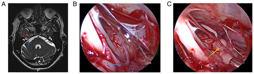
2                                           JIANG et al: FULLY ENDOSCOPIC MVD OF HFS

Patients                                                             order to make the bone window as close to the sigmoid sinus as
Patient 1. A 57‑year‑old male had suffered from left‑sided           possible, the mastoid air chamber can be opened if necessary,
facial twitches for 5 years. His left facial twitch was parox‑       but it must be blocked with bone wax in time. After suspending
ysmal and could be resolved spontaneously. The patient had           and cutting the dura mater, the arachnoid membrane was cut
been treated with acupuncture and moxibustion, but with little       to slowly release cerebrospinal fluid, so that the cerebellar
effect. He was admitted to hospital on May 5, 2020. A physical       hemisphere collapsed naturally and formed enough space
examination showed that the left‑sided facial twitches were          for surgery. Next, under the neuroendoscope, the arachnoid
involuntary, and that hearing and extremity muscle tone were         surrounding the trigeminal nerve, the acoustic‑facial bundle
normal.                                                              and the lower cranial nerves was completely cleared and
                                                                     dissected, and the neurovascular conflict area was identified.
Patient 2. A 46‑year‑old female had experienced right‑sided          The offending blood vessels in contact with the facial nerve
facial twitches for 13 years. The initial clinical manifestation     roots were separated and shifted, and decompressed using a
of the patient was twitching of the right eyelid, and later the      Teflon pad. Meanwhile, a small piece of Teflon pad cotton was
clinical symptoms gradually became the whole right facial            placed parallel to the root outlet zone (REZ) along the nerve to
twitching. The patient had been treated with BT injection four       prevent blood vessels from compressing the REZ. Arteries with
times, and after each treatment, the symptoms were relieved          severe atherosclerosis should be avoided during the operation,
for several months before relapsing again. She was admitted to       so as not to distort arteries and cause blood flow obstruction.
hospital on May 25, 2020.                                            Next, under the neuroendoscope, checks were performed to
                                                                     confirm that no offending blood vessels were missed, that
Patient 3. A 54‑year‑old male had experienced involuntary            decompression was sufficient, and that the size and location of
bilateral lateral twitching for 2 months. His bilateral facial       the Teflon pads were appropriate. Finally, the dura mater was
twitches were paroxysmal, and these clinical manifestations          sutured and the skull defect was repaired with titanium plates
aggravated when he was tense and disappeared when he slept.          and screws, and sutured closed layer by layer.
The patient was admitted to the hospital on May 7, 2020 and
November 23, 2020. He received no treatment prior to his first       Results
admission to the hospital.
                                                                     Overall results. Figs. 1‑5 show the relevant images of cases 1‑5,
Patient 4. A 50‑year‑old male had suffered from left‑sided           respectively, including the preoperative MRI and intraoperative
facial twitches for 6 years. The patient had received acupunc‑       images. From the intraoperative images, the offending blood
ture and moxibustion therapy with poor results. The patient          vessels are clearly visible in all 5 cases, including 3 cases in
had also received BT injections several times, and after each        which the anterior inferior cerebellar artery was affected (Figs. 1,
treatment, the symptoms were only relieved for a few months          3 and 5), 2 cases in which the posterior inferior cerebellar artery
before the condition relapsed again. He was admitted to              was affected (Figs. 2 and 4) and 1 case where the small arteries
hospital on July 1, 2020.                                            were affected (Fig. 3). Notably, the offending blood vessels were
                                                                     found on both sides of the left and right face of the same patient,
Patient 5. A 64‑year‑old male had experienced right‑sided            with the left side affected by the small arteries and the right side
facial twitches for 2 years. His left facial twitch was parox‑       by the inferior cerebellar artery. (Fig. 3). Generally, the criteria
ysmal and could be resolved spontaneously. The patient was           for judging the curative effect of HFS after surgery are divided
admitted to hospital on July 1, 2020. He had not received any        into four levels using a postoperative efficacy classification based
treatment prior to his admission to the hospital.                    on previous literature and clinical symptoms (12): i) Excellent:
                                                                     The symptoms of HFS completely disappear. ii) Good: The
MVD treatment. All operations were performed via the                 symptoms of HFS basically disappear and can only be induced
suboccipito‑retrosigmoid approach under full endoscopy.              occasionally when the patient is emotionally intense or during
The patients were placed in an upside‑down position on the           certain facial movements. In addition, when the symptoms
surgical site, with the head drooping 15˚ and rotating 10˚ to        have basically disappeared and the patient feels subjectively
the opposite side. The neck was slightly forward, the mandible       satisfied. iii) Fair: The symptoms of HFS are relieved but still
was ~2 transverse fingers from the sternum, and the mastoid          frequent, and the patient feels subjectively dissatisfied. iv) Poor:
on the surgical side was roughly parallel to the operating table     The symptoms of HFS did not change and even worsened.
and in the highest position. The head was fixed and slightly         According to the aforementioned classification, the surgical
turned to the surgical site, in order to help the cerebellum leave   efficacy of the 5 cases was evaluated, from which 2 cases were
the petrous bone due to its own gravity, without the use of a        graded as excellent and 3 cases were graded as good, with an
brain plate. A straight incision parallel to the inner edge of the   effective rate of 100%. This showed that fully endoscopic MVD
hairline or a transverse incision through the root of the mastoid    was a safe and effective method for the treatment of HFS. In
was performed, at a length of 4‑6 cm. The upper and the outer        addition, 4 cases had no postoperative complications, while only
edges of the bone window were located under the transverse           1 patient experience postoperative aseptic meningitis, but fully
sinus and exposed the edge of the sigmoid sinus. Usually,            recovered after follow‑up treatment.
the diameter of the bone window is only 2‑3 cm. In order to
prevent damage to the venous sinus, drill a hole farthest from       Individual patient results
the venous sinus, then grind the skull, and gradually expand         Patient 1. The symptoms of left‑sided HFS were significantly
the bone window to the transverse sinus and sigmoid sinus. In        improved after fully endoscopic MVD, but twitching around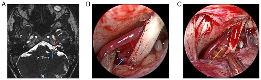
EXPERIMENTAL AND THERAPEUTIC MEDICINE 24: 483, 2022 3
Figure 1. Patient 1. (A) Preoperative head magnetic resonance imaging of the patient. (B) The anterior inferior cerebellar artery and compressed cranial nerves
were clearly visible under the endoscopic view. (C) The Teflon pad was placed between the root exit zone of cranial nerve 7 and the anterior inferior cerebellar
artery under the endoscopic view. Red arrows indicate the compressed cranial nerve vessels, blue arrows indicate the anterior inferior cerebellar artery and
the yellow arrow indicates the Teflon pad.
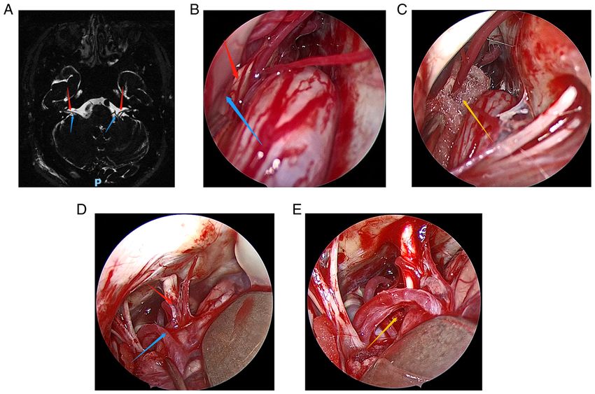
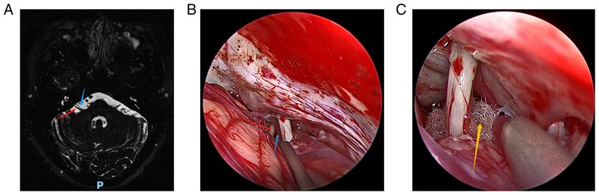
Figure 2. Patient 2. (A) Preoperative head magnetic resonance imaging of the patient. (B) The posterior inferior cerebellar artery and compressed cranial
nerves were clearly visible under the endoscopic view. (C) The Teflon pad was placed between the root exit zone of cranial nerve 7 and the posterior inferior
cerebellar artery under the endoscopic view. Red arrows indicate the compressed cranial nerve vessels, blue arrows indicate the posterior inferior cerebellar
artery and the yellow arrow indicates the Teflon pad.
the eyes occasionally occurred. The patient also suffered from blood vessels were identified as the anterior inferior cerebellar
aseptic meningitis after the surgery, but recovered well after artery on the right side and the small artery on the left side.
lumbar puncture and drainage. After 12 months of follow‑up, The postoperative curative effect was evaluated as excellent.
the clinical symptoms disappeared without recurrence and
complications. The offending blood vessel was identified as Patient 4. The patient underwent fully endoscopic MVD and
the anterior inferior cerebellar artery during the operation, and the symptoms on the left side of the face were significantly
the postoperative curative effect was evaluated as good. improved. There was no recurrence during a follow‑up period
of 12 months. The offending blood vessel was identified as the
Patient 2. The symptoms of HFS basically disappeared after posterior inferior cerebellar artery during the operation, and
fully endoscopic MVD, and there was no recurrence during the postoperative curative effect was evaluated as good.
12 months of follow‑up. The offending blood vessel was
identified as the posterior inferior cerebellar artery during the Patient 5. The patient underwent fully endoscopic MVD and
operation, and the postoperative curative effect was evaluated the symptoms were completely relieved. After 12 months
as good. of follow‑up, there was no recurrence. The offending blood
vessel was identified as the anterior inferior cerebellar artery
Patient 3. After the first fully endoscopic MVD, the during the operation, and the postoperative curative effect was
right‑sided facial symptoms were completely relieved, but the evaluated as excellent.
left facial symptoms were only slightly improved. At 5 months
post‑surgery, the patient underwent a second fully endoscopic Discussion
MVD and the left‑sided facial symptoms were also completely
relieved. After 12 months of follow‑up, there was no recurrence HFS is a common clinical cranial nerve disease that mani‑
on either side of the face. In the two operations, the responsible fests as paroxysmal involuntary facial muscle twitching.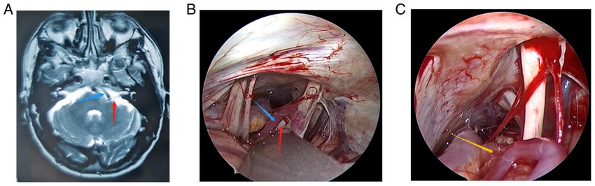
4                                                  JIANG et al: FULLY ENDOSCOPIC MVD OF HFS

Figure 3. Patient 3. (A) Preoperative head magnetic resonance imaging of the patient. (B) In the first operation, the anterior inferior cerebellar artery and
compressed cranial nerves were clearly visible under the endoscopic view. (C) The Teflon pad was placed between the root exit zone of cranial nerve 7 and the
anterior inferior cerebellar artery under the endoscopic view in the first operation. (D and E) They are the images observed under the endoscope during the
second operation, the offending blood vessel is the small artery. Red arrows indicate the compressed cranial nerve vessels, blue arrows indicate the offending
blood vessels and yellow arrows indicate the Teflon pad.

Figure 4. Patient 4. (A) Preoperative head magnetic resonance imaging of the patient. (B) The the posterior inferior cerebellar artery and compressed cranial
nerves were clearly visible under the endoscopic view. (C) The Teflon pad was placed between the root exit zone of cranial nerve 7 and the posterior inferior
cerebellar artery under the endoscopic view. Red arrows indicate the compressed cranial nerve vessels, blue arrows indicate the posterior inferior cerebellar
artery and the yellow arrow indicates the Teflon pad.

When the symptoms are relatively severe, it can cause                            artery were both observed and separated from the facial
serious psychological obstacles to patients, and even affect                     nerve root during surgery. The involuntary facial convul‑
their QOL, work and social communication. Therefore, the                         sion symptoms were relieved to a certain extent after the
treatment desire of patients is strong. In 1947, Campbell and                    surgery. In 1962, Gardner (3) first proposed that the HFS was
Keedy (13) reported 2 cases of HFS caused by abnormal                            caused by vascular compression of the facial nerve roots.
blood vessels in the posterior fossa compressing the facial                      In 1966, Jannetta and Rand (14) hypothesized that the REZ
nerve roots. Both patients underwent surgery through the                         of the facial nerve was compressed by the offending blood
suboccipital approach, and cirsoid aneurysms of the basilar                      vessels, resulting in demyelination of the facial nerve and
EXPERIMENTAL AND THERAPEUTIC MEDICINE 24: 483, 2022                                                             5

Figure 5. Patient 5. (A) Preoperative head magnetic resonance imaging of the patient. (B) The the anterior inferior cerebellar artery and compressed cranial
nerves were clearly visible under the endoscopic view. (C) The Teflon pad was placed between the root exit zone of cranial nerve 7 and the anterior inferior
cerebellar artery under the endoscopic view. Red arrows indicate the compressed cranial nerve vessels, blue arrows indicate the anterior inferior cerebellar
artery and the yellow arrow indicates the Teflon pad.

an impulse short circuit between the afferent and efferent                      relieved after the operation. Cheng et al (23) reported that
nerve fibers, which was the root cause of HFS. The vascular                     10 cases with HFS were cured by fully endoscopic MVD
compression etiology theory was thus further improved.                          without serious complications. Flanders et al (24) reported
Based on this, MVD was pioneered to treat HFS (14). At                          27 cases of fully endoscopic MVD treatment for HFS, with
present, MVD is mainly performed using a microscope                             the cure rate and postoperative complication rate recorded
(microscopic MVD), a microscope combined with an endo‑                          as 60.7 and 26.0%, respectively. Feng et al (11) reported
scope (endoscope‑assisted MVD) or using an endoscope                            45 cases of HFS that were treated with fully endoscopic
only (fully endoscopic MVD). Among these methods,                               MVD, for which the cure rate was 93.3%, the total effective
microscopic MVD is the most traditional and is commonly                         rate was 97.8% and the postoperative complication rate was
used. However, the straight viewing field of the microscope                     only 4.4%. At total of two case series with complete data,
limits the observation of deep structures and the angle,                        including 56 cases of HFS treated with fully endoscopic
making it difficult for doctors to accurately determine the                     MVD, were reviewed (11,25), for which the postoperative
compression points of all offending blood vessels. A study                      effective rate reached ~92.8% and the postoperative compli‑
has highlighted that ~23% of double vessel compression is                       cation rate was ~7.2% (including 2 cases of temporary facial
difficult to detect under the microscope (15). With the rapid                   paralysis, 1 case of mild but significant hearing loss and
development of endoscopic technology, endoscopy now has                         1 case of intracranial infection). In the present study, all 5
advantages such as the wide field of vision, bright light,                      HFS patients underwent fully endoscopic MVD, and the
no obstruction of the field of vision and flexible operation,                   offending blood vessels were accurately identified during
and so has become a standard operative tool for minimally                       the operation. The postoperative effective rate was 100%,
invasive neurosurgery of the sellar and parasellar regions,                     and no serious complications occurred. Only 1 patient devel‑
and the ventricular system (16‑18). As a representative of                      oped aseptic meningitis, but fully recovered after treatment.
minimally invasive neurosurgery, endoscopy‑assisted MVD                         There was no hearing loss, facial paralysis or cerebrospinal
has been applied in clinical treatment. For MVD, compared                       fluid leakage recorded.
with the microscope, the endoscope has the greatest advan‑                          The manner in which complications can be reduced and
tage of accurately identifying the offending blood vessels.                     the efficiency of surgery can be improved is a concern for
Magnan et al (19) reported the first 43 cases of patients                       all neurosurgeons. The complications of MVD treatment for
who received endoscopy‑assisted MVD and suggested the                           HFS mainly include cranial nerve dysfunction, cerebellar
endoscopy could accurately identify the offending blood                         brainstem injury, intracranial hypotension syndrome,
vessels causing the facial nerve compression. It was then                       cerebrospinal fluid leakage, vertebral artery injury, aseptic
reported that for the same 60 patients, the confirmation                        meningitis and wound infection (26‑30). Specifically, when
rate of vascular nerve compression location by endoscopy                        MVD is used to treat HFS, whether it is microscopic MVD,
was 65% higher than that by the microscope, which may be                        endoscopic‑assisted MVD or fully endoscopic MVD, it is
due to the complexity of local neurovascular anatomy, local                     necessary to first identify the offending blood vessels, and
vessels abnormalities or even branch entanglement, and the                      then place a Teflon pad between the seventh and eighth
offending blood vessels being located in the deep part of the                   pairs of cranial nerves (facial and auditory nerves) and the
abnormal vascular plexus (20). Teo et al (21) also reported                     offending blood vessels to relieve vascular compression.
that in endoscopy‑assisted MVD, endoscopy accurately iden‑                      Among the three types of MVD, fully endoscopic MVD has
tified 8% of offending blood vessels missed by microscope.                      many advantages in the treatment of HFS. Firstly, the excel‑
In the same period, fully endoscopic MVD was also trialed                       lent visual field of endoscopy helps surgeons to accurately
to treat HFS. The first report on 3 cases of HFS treated with                   identify the blood vessels and nerves in the surgical area,
fully endoscopic MVD was published by Eby et al (22) in                         so as to avoid damaging the surrounding brain tissue, blood
2001, and the HFS symptoms of the patients were completely                      vessels and nerves, and to reduce postoperative complications.
6                                           JIANG et al: FULLY ENDOSCOPIC MVD OF HFS

In addition, during the operation, endoscopy is helpful to           Ethics approval and consent to participate
evaluate the position of the Teflon pad, the decompression
effect and other injuries in a timely manner, so that surgeons       This study was approved by the Ethics Committee of
can adjust the operation strategy according to the intraop‑          Chongqing General Hospital (Chongqing, China).
erative situation. Finally, fully endoscopic MVD can shorten
the length of the surgical incision, eliminate excessive crani‑      Patient consent for publication
otomy exposure, avoid excessive separation of arachnoids,
and reduce the traction of brain tissues and cranial nerves. In      Written informed consent was obtained from the patients for
addition, the average operation time of fully endoscopic MVD         the publication of anonymized data and any accompanying
is equivalent to that of microscopic MVD, which helps to             images.
avoid the wasted time of endoscopic‑assisted MVD in terms
of changing from the microscope to the endoscope to the              Competing interests
microscope again. In addition to the aforementioned advan‑
tages, fully endoscopic MVD also has some disadvantages.             The authors declare that they have no competing interests.
For example, the endoscope occupies a certain operational
space during surgery and can only provide two‑dimensional            References
images. Intraoperative bleeding may also pollute the endo‑
scope lens, affecting the image quality and the continuity of         1. Blue R, Li C, Spadola M, Saylany A, McShane B and Lee JYK:
the operation. Furthermore, it should be mentioned that the              Complication rates during endoscopic microvascular decom‑
                                                                         pression surgery are low with or without petrosal vein sacrifice.
present study has certain limitations, as the follow‑up time             World Neurosurg 138: e420‑e425, 2020.
was relatively short and only 5 patients were studied. At the         2. Jannetta PJ: Neurovascular compression in cranial nerve and
same time, it was designed as a non‑randomized retrospec‑                systemic disease. Ann Surg 192: 518‑525, 1980.
                                                                      3. Gardned WJ: Concerning the mechanism of trigeminal neuralgia
tive study and does not completely rule out potential selection          and hemifacial spasm. J Neurosurg 19: 947‑958, 1962.
biases.                                                               4. McLaughlin MR, Jannetta PJ, Clyde BL, Subach BR, Comey CH
    In conclusion, with the rapid development of endoscopic              and Resnick DK: Microvascular decompression of cranial
                                                                         nerves: Lessons learned after 4400 operations. J Neurosurg 90:
technology, fully endoscopic MVD for the treatment of                    1‑8, 1999.
HFS is widely recognized, and its advantages are gradually            5. Teton ZE, Blatt D, Holste K, Raslan AM and Burchiel KJ:
obvious compared with other technologies. The present study              Utilization of 3D imaging reconstructions and assessment of
                                                                         symptom‑free survival after microvascular decompression of the
reviewed 5 cases of HFS that were successfully treated using             facial nerve in hemifacial spasm. J Neurosurg: 1‑8, 2019 (Epub
a fully endoscopic MVD technique. Fully endoscopic MVD                   ahead of print).
should be considered as safe and effective to treat HFS. For          6. Felício AC, Godeiro‑Junior Cde O, Borges V, Silva SM and
                                                                         Ferraz HB: Bilateral hemifacial spasm: A series of 10 patients with
surgeons, fully endoscopic MVD technology has certain chal‑              literature review. Parkinsonism Relat Disord 14: 154‑156, 2008.
lenges, but sufficient training, rich experience and constantly       7. Haller S, Etienne L, Kövari E, Varoquaux AD, Urbach H and
updated equipment can help them to successfully master this              Becker M: Imaging of neurovascular compression syndromes:
                                                                         Trigeminal neuralgia, hemifacial spasm, vestibular paroxysmia,
technology.                                                              and glossopharyngeal neuralgia. AJNR Am J Neuroradiol 37:
                                                                         1384‑1392, 2016.
Acknowledgements                                                      8. Lawrence JD, Frederickson AM, Chang YF, Weiss PM,
                                                                         Gerszten PC and Sekula RF: An investigation into quality of life
                                                                         improvement in patients undergoing microvascular decompres‑
Not applicable.                                                          sion for hemifacial spasm. J Neurosurg 128: 193‑201, 2018.
                                                                      9. Mizobuchi Y, Nagahiro S, Kondo A, Arita K, Date I, Fujii Y,
                                                                         Fujimaki T, Hanaya R, Hasegawa M, Hatayama T, et al:
Funding                                                                  Prospective, multicenter clinical study of microvascular decom‑
                                                                         pression for hemifacial spasm. Neurosurgery 88: 846‑854, 2021.
No funding was received.                                             10. Artz GJ, Hux FJ, Larouere MJ, Bojrab DI, Babu S and Pieper DR:
                                                                         Endoscopic vascular decompression. Otol Neurotol 29: 995‑1000,
                                                                         2008.
Availability of data and materials                                   11. Feng BH, Zhong WX, Li ST and Wang XH: Fully endoscopic
                                                                         microvascular decompression of the hemifacial spasm: Our
                                                                         experience. Acta Neurochir (Wien) 162: 1081‑1087, 2020.
The datasets used and/or analyzed during the current study           12. Feng BH, Zheng XS, Wang XH, Ying TT, Yang M, Tang YD and
are available from the corresponding author on reasonable                Li ST: Management of vessels passing through the facial nerve in
request.                                                                 the treatment of hemifacial spasm. Acta Neurochir (Wien) 157:
                                                                         1935‑1940, 2015.
                                                                     13. Campdell E and Keedy C: Hemifacial spasm; a note on the
Authors' contributions                                                   etiology in two cases. J Neurosurg 4: 342‑347, 1947.
                                                                     14. Jannetta PJ and Rand RW: Transtentorial retrogasserian
                                                                         rhizotomy in trigeminal neuralgia by microneurosurgical tech‑
HTJ, PW, DWZ, LWZ, BL and NW participated in the concep‑                 nique. Bull Los Angeles Neurol Soc 31: 93‑99, 1996.
tion, design and data acquisition of the article. HTJ participated   15. Dubey A, Yadav N, Ratre S, Parihar VS and Yadav YR: Full
in drafting and revising the manuscript. PW critically revised           endoscopic vascular decompression in trigeminal neuralgia:
                                                                         Experience of 230 patients. World Neurosurg 113: e612‑e617, 2018.
the article. NW ensured that questions related to the integrity      16. Jho HD and Carrau RL: Endoscopy assisted transsphenoidal
of any part of the work were appropriately investigated and              surgery for pituitary adenoma. Technical note. Acta Neurochir
resolved. HTJ, PW, DWZ, LWZ, BL and NW confirm the                       (Wien) 138: 1416‑1425, 1996.
                                                                     17. Adappa ND, Learned KO, Palmer JN, Newman JG and Lee JY:
authenticity of all the raw data. All authors read and approved          Radiographic enhancement of the nasoseptal flap does not predict
the final version of the manuscript.                                     postoperative cerebrospinal fluid leaks in endoscopic skull base
                                                                         reconstruction. Laryngoscope 122: 1226‑1234, 2012.
EXPERIMENTAL AND THERAPEUTIC MEDICINE 24: 483, 2022 7
18. Nagata Y, Watanabe T, Nagatani T, Takeuchi K, Chu J and 25. Cai Q, Li Z, Guo Q, Wang W, Ji B, Chen Z, Dong H and Mao S:
Wakabayashi T: The multiscope technique for microvascular Microvascular decompression using a fully transcranial neuro‑
decompression. World Neurosurg 103: 310‑314, 2017. endoscopic approach. Br J Neurosurg: 1‑4, 2021 (Epub ahead of
19. Magnan J, Chays A, Lepetre C, Pencroffi E and Locatelli P: print).
Surgical perspectives of endoscopy of the cerebellopontine 26. Huh R, Han IB, Moon JY, Chang JW and Chung SS:
angle. Am J Otol 15: 366‑370, 1994. Microvascular decompression for hemifacial spasm: Analyses
20. Magnan J, Caces F, Locatelli P and Chays A: Hemifacial spasm: of operative complications in 1582 consecutive patients. Surg
Endoscopic vascular decompression. Otolaryngol Head Neck Neurol 69: 153‑157; discussion 157, 2008.
Surg 117: 308‑314, 1997. 27. Wilkins RH: Hemifacial spasm: A review. Surg Neuro 36:
21. Teo C, Nakaji P and Mobbs RJ: Endoscope‑assisted microvas‑ 251‑277, 1991.
cular decompression for trigeminal neuralgia: Technical case 28. Barker FG II, Jannetta PJ, Bissonette DJ, Shields PT,
report. Neurosurgery 59 (4 Suppl 2): ONSE489‑90; discussion Larkins MV and Jho HD: Microvascular decompression for
ONSE490, 2006. hemifacial spasm. J Neurosurg 82: 201‑210, 1995.
22. Eby JB, Cha ST and Shahinian HK: Fully endoscopic vascular 29. Jannetta PJ: Typical or atypical hemifacial spasm. J Neurosurg 89:
decompression of the facial nerve for hemifacial spasm. Skull 346‑347, 1998.
Base 11: 189‑197, 2001. 30. Wang A and Jankovic J: Hemifacial spasm: Clinical findings and
23. Cheng WY, Chao SC and Shen CC: Endoscopic microvas‑ treatment. Muscle Nerve 21: 1740‑1747, 1998.
cular decompression of the hemifacial spasm. Surg Neurol 70
(Suppl 1): S40‑S46, 2008. This work is licensed under a Creative Commons
24. Flanders TM, Blue R, Roberts S, McShane BJ, Wilent B, Attribution-NonCommercial-NoDerivatives 4.0
Tambi V, Petrov D and Lee JYK: Fully endoscopic microvas‑ International (CC BY-NC-ND 4.0) License.
cular decompression for hemifacial spasm. J Neurosurg 131:
813‑819, 2018.You can also read