LUMINOSITY AND CONTRAST ADJUSTMENT BASED ENHANCEMENT OF DIABETIC RETINAL IMAGES
←
→
Page content transcription
If your browser does not render page correctly, please read the page content below
Turkish Journal of Physiotherapy and Rehabilitation; 32(3)
                                                                  ISSN 2651-4451 | e-ISSN 2651-446X

LUMINOSITY AND CONTRAST ADJUSTMENT BASED ENHANCEMENT OF
                 DIABETIC RETINAL IMAGES

                         Satish Tunga1, D Jayadevappa2, C Gururaj3, H R Harshitha4
                        1,4
                          Ramaiah Institute of Technology, Bengaluru, Karnataka 560054,
                               India 2JSS Academy of Technical Education NOIDA,
                               DISTT.U.P., INDIA & 3BMS College of Engineering,
                               Basavanagudi, Bengaluru, Karnataka 560019, India
                                1
                                  Satish.tunga@msrit.edu, 2devappa22@gmail.com,
                                4
                                  harshithahr16@gmail.com, 3gururaj.c@gmail.com

                                                    ABSTRACT

    Enhancement of an image plays a crucial role in image analysis and pattern recognition. Retinal images are
    examined by the ophthalmologists for early diagnosis of common retinal disorders. This includes age-related
    macular degeneration and diabetic retinopathy. The intention of a paper is to design image enhancement
    technique to provide better color retinal image contrast and luminosity. Various methods like luminance gain
    matrix obtained by gamma correction of the value channel in the HSV color space is used to enhance the R,
    G, and B (Red, Green and Blue) channels, respectively. Contrast enhancement in the luminosity channel of
    L*a*b* color space is carried by CLAHE. The images obtained after enhancement of luminosity and contrast
    is further undergoes segmentation for further analysis of the retinal image.

    Keywords—Retinopathy, Segmentation, Enhancement, HSV, Luminosity, Fundus image.

                                               I.    INTRODUCTION
Inaccurate image processing, classification may decrease the ophthalmologists' ability to detect significant eye
characteristics or distinguish retinal diseases. Retinal images with poor contrast makes it impossible to segment
the desired object and also difficult to diagnose retinal diseases by computer aided diagnosis systems [1], which
are helpful to automate to assist the eye specialists and also for the interpretation. Hence, it is necessary to
overcome the issues with respect to poor quality of images. One of the effective technique is the use of image
enhancement method to cater better vision of retinal anatomical structure. Various new enhancement techniques
have been proposed in the recent year’s for retinal images like histogram equalization, including luminosity of
image and contrast image normalization and contourlet transform based multi-scale methods. The Contrast
Limited Adaptive Histogram Equalization (CLAHE), enhancement of blood vessel by top-hat transform and
linear stretching with histogram Gaussian curve fitting, combinational methods with coding. Most of these
techniques concentrate on retinal blood vessels enhancement to obtain the contrast between retinal background
and blood vessels [2]. This helps to achieve better vessel segmentation both grayscale and colour images of
retina. This technique is mainly helpful for colour images of retina. In general, a high contrast between the
vessels and the background is displayed by green channel. Any enhancement techniques may lead to lose colour
details or other important image features like macula luteaq, optic discs and lesions. This results in deteriorating
the improvisation of present status of diagnosis by ophthalmologists. According to World Health Organization
(WHO) diabetes is considered one of the deadliest diseases in India. Therefore, immediate and fast diagnosis can
help to treat the patient in a better way and this reduces the cost as compared to advanced phase which may later
leads to severe disease.

                                             II.    RELATED WORK
Image enhancement is a technique to enhance the image and hold and display the same information but in a more
meaningful and understandable way. No new information is added during the enhancement of the images. These
operations involve improving the qualities of an image by improvising the image’s contrast and luminance

www.turkjphysiotherrehabil.org                                                                              1842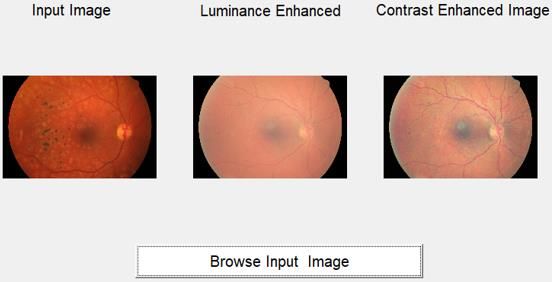
Turkish Journal of Physiotherapy and Rehabilitation; 32(3)
                                                                  ISSN 2651-4451 | e-ISSN 2651-446X

characteristics. It also includes reducing the noise content, or sharpening the details. Here are some of the work
carried out and their details regarding the related work progressed in the field of image processing on diabetic
retinopathy.

Geetha Ramani and L. Balasubramanian [3] have proposed the segmentation of retinal blood vessel using data
mining technique and image processing for computerized retinal image analysis. In this method, the retinal
fundus images allows us to identify most of the retinal diseases. Therefore the segmentation of retinal images is
very essential to find the irregularities in the retinal structure. The vessel segmentation by manually doing is time
consuming and needs experts. Computerized approaches for this task are employed that would help in efficient
retinal analysis.      In the proposed technique that involves sequential image pre-processing, Gabor filtering [4]
and half wave rectification are sequentially applied. The proposed methodology was able to achieve an Accuracy
of 95.36%, Sensitivity of 70.79%, Specificity of 97.78% and PPV of 75.76%.

S. You et al. [5], presented retinal vessel segmentation based on principle curve towards diagnosis of retinal
diseases. This comprises of three stages: First step is Pre-processing of the image is done to remove the noise,
remove the unwanted background and enhance the vessel structure. In second step, the tabular structures of
vessels are enhanced using Frangi filters and the radius of the vessels are estimated using isotropic Gaussian
kernels. The third step is, a kernel interpolation of intensity, later algorithm for principal curve projection is used
to move the pixels onto the ridges of the Gaussian kernel. The vessel trees are recursively traced by applying
principal curve tracing algorithm. The results recorded were Accuracy of 94.56%, Sensitivity of 80.33%,
Specificity of 95.54%.

M. Usman Akram et al. [6] have presented a system that was developed for the detection of neovascularization.
The input retinal images are made free from background by applying a pre-processing algorithm. The inverted
green channel from RGB is considered for enhancement of blood vessels by 2-D Gabor wavelet. Later a binary
mask for the segmentation of vessel is achieved by applying multilayered thresholding and adaptive thresholding
techniques. The abnormal blood vessels are then found by the sliding window. The results were evaluated on
DRIVE and STARE databases. An average Accuracy of 94.69% and standard deviation of 0.0053 was recorded
for DRIVE database, and an average Accuracy of 95.02% and standard deviation of 0.0253 was recorded for
STARE database.

C. Nivetha et al. [7] have introduced the extraction of blood vessels and identification of exudates by PNN and
wavelet transform approach for the diabetic retinopathy. Assessment. They have come up with a novel method to
identify the exudates in retinal images, classify normal and abnormal blood vessels. In order to improve contrast
of images and remove noise, Daubechies wavelet transform was employed [8].The feature extracted from
Daubechies wavelet transform is compared with the database images using PNN. The PNN classifier had
classified the images into abnormal and normal. The abnormal images was then subjected to morphological
process, where the blood vessels are extracted. Later the Fuzzy C-means logic is used to determine the presence
of exudates in the images. An Accuracy of 97.76% and Sensitivity of 96.77% was achieved by this method.

Syna Sreng et al. [9] have proposed a diabetic retinopathy screening on the basis of including morphology and
SVM. In this method, to differentiate DR and non-DR depends on both red and bright lesions. In this method
total 90 images were trained and 229 image were tested. The computational time was 8 sec. The results recorded
were

Accuracy- 90%, Sensitivity- 86.33%, Specificity- 98.55%.

P. Furtado et al [10] have presented the fundus image segmentation by density clustering in DR. In their
technique, they gave a novel methodology to detect lesions which are one of the symptoms of DR. It involves a
pre-processing step where the median filter is used to remove the noises and smoothen the images. The next step
involves the bright and dark lesion segmentation and the bright lesions are detected using Kirsch edge detection.
The red lesions are detected by a filtering method called top-hat morphological method. Later the dark and bright
lesions are combined using AND operator. The noises remaining after the processing is removed by bob analysis.
At the end the features obtained are fed to the SVM classifiers. This method got 90% of accuracy, 86.33% of
sensitivity and 98.55% specificity 8 second per image of average computational time.

www.turkjphysiotherrehabil.org                                                                                1843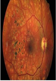
Turkish Journal of Physiotherapy and Rehabilitation; 32(3)
ISSN 2651-4451 | e-ISSN 2651-446X
Mei Zhou et al. [11] have introduced an enhancement for color retinal images based on contrast and luminosity
adjustment. In this technique improvement in retinal image processing by an effective method which considered
the luminosity enhancement and contrast adjustment of images have been carried out. Enhancement of luminosity
is achieved by gamma correction of luminance channel. The image is converted to L*a*b color space over which
the CLAHE (Contrast limited adaptive histogram equalization) is used to adjust contrast. The results recorded
were assessed scores averages 0.1957 with standard deviation 0.0819 and for enhanced images are 0.4610 with
0.1152 standard deviation.
Ashim Chakraborty et al. [12] have presented decision scheme for mobile detection of early stage DR. In this
method, they have come up with a system and proposed to enable the users examine their own conditions and
detect DR at the earliest stage. This system extracted two features namely hard exudates and blood vessels. It
involved combining image processing methods like morphological opening and closing, black top hat, blob
function canny edge detection and color thresholding. The illumination problems was overcome by adaptive
algorithm. The results were verified and was recorded 92% accuracy for blood vessels and 90.3% accuracy for
the extraction of hard exudates. Their methodology took only 0.6 seconds for analysis of 1 image.
Arisha Roy et al. [13] presented a filter and FCM based extraction of features and classification of DR using
SVMs. In this method, retinal vessel extraction and exudates extraction which involved Fuzzy means technique.
The optical disk removal was carried out by convex Hull technique as optic nerves are misinterpreted as exudates
sometimes[33][34][35]. The retinal vessel extraction is carried out by using cascaded Gaussian and Median Filter
and later followed by a top hat filter of specific size. The output image undergoes binarization and morphological
operators was applied which made the blood vessels more prominent. The two features obtained were classified
as normal or effected by diabetic retinopathy by training it through 2-fold SVM classifiers. The SVM showed an
efficiency of 96.23% for classifying the images correctly [21] – [32].
III. PROPOSED METHODOLOGY
A. Luminosity Enhanacement
Since inadequate or uneven luminance clouds visual view of retinal pictures, making analytic subtleties
imperceptible, it is fundamental to upgrade the luminance impact first. Be that as it may, for a shading image, the
shading ought not to change for any pixel, to forestall picture mutilation. When all is said in done, shading retinal
pictures are put away and seen utilizing RGB shading space. The R, G, and B channels at the same time contain
the iridescence data and the shading data, which are associated with one another. To improve the iridescence and
safeguard the shading, the R, G, and B channels ought to be balanced by a similar extent. Our answer is to get a
luminance gain framework G(x, y) which is characterized as follow in condition 1.
r ' ( x, y ) g ' ( x, y ) b' ( x, y )
G( x, y)
r ( x, y) g ( x, y ) b( x, y )
(1)
here, r'(x, y), g'(x, y), and b'(x, y) R, G, and B enhanced values of pixel at (x, y) and the r(x, y), g(x, y), and b(x,
y) are the original R, G, and B values.
V ' ( x, y) V ' ( x, y)
G( x, y)
V ( x, y) max[r ( x, y).g ( x, y).b( x, y )
(2)
here, V(x, y) is the luminance intensity of a pixel at (x, y) position, and V'(x, y) is the function of V(x, y), which
determines the effect of luminosity enhancement, We can see that the processing can be directly done in the RGB
color space, which reduces the computational complexity.
B. Performance Metrics
Using Messidor dataset, the retinal images were processed by they are also evaluated by the same assessment
technique. The mean/average values are 0.1957 and 0.4610 for the original and enhanced images respectively.
The performance of the proposed method shows that, the proposed methodology can improve the normal retinal
images, too. Additionally calculated MSE and PSNR proves the superiority of our method.
www.turkjphysiotherrehabil.org 1844
Turkish Journal of Physiotherapy and Rehabilitation; 32(3)
                                                                 ISSN 2651-4451 | e-ISSN 2651-446X

Mean Square Error: The Mean Square Error (MSE) is used to calculate the deviations or average of the squares
of the errors. MSE is depicted in the following equation.

                                                  (|        |)
                                       ∑    ∑                                       (3)

Peak Signal to Noise Ratio: The PSNR (Peak Signal to Noise Ratio) is generally used to analyze quality of image
and video files. The PSNR computation of two images, presents the how far two images are equal.

                                   (   )               (    )                        (4)

C. Design of Channel Conversion
Design diagram for channel conversion depicting the RGB conversion to individual channels and RGB to LAB
conversions is shown in figure 1.

                                    Fig. 1. Block diagram for channel conversion.

D. Design for Blood Vessel Segmentation
The retinal images blood vessels emit from the Optic Disc (OD). Appropriate evacuation of blood vessels and OD
is important in sore identification since blood vessels and OD are the critical wellsprings of bogus positives for
dull and brilliant injury location, individually. Hence blood vessel extraction and OD discovery are to be done
appropriately so the general sore recognition execution isn't influenced. The current work separates the blood
vessels from a retina picture utilizing the blend of normal sifting and Contrast Limited Adaptive Histogram
Equalization (CLAHE) algorithm. The smoothened picture is deducted with the upgraded picture. From that, the
blood vessel area will be featured. At that point we apply thresholding algorithm and we fragment the blood
vessel area. To expel the undesirable articles, we are further utilizing morphological tasks. The blood vessels are
gotten at long last experiences segmentation. (Fig 2)

www.turkjphysiotherrehabil.org                                                                             1845
Turkish Journal of Physiotherapy and Rehabilitation; 32(3)
ISSN 2651-4451 | e-ISSN 2651-446X
Figure 2. Block diagram for the blood vessel segmentation.
E. CLAHE
In biomedical image analysis, it is a very popular method. The image is divided into various disjoint parts and in
each part local histogram equalization is applied. Then, the boundaries between the regions are eliminated with a
bilinear interpolation. Primary goal of this method is to characterize a point change inside a neighborhood. The
point change appropriation is restricted around the mean power of window and it covers the entire force scope of
picture.
Assume an image W which is N X N pixels at the center of a pixel P (i, j).This image is filtered to get another
sub- image P of (N X N) pixels.
[ ( ) ( )]
([ ( ) (
)
)]
(5)
here,
( ) * ( )+ (6)
and Max, Min = maximum and minimum intensity values of the entire image, while and shows the local
window mean and SD which are expressed as,
∑( ) ( ) ( ) (7)
√ ∑( ) ( )( ( ) ) (8)
IV. RESULTS AND DISCUSSION
Proposed methodology is experimented in Matlab environment. The experiment was carried out with standard
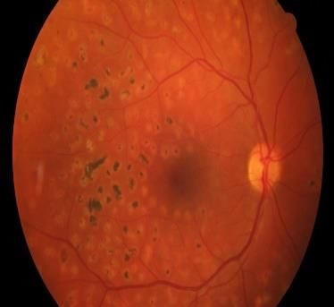
data sets of fundus images. The experimental results at various stage is recorded. Figure 3 shows the input fundus
image used for the experiment.
www.turkjphysiotherrehabil.org 1846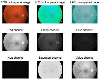
Turkish Journal of Physiotherapy and Rehabilitation; 32(3)
                                                                    ISSN 2651-4451 | e-ISSN 2651-446X

                                               Fig.3. Fundus image for input.

                                       (a)                  (b)                   (c)

                                 Fig. 4. Original Image Splitted to R, G and B Images.

                                             (a)                            (b)

                                 Fig. 5. (a) Background removal and (b) HSV image.

We aim to increase the dynamic range of the low gray level region significantly to slightly increase the moderate
gray level region and to maintain or compress the high gray level region. Gamma correction, a popular imaging
processing methods, is used to transform luminance nonlinearly. Gamma ranges from 0 to 1 denote the
normalized pixel value of the luminosity channel.(Fig4-6)

                                   Fig. 6. RGB, HSV and LAB color space images.

www.turkjphysiotherrehabil.org                                                                              1847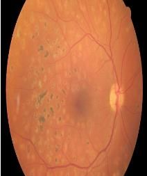
Turkish Journal of Physiotherapy and Rehabilitation; 32(3)
                                                                   ISSN 2651-4451 | e-ISSN 2651-446X

After Gamma correction applied to image and the output luminosity channel is as shown in

ure 7(a). The channel from LAB color space is subjected to CLAHE conversion to obtain the resultant image as
shown in figure 7(b).

                                        (a)                 (b)               (c)

Fig. 7. (a) Gamma correction applied RGB image, Luminosity channel output image and contrast enhanced RGB
image.

The contrast enhanced image is subjected to average filtering as shown in figure 8(a). The binarised image is
carried out with vessel segmentation which is shown in figure 8(b) and the vessel segmentation image is applied
with inverse vessel segmentation which is as shown in figure 8(c).

The vessel segmentation image is applied with inverse vessel segmentation which is shown in figure 9(a).

The inverse vessel segmentation undergoes the removal of circle and converted to colored image which is as
shown in figure 9(b). The blood vessel segmented image converted to gray scale image which is shown in figure
9(c).

                                         (a)                (b)               (c)

            Fig. 8. (a) Average Filter Image, (b) Binarised Image and (c) Vessel segmented image with circles.

                                       (a)                  (b)                (c)

   Fig. 9. (a) Inverse vessel segmented image with circles, (b) Blood vessel segmented color image and (c) Blood vessel
                                                  segmented BW image.

www.turkjphysiotherrehabil.org                                                                                    1848
Turkish Journal of Physiotherapy and Rehabilitation; 32(3)
ISSN 2651-4451 | e-ISSN 2651-446X
Fig. 10. GUI of input and output images.
The GUI shown in figure 10 depicting the input image, luminance enhanced image and contrast enhanced image.
The performance parameters such as Mean, and standard deviation for input images are shown in table 1. The
performance parameters such as MSE, PSNR, Mean, and standard deviation for LAB, HSI, HSV images are
shown in table 2. The performance parameters such as MSE, PSNR, Mean, and standard deviation for enhanced
images are shown in table 3.
Table1. Input image performance measures.
Table 2. L*A*B, HSI and HSV image performance measures.
Sl. MSE PSNR Mean Standard
No Deviation
1 0.0025 74.1351 0.4711 0.2872
2 0.0041 71.9816 0.4549 0.2821
3 0.0018 75.5938 0.5063 0.2844
4 0.0094 68.4103 0.4368 0.2628
5 0.0040 72.0627 0.4621 0.2824
www.turkjphysiotherrehabil.org 1849Turkish Journal of Physiotherapy and Rehabilitation; 32(3)
ISSN 2651-4451 | e-ISSN 2651-446X
6 0.0037 72.4846 0.4597 0.2836
7 0.0124 67.1925 0.4421 0.2586
8 0.0017 75.8051 0.5347 0.2933
9 0.0076 69.3102 0.4912 0.2579
10 0.0083 68.9403 0.4253 0.2726
Table 3. Performance measures of enhanced image.
Sl. MSE PSNR Mean Standard
No Deviation
1 0.0025 74.1351 0.4711 0.2872
2 0.0041 71.9816 0.4549 0.2821
3 0.0018 75.5938 0.5063 0.2844
4 0.0094 68.4103 0.4368 0.2628
5 0.0040 72.0627 0.4621 0.2824
6 0.0037 72.4846 0.4597 0.2836
7 0.0124 67.1925 0.4421 0.2586
8 0.0017 75.8051 0.5347 0.2933
9 0.0076 69.3102 0.4912 0.2579
10 0.0083 68.9403 0.4253 0.2726
V. CONCLUSION
This paper presented an effective technique of enhancement based on luminosity and contrast adjustment for
color retinal images. For image enhancement, very simple and effective system that includes various
enhancement techniques such as luminosity enhancement and contrast enhancement using CLAHE was
introduced. Results shows that in comparison with enhancement in other approaches the proposed technique is
able to exhibit better improvements in color images of the retinal. This effective method will largely help
ophthalmologists for diagnosis of various disease through retinal image analysis.
CONSENT FOR PUBLICATION
Not applicable.
FUNDING
None
CONFLICTS OF INTEREST
Not applicable.
ACKNOWLEDGEMENTS
www.turkjphysiotherrehabil.org 1850Turkish Journal of Physiotherapy and Rehabilitation; 32(3)
ISSN 2651-4451 | e-ISSN 2651-446X
Not applicable.
REFERENCES
1. F. M. Hani and H. A. Nugroho, ―Retinal vasculature enhancement using independent component analysis‖, J. Biomed. Sci. Eng., vol. 2, no. 7, pp. 543-
549, Nov. 2005.
2. J. V. B. Soares, J. J. G. Leandro, R. M. Cesar, H. F.Jelinek and M. J. Cree, ―Retinal vessel segmentation using the 2-D Gabor wavelet and supervised
classification‖, IEEE Trans. on Med. Imag., vol. 25, no. 9, pp. 1214-1222, Sep. 2006.
3. R. GeethaRamani and L. Balasubramanian, ―Retinal blood vessel segmentation employing image processing and data mining techniques for
computerized retinal image analysis‖, Biocybern. Biomed. Eng., vol. 36, no. 1, pp. 102-118, Jan. 2016.
4. J. V. B. Soares, J. J. G. Leandro, R. M. Cesar, H. F.Jelinek and M. J. Cree, ―Retinal vessel segmentation using the 2-D Gabor wavelet and supervised
classification‖, IEEE Trans. on Med. Imag., vol. 25, no. 9, pp. 1214-1222, Sep. 2006.
5. S. You, E. Bas, D. Erdogmus, J. Kalpathy-Cramer, ―Principal Curve Based Retinal Vessel Segmentation Towards Diagnosis of Retinal Diseases‖,
2011 First IEEE International Conference on Healthcare Informatics, Imaging and Systems Biology.
6. M. Usman Akram, Ibaa Jamal, Anam Tariq and Junaid Imtiaz, ―Automated Segmentation of Blood Vessels for Detection of Proliferative Diabetic
Retinopathy‖, Proceedings of the IEEE-EMBS International Conference on Biomedical and Health Informatics (BHI 2012), Hong Kong and
Shenzhen, China, 2-7 Jan 2012. [7]. C. Nivetha, S. Sumathi, and M. Chandrasekaran,‖Retinal Blood Vessels Extraction and Detection of Exudates
Using Wavelet Transform and PNN Approach for the Assessment of Diabetic Retinopathy‖, International Conference on Communication and Signal
Processing, April 6-8, 2017.
7. J. Staal, M. D. Abramoff, M. Niemeijer, M. A. Viergever and B. van Ginneken, ―Ridge-based vessel segmentation in color images of the
retina‖, IEEE Trans. Med. Imag. 23(4). Pp. 501-509, 2004.
8. Syna Sreng, Noppadol Maneerat, Don Isarakorn, Kazuhiko Hamamoto, Ronakorn Panjaphongse, ―Primary Screening of Diabetic Retinopathy Based
on Integrating Morphological Operation and Support Vector Machine‖, ICIIBMS 2017, Track 3: Bioinformatics, Medical Imaging and Neuroscience,
Okinawa, Japan.
9. P. Furtado , C. Travassos , R. Monteiro , S. Oliveira, C. Baptista, F. Carrilho, ―Segmentation of Eye Fundus Images by Density Clustering in Diabetic
Retinopathy‖, Biomedical and Health Informatics (BHI), 2017 IEEE EMBS International Conference, 2017.
10. [Mei Zhou, Kai Jin, Shaoze Wang, Juan Ye, and Dahong Qian, ―Color Retinal Image Enhancement Based on Luminosity and Contrast Adjustment‖,
IEEE Transactions on Biomedical Engineering, Vol. 65, No. 3, March 2017.
11. Ashim Chakraborty, David Chik, Matilda Biba M A Hossain, ―A Decision Scheme based on Adaptive Morphological Image Processing for Mobile
Detection of Early Stage Diabetic Retinopathy‖, 11th International Conference on Software, Knowledge, Information Management and Applications
(SKIMA), 2017.
12. Arisha Roy, Debasmita Dutta, Pratyusha Bhattacharya and Sabarna Choudhury, ―Filter and Fuzzy C Means Based Feature Extraction and
Classification of Diabetic Retinopathy using Support Vector Machines‖, International Conference on Communication and Signal Processing, April 6-
8, 2017.
13. E.A.M Yasir, and H.I. Bashir, "Classification of Diabetic Retinopathy using Stacked Autoencoder-Based Deep Neural Network", J Comput. Sci.
Intell. Technol., vol. 1, no. 1, pp. 09-14, 2020.
14. T. Anitha, N. Shanthi, R. Sathiyasheelan, G. Emayavaramban, and T. Rajendran, "Brain-Computer Interface for Persons with Motor Disabilities - A
Review", Open Biomed. Eng. J., vol. 13, no. Suppl-1, M5, pp. 127-133, 2019.
15. T. Rajendran, K. P. Sridhar, S. Manimurugan, and S. Deepa, "Advanced Algorithms for Medical Image Processing", Open Biomed. Eng. J., vol. 13,
no. Suppl-1, M1, p. 102, 2019.
16. T. Rajendran, K.P. Sridhar, S. Manimurugan, and S. Deepa, "Recent Innovations in Soft Computing Applications", Curr. Signal Transduct. Ther., vol.
14, no. 2, pp. 129-130, 2019.
17. M. Sreerangappa, M. Suresh, and D. Jayadevappa, "Segmentation of Brain Tumor and Performance Evaluation Using Spatial FCM and Level Set
Evolution", Open Biomed. Eng. J., vol. 13, no. Suppl-1, M6, pp. 134-141, 2019.
18. Kumar, CV Arul, D. Manoj Kumar, and P. Prithiviraj. "Privacy Policy Multiparty Access Control On Content Sharing Sites." Imperial Journal of
Interdisciplinary Research 2.6 (2016).
19. V Arulkumar, Charlyn Puspha Latha, Daniel Jr Dasig, "Concept of Implementing Big Data In Smart City: Applications, Services, Data Security In
Accordance With Internet of Things and AI" International Journal of Recent Technology and Engineering 8, no. 3 (September 2019): 237-49. 2277-
3878.
20. Balakrishnan, S., Janet, J., Sujatha, K., & Rani, S. (2018). An Efficient and Complete Automatic System for Detecting Lung Module. Indian Journal
Of Science And Technology, 11(26). doi:10.17485/ijst/2018/v11i26/130559
21. Sujatha, K., Krishnan, S., Rani, S., & Bhuvana, M. (2018). Biometric Authentication System with Hand Vein Features using Morphological
Processing. Indian Journal Of Science And Technology, 11(26). doi:10.17485/ijst/2018/v11i26/130558
22. P.Palanikumar, S.Geofrin Shirly, Balakrishnan S, ―An Effective Two Way Classification of Breast Cancer Images―, International Journal of Applied
Engineering Research, ISSN 0973-4562, Volume 10, Number 21 (2015) pp 42472-42475.
23. D. Dharunya Santhosh, S. Balakrishnan, C.K.Dhivyasshri, N.Prakash, ―Biometric Authentication System Using Matlab‖, International Journal of
Engineering and Technology (UAE). Vol. 7, (4.19) (2018), pp. 101-103.
24. Balaji, B.S., Balakrishnan, S., Venkatachalam, K. et al. Automated query classification based web service similarity technique using machine
learning. J Ambient Intell Human Comput (2020). https://doi.org/10.1007/s12652-020-02186-6.
25. Balakrishnan S, K.Aravind, A. Jebaraj Ratnakumar, ―A Novel Approach for Tumor Image Set Classification Based On Multi-Manifold Deep Metric
Learning‖, International Journal of Pure and Applied Mathematics, Vol. 119, No. 10c, 2018, pp. 553-562.
26. S. Balakrishnan, D.Deva, ‖Machine Intelligence Challenges in Military Robotic Control‖, CSI Communications magazine, Vol. 41, issue 10, January
2018, pp. 35-36.
27. Jebaraj Rathnakumar, S.Balakrishnan, ―Machine Learning based Grape Leaf Disease Detection‖, Jour of Adv Research in Dynamical & Control
Systems, Vol. 10, 08-Special Issue, 2018. Pp. 775-780.
28. D.Prabha, R. Siva Subramanian, S.Balakrishnan, M.Karpagam, Performance Evaluation of Naive Bayes Classifier with and without Filter Based
Feature Selection, International Journal of Innovative Technology and Exploring Engineering (IJITEE) ISSN: 2278-3075, Volume-8 Issue-10, August
2019, pp. 2154-2158.
29. S. Vasu, A.K. Puneeth Kumar, T. Sujeeth, Dr.S. Balakrishnan, ―A Machine Learning Based Approach for Computer Security‖, Jour of Adv Research
in Dynamical & Control Systems. Vol.10, 11-Special issue, 2018, pp. 915- 919.
30. D. Winston Paul, S. Balakrishnan, A.Velusamy, Rule Based Hybrid Weighted Fuzzy Classifier For Tumor Data, International Journal of Engineering
and Technology (UAE). Vol. 7, (4.19) (2018), pp. 104-108 Doi: https://doi.org/10.14419/ijet.v7i4.19.
31. Balakrishnan S, M.Syed Shahul Hameed, Shameer Mohammed, Shimaz Khan Shaik, Aswin G, SARS-CoV-2 Trauma Handling and Hazard Rescue
Through Machine Learning, International Journal of Pharmaceutical Research. - Mar 2021, Vol. 13, Issue 1, pp. 3302 – 3308.
https://doi.org/10.31838/ijpr/2021.13.01.500
www.turkjphysiotherrehabil.org 1851Turkish Journal of Physiotherapy and Rehabilitation; 32(3)
ISSN 2651-4451 | e-ISSN 2651-446X
32. P. Prabu, Ahmed Najat Ahmed, K. Venkatachalam, S. Nalini, R. Manikandan,Energy efficient data collection in sparse sensor networks using multiple
Mobile Data Patrons, Computers & Electrical Engineering,Volume 87,2020,
33. V.R. Balaji, Maheswaran S, M. Rajesh Babu, M. Kowsigan, Prabhu E., Venkatachalam K,Combining statistical models using modified spectral
subtraction method for embedded system,Microprocessors and Microsystems, Volume 73,2020.
34. Malar, A.C.J., Kowsigan, M., Krishnamoorthy, N. S. Karthick, E. Prabhu & K. Venkatachalam (2020). Multi constraints applied energy efficient
routing technique based on ant colony optimization used for disaster resilient location detection in mobile ad-hoc network. Journal of Ambient
Intelligence and Humanized Computing, 01767-9.
www.turkjphysiotherrehabil.org 1852You can also read