Fungi in the Antarctic cryosphere: using DNA metabarcoding to reveal fungal diversity in glacial ice from the Antarctic Peninsula region
←
→
Page content transcription
If your browser does not render page correctly, please read the page content below
Fungi in the Antarctic cryosphere: using DNA
metabarcoding to reveal fungal diversity in glacial
ice from the Antarctic Peninsula region
Graciéle de Menezes
Departamento de Microbiologia, Instituto de Ciências Biológicas, Universidade Federal de Minas
Paulo Câmara
Departamento de Botânica, Universidade de Brasília
Otávio Pinto
Departamento de Biologia Celular, Universidade de Brasília
Peter Convey
British Antarctic Survey, NERC, High Cross
Micheline Carvalho-Silva
Departamento de Botânica, Universidade de Brasília
Jefferson Simões
Centro Polar e Climático, Universidade Federal do Rio Grande do Sul
Carlos Rosa
Departamento de Microbiologia, Instituto de Ciências Biológicas, Universidade Federal de Minas
Luiz Rosa ( lhrosa@icb.ufmg.br )
Departamento de Microbiologia, Instituto de Ciências Biológicas, Universidade Federal de Minas
https://orcid.org/0000-0001-9749-5182
Research Article
Keywords: Antarctica, ecology, environmental DNA, extremophiles
Posted Date: April 21st, 2021
DOI: https://doi.org/10.21203/rs.3.rs-423825/v1
License: This work is licensed under a Creative Commons Attribution 4.0 International License.
Read Full License
Version of Record: A version of this preprint was published at Microbial Ecology on July 6th, 2021. See
the published version at https://doi.org/10.1007/s00248-021-01792-x.
Page 1/18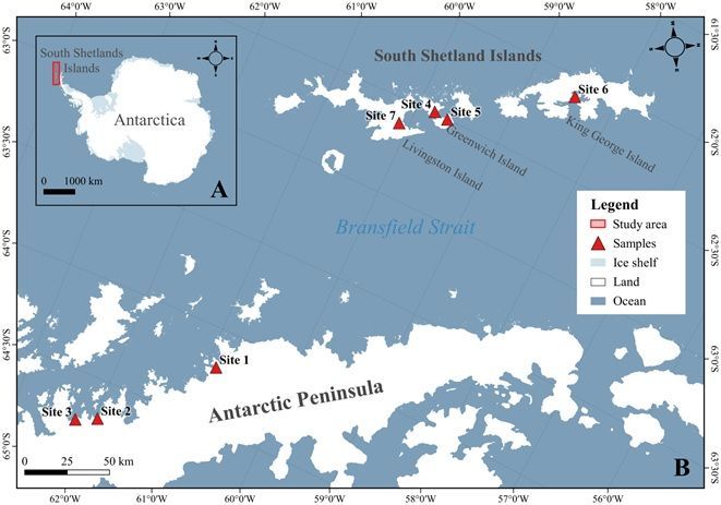
Abstract
We assessed fungal diversity present in glacial of the Antarctic Peninsula using DNA metabarcoding
through high throughput sequencing (HTS). We detected a total of 353,879 fungal DNA reads,
representing 94 genera and 184 taxa, in glacial ice fragments obtained from seven sites in the north-west
Antarctic Peninsula and South Shetland Islands. The phylum Ascomycota dominated the sequence
diversity, followed by Basidiomycota and Mortierellomycota. Penicillium sp., Cladosporium sp.,
Penicillium atrovenetum, Epicoccum nigrum, Pseudogymnoascus sp. 1, Pseudogymnoascus sp. 2,
Phaeosphaeriaceae sp. and Xylaria grammica were the most dominant taxa, respectively. However, the
majority of the fungal diversity comprised taxa of rare and intermediate relative abundance,
predominately known mesophilic fungi. High indices of diversity and richness were calculated, along with
moderate index of dominance, which varied among the different sampling sites. Only 26 (14%) of the
total fungal taxa detected were present at all sampling sites. The identified diversity was dominated by
saprophytic taxa, followed by known plant and animal pathogens and a low number of symbiotic fungi.
Our data suggest that Antarctic glacial ice may represent a hotspot of previously unreported fungal
diversity; however, further studies are required to integrate HTS and culture approaches to confirm
viability of the taxa detected.

Introduction
Despite its generally extreme conditions, Antarctica hosts diverse environments. Microorganisms
dominate many of the most extreme environments of the continent [1, 2, 3]. Antarctica’s continental ice
sheets contain the largest volume of glacial ice, inherently characterized by unfavorable conditions to life,
including low temperatures, low water activity, low nutrient availability and, in their surface layers,
exposure to high levels of solar radiation [4, 5].

Fungi are amongst the microorganisms reported from the Antarctic cryosphere [6]. However, despite their
recognised importance for ecosystem functioning in Antarctica and elsewhere, few studies have
attempted to recover and identify fungal species from glacial ice and, until now, few species, mainly
representing the phyla Ascomycota, Basidiomycota and Mortirellomycota, have been characterized from
this environment [7, 8, 9, 10, 11, 12].

Glacial ice is formed through the precipitation, accumulation, compaction and recrystallization of snow. It
can contain spores and mycelial fragments of fungi deposited from the air column, both on
contemporary and palaeo timescales [5, 12, 13, 14]. Sonjak et al. [15] suggested that viable fungal cells
obtained from Arctic and Antarctic glacial ice may range in age from 10,000 to 140,000 years, which
represent aeolian transport of propagules of both local and distant origin. In addition, Rosa et al. [16]
studied air samples in the Antarctic Peninsula region and showed the presence of fungi in the airspora,
supporting the possibility of dispersion in different geographic scales around Antarctica. To date, very
few studies have addressed fungal diversity present in Antarctic glacial ice, with those available being
based on cultivation techniques [5]. Furthermore, fungal diversity present in Antarctic glacial ice has not

                                                     Page 2/18
been assessed using cutting edge modern DNA metabarcoding techniques. In the current study, we
assessed the fungal diversity, richness, abundance and distribution in glacial ice sampled in the different
Antarctic sites using DNA metabarcoding high throughput sequencing (HTS).
Methods
Ice sampling
Three ‘bergy bits’ (glacial ice fragments) each of approximately 20 kg mass, were collected adjacent to
the ice fronts of seven marine terminating glaciers in the South Shetland Islands and the north-west
Antarctica Peninsula during the austral summer season in December 2015 and December 2016 (Fig. 1).
Each was collected using sterile suits and gloves to minimize contamination risk. In the microbiology
laboratory on board the Brazilian polar research vessel Admiral Maximiano, each sample was broken into
smaller pieces, and surface decontamination carried out using 5% sodium hypochlorite (10 s), sterilized
distilled water (10 s), and exposure to ultraviolet radiation (10 min) [12, 17]. The samples were melted and
a total of 12–15 L of the resulting water filtered through 47 mm diameter (Millipore) membranes (three
membranes per sampling site, each using 4–5 L) until each membrane became saturated. Membranes
were then stored at -20°C until DNA extraction in the laboratory of Polar Microbiology and Tropical
Connections of Universidade Federal of Minas Gerais, Brazil. Physico-chemical parameters (temperature,
conductivity, resistivity, total dissolved solids, oxidation-reduction potential, pH, salinity) of the melted
samples from each site were measured using a Hanna multi-parameter probe HI 9828 (Hanna
Instruments, USA). A map showing the sample collection locations was generated using QGIS software
(version 3.14.15; https://www.QGIS.org) and the SCAR Antarctic Digital Database (ADD version 7.0;
http://www.add.scar.org).
DNA extraction, data analyses and fungal identification
The three membranes resulting from filtering the melted ice from each sampling site were processed
together in order to increase DNA yield. Total DNA was extracted using 0.5 mL extraction buffer [sodium
dodecyl sulfate (SDS) 10%], left at 55°C for 18 h, followed by 165 µL NaCl (5 M) and 165 µL
cetyltrimethylammonium bromide (CTAB, 10%), then 600 µL chloroform was added and the mixture
centrifuged (Eppendorf/Germany) at 13,000 rpm for 10 min. The supernatant was cleaned using the
QIAGEN DNeasy PowerClean cleanup Kit. Extracted DNA was used as a template for generating PCR-
amplicons. The internal transcribed spacer 2 region (ITS2) of the nuclear ribosomal DNA was used as a
DNA barcode for molecular species identification [18, 19]. PCR-amplicons were generated using the
universal primers ITS3 and ITS4 [20] and were sequenced at Macrogen Inc. (South Korea) on an Illumina
MiSeq sequencer, using the MiSeq Reagent Kit v3 (600-cycle) following the manufacturer’s protocol.
Raw fastq files were filtered using BBDuk version 38.34 (BBMap – Bushnell B. –
sourceforge.net/projects/bbmap/) to remove Illumina adapters, known Illumina artefacts, and the PhiX
Control v3 Library. Quality read filtering was carried out using Sickle version 1.33 -q 30 -l 50 [21], to trim 3’
or 5’ ends with low Phred quality score, and sequences shorter than 50 bp were also discarded. The
Page 3/18remaining sequences were imported to QIIME2 version 2019.10 (https://qiime2.org/) for bioinformatics
analyses [22]. The qiime2-dada2 plugin is a complete pipeline that was used for filtering, dereplication,
turn paired-end fastq files into merged, and removal of chimeras [23]. Taxonomic assignments were
determined for amplicon sequence variants (ASVs = taxa) using the qiime2-feature-classifier [24] classify-
sklearn against the UNITE fungal ITS database version 8.2 [25] and trained with Naive Bayes classifier
and a confidence threshold of 98.5%. Fungal classification followed Kirk et al. [26], Tedersoo et al. [27],
MycoBank (http://www.mycobank.org) and the Index Fungorum (http://www.indexfungorum.org).
Diversity, distribution and ecological analysis
To quantify species diversity, richness, and dominance, we used the following indices: (i) Fisher’s α, (ii)
Margalef’s, and (iii) Simpson’s, respectively, to assess alpha diversity. In addition, the Sorensen and Bray-
Curtis similarity indices were used to assess beta diversity among the fungal assemblages. The relative
abundance of the ASVs was used to quantify the fungal taxa present in the glacial ice sampled as
described by Rosa et al. [28]. Fungal ASVs with relative abundance > 10% were considered dominant,
ASVs with relative abundance of 1–10% intermediate and ASVs with < 1% minor (rare) components of the
fungal community. All of the results were obtained with 95% confidence, and bootstrap values were
calculated from 1,000 iterations. Taxon accumulation curves were obtained using the Mao Tao index. All
diversity index calculations and t test were performed using PAST, version 1.90 [29]. To prepare Kronar
charts, QIIME2 taxonomy classifications and the table of taxa abundance were converted to tsv and biom
format, respectively. The table of fungal abundance was converted to tsv by using biom convert and
combined with taxonomy classification with a custom script krona_qiime.py
(https://github.com/lokeshbio/Amplicon_course/blob/master/krona_qiime.py). The Krona Tools (v.
2.7.1) [30] program, ktImportText.pl, was used to provide interactive visualization of identified fungi
species. Venn analysis to compare the fungal diversity obtained from the different sampling locations
was carried out using the program available at http://bioinformatics.psb.ugent.be/webtools/Venn/. The
functional assignments of fungal ASVs at species and generic levels were assessed using FunGuild [31].
Results
Fungal taxonomy
We detected a total of 353,879 fungal DNA reads, representing 94 genera and 184 distinct taxa in glacial
ice obtained from the seven sampling locations in the South Shetland Islands and north-west Antarctic
Peninsula (Suppl. Table 1). The phylum Ascomycota was dominant in all fungal assemblages, followed
by Basidiomycota and Mortierellomycota. A single Mucoromycota taxon (Rhizopus arrhizus) was
detected at low abundance (Fig. 2). Penicillium sp., Cladosporium sp., Penicillium atrovenetum,
Epicoccum nigrum, Pseudogymnoascus sp. 1, Pseudogymnoascus sp. 2, Phaeosphaeriaceae sp. and
Xylaria grammica (Ascomycota) were the most dominant taxa (all with > 10% of DNA reads), in rank
order. Further 30 taxa were detected at intermediate abundance (< 1% DNA reads). The majority of the
fungal ASVs detected (146 taxa; 79.3%), were classified as rare. Thirty-seven taxa could only be assigned
to higher taxonomic levels (phylum, class, order, family).
Page 4/18Diversity, distribution and ecology
The Mao Tao's rarefaction curves approached a plateau for all sampling locations, indicating that the
DNA reads obtained gave a good representation of the fungal sequence diversity present at each
(Supplementary Fig. 1). Alpha diversity indices across the sampled locations indicated generally high
diversity (Fisher α) and richness (Margalef) and moderate dominance (Simpson) indices (Table 1),
varying between the different sites. The sequence diversity detected in the Leonardo-Blanchard region
(Antarctic Peninsula) was most diverse and rich, and included a wider range of dominant taxa, followed
by those of Greenwich Island, when compared with the other sampling locations. The sequence diversity
detected in the Sikorsky region (Antarctic Peninsula) displayed the lowest diversity indices. The
Leonardo/Blanchard (Antarctic Peninsula) location had the lowest values of conductivity and salinity.
Table 1
Physicochemical parameters of melted glacial ice and diversity indices of fungal assemblages at the
different sampling locations in the north-west Antarctic Peninsula and South Shetland Islands.
Parameters/diversity Sampling locations
indices/density
KG- GI-FA GI-T AP-S AP-LB AP-RW LI-H
ASH
Temperature melted ice (°C) 15.6 16 9.6 15.6 14.5 16.1 10.1
Conductivity (µS cm− 1) 25 23.5 21 50 6 23 18
Resistivity (MΩ cm− 1) 0.04 0.06 0.04 0.08 0.16 0.06 0.10
Total dissolved solids (ppm) 12 11.5 11 25.5 3 11.5 9
Oxidation-reduction potential 520.1 540.7 750.4 509.5 188.8 540.7 494.7
(mV)
pH 6.51 6.7 6.7 7.4 6.5 6.65 6.9
Salinity (ppt) 0.01 0.01 0.02 0.02 0 0.01 0
Total number of reads 47,622 47,560 44,715 48,164 34,608 93,545 37,665
Number of taxa 82 88 61 54 95 87 64
Fisher’s α 9.64 10.45 6.96 6.01 11.91 9.46 7.51
Margalef 7.52 8.08 5.60 4.92 8.99 7.51 5.98
Simpson 0.84 0.84 0.73 0.80 0.85 0.78 0.78
KG-ASH = King George Island, Ajax-Stenhouse Glacier; GI-FA = Greenwich Island, Fuerza Aérea; GI-T =
Greenwich Island, Troub; AP-S = Antarctic Peninsula, Sikorsky; AP-LB = Antarctic Peninsula, Leonardo-
Blanchard; AP-RW = Arctowski Peninsula, Rozier-Woodbury; LI-H = Livingston Island, Huron.
Page 5/18The beta diversity of the fungal assemblages varied across the different sampling locations (Fig. 3). The
presence-absence-based Sorensen index showed that the most similar fungal assemblages were found
at Greenwich Island, Traub and Livingston Island, Huron, followed by Antarctic Peninsula, Leonardo-
Blanchard and King George Island, Ajax-Stain House. However, the abundance-related Bray-Curtis index
indicated that the fungal assemblages from Greenwich Island, Fuerza Aérea and Antarctic Peninsula,
Sikorsky showed the highest similarity.
The physicochemical properties of all the ice samples were generally similar, except for those from
Antarctic Peninsula, Leonard-Blanchard and Antarctic Peninsula, Sikorsky, which displayed the extreme
values of conductivity and total dissolved solids (Table 1; Supplementary Fig. 2). The fungal assemblage
detected in the ice sampled in the Antarctic Peninsula, Leonard-Blanchard site, which had the lowest
physicochemical parameters, included the lowest number of DNA reads and the highest number of ASVs,
and had the highest diversity (Fisher α) and richness (Margalef) indices. In contrast, the fungal
assemblage detected in the ice of Antarctic Peninsula, Sikorsky displayed the lowest values of the same
diversity parameters. PCA analysis indicated that the conductivity, total dissolved solids, oxidation-
reduction potential, pH and salinity showed negative correlation with the number of taxa, Fisher α,
Margalef and Simpson indices. Twenty-six of the 186 fungal taxa detected were present at all sampling
locations (Suppl. Table 2), while 82 taxa were detected at only a single location. Ecological functional
assignments of the taxa detected at generic level are given in in Suppl. Table 3. Taxa of 94 genera were
detected, with the most common group being saprophytic fungi, followed by plant and animal pathogens
and a small number of symbiotic fungi.
Discussion
Fungal taxonomy
Glacial ice is considered an extreme and ultra-oligotrophic environment and one of the most challenging
natural environments for life globally [2]. Representatives of Bacteria, Archaea and Fungi have been
detected in glacial ice from different cold regions of the planet [15, 32]. However, among the
microorganisms present in the glacial ice, fungi remain poorly known and few taxa have been reported to
date [6, 14] in studies based on traditional culturing methods.
Many factors, such as extraction, PCR and primer bias, can influence the outcomes of metabarcoding
studies and the numbers of reads obtained [33], thus leading to misinterpretation of absolute abundance
[34]. However, Giner et al. [35] concluded that such biases did not affect the proportionality between reads
and cell abundance, implying that more reads are linked with higher abundance [36, 37]. Therefore, for
comparative purposes, we consider here the number of reads as a proxy for relative abundance. The
current study, using a state-of-the-art HTS metabarcoding approach, focused on the detection of fungal
DNA in environmental samples.
Our data revealed the presence of rich and diverse fungal sequence diversity in glacial ice collected from
the seven different sampling locations. The total sequence diversity was dominated by a relatively small
Page 6/18number of taxa of the genera Penicillium, Cladosporium, Epicoccum, Pseudogymnoascus and Xylaria, all
members of the Ascomycota. However, the majority of the diversity identified comprised intermediate and
rare members of the phyla Basidiomycota, Mortierellomycota and Mucoromycota.
The genera Cladosporium and Penicillium include well-known cosmopolitan species often detected in the
airspora. In Antarctica, different species of Cladosporium have been detected in association with plants
and soil [3]. Species of Penicillium are widespread across Antarctica and have been reported in studies of
multiple terrestrial substrates including soils [38, 39, 40], permafrost [41, 42], associated with marine
macroalgae [43], invertebrates [44], sediments [45, 46] and seawater [47]. 2017). Penicillium atrovenetum
was detected as the dominant fungal sequence present in the gypsum encrustations and carbonate veins
of rocks in a polar desert region of continental Antarctica [48]. Members of Pseudogymnoascus occur
widely in cold polar, alpine, and temperate environments [49, 50, 51, 52]. In Antarctica, they have been
reported from soils [49, 53, 54], associated with plants [55, 56, 57] and marine macroalgae [58], in
freshwater lakes [45], and associated with lichens [59]. Cladosporium, Penicillium and
Pseudogymnoascus sequences were detected as dominant fungal sequences present in air and snow
samples from the South Shetland Islands [16, 28, 60]. The dominance of sequences in these genera in the
glacial ice examined here is consistent with these fungi being abundant in the air, being deposited
(possibly facilitated by snow precipitation) on the glacier surface and progressively incorporated in the
glacial ice as it becomes compacted overtime. It is important, however, to note that metabarcoding
methodologies detect the presence of DNA sequences, with identification still limited by the available
sequence databases, and do not provide any confirmation of viability. Therefore, further specific studies
are necessary to determine whether the fungal taxa detected are present in a viable form.
Members of the genus Epicoccum are commonly present in air, soil, decaying vegetation and as
endophytes in living plant tissues, with some also being documented producers of bioactive compounds
[61]. In Antarctica, species of Epicoccum have been documented in aerobiological studies on Signy
Island, South Orkney Islands [62] and associated with Antarctic marine sponges [63]. The DNA of
Epicoccum nigrum was also recently detected in rock surface gypsum encrustations and carbonate veins
in the Ellsworth Mountains [48].
The genus Xylaria contains between 570–670 recognized species [64], but may include many more, yet to
be described [65]. Species of Xylaria are important saprophytic fungi, found on decomposing wood in
temperate and tropical ecosystems [66] and also as plant endophytes [67]. Additionally, members of the
genus are amongst the most prolific secondary metabolite producers [64]. In Antarctica, Xylaria has been
reported from soil exposed by glacial retreat on King George Island [68].
Diversity, distribution and ecology
Aside from the eight Ascomycota taxa classified as dominant in the current study, the majority of taxa
were of rare or intermediate abundance, and were mostly known as mesophilic fungi. de Menezes et al.
[12] reported culturable fungal diversity from the same glacial ice samples as examined here. They
documented the presence of 27 taxa belonging to 14 genera. The number of taxa detected and diversity
Page 7/18ecological indices calculated using the metabarcoding approach in the current study were approximately
seven times greater than those reported by de Menezes et al. [12] (Table 2). The use of metabarcoding
revealed sequence diversity potentially representing a much richer and more diverse fungal community
than previously appreciated, including 184 taxa belonging to the 98 genera, amongst which were fungi
not previously reported from Antarctica. The fact that 37 taxa could only be assigned at higher taxonomic
levels (phylum, class, order, family), provides reinforce the evidence that Antarctic environments are likely
to host new and/or previously unreported fungal diversity.
Table 2
Comparison of diversity indices obtained in studies of fungal diversity detected by traditional culturing
methods and DNA metabarcoding from glacial ice samples obtained from the north-west Antarctic
Peninsula and South Shetland Islands
Sampling location Number of Fisher α Margalef Simpson
taxa/ASVs
TCMa HTS b TCM HTS TCM HTS TCM HTS
King George Island, 4 82 1.12 9.64 0.82 7.52 0.61 0.84
Ajax-Stenhouse
Arctowski 1 87 - 9.46 - 7.51 - 0.78
Peninsula, Rozier-
Woodbury
Livingston Island, 8 64 12.98 7.51 3.36 5.98 0.82 0.78
Huron
Greenwich Island, 1 61 - 6.96 - 5.60 - 0.73
Traub
Greenwich Island, 5 88 2.52 10.45 1.51 8.08 0.73 0.84
Fuerza Aerea
Antarctic Peninsula, 6 54 1.78 6.01 1.28 4.92 0.33 0.80
Sikorsky
Antarctic Peninsula, 3 95 0.95 11.91 0.65 8.99 0.55 0.85
Leonardo-Blanchard
a
Traditional culturing methods [12]; bDNA metabarcoding (current study).
Sorensen and Bray-Curtis similarity indices indicated that the beta diversity of the fungal assemblages
varied across the different sampling sites, which may be related with ice physicochemical properties. The
fungal assemblage detected in the ice sampled in the Antarctic Peninsula, Sikorsky location formed an
isolated group based on the fungal taxa present, as well as having the highest values of conductivity and
total dissolved solids and the lowest number of ASVs, Fisher’s α (diversity) and Margalef index values
(richness). However, when the Bray-Curtis index was compared with the physicochemical parameters and
alpha diversity indices no other correlations were detected. Our alpha and beta diversity data differed
Page 8/18from those reported by de Menezes et al. [12] in their study of culturable fungal diversity from the same
glacial ice samples as examined here. The PCA analysis reported by de Menezes et al. [12] showed a
positive correlation between pH and the diversity indices only at two sampling sites, differing from the
current analyses, which identified a positive correlation between the lowest values of the physicochemical
parameters and the highest diversity values.
Conclusions
Our data suggest that Antarctic glacial ice may host a hotspot of as yet unreported fungal diversity. Use
of a DNA metabarcoding approach revealed the presence of high fungal sequence diversity including a
small number of dominant fungi with capabilities for aerial dispersal and a high number of taxa of rare or
intermediate abundance. The sequence diversity detected was dominated by saprophytic taxa, followed
by known plant and animal pathogens and a small number of symbiotic fungi. The potentially high
fungal diversity detected here in glacial ice samples emphasizes the need for further studies
characterizing fungal communities of this extreme ecosystem using a combination of culturing and
metabarcoding approaches.
Declarations
Acknowledgements
We acknowledge financial support from PROANTAR CNPq (442258/2018-6), INCT Criosfera, FAPEMIG,
CAPES and FNDCT. GCA de Menezes’ scholarship was supported by CNPq (151195/2019-6). We are also
grateful for the generous support of MSc. Rodrigo Paidano Alves in preparation of Figure 1. P. Convey is
supported by NERC core funding to the British Antarctic Survey’s ‘Biodiversity, Evolution and Adaptation’
Team. We also thank congresswoman Jô Moraes and the Biological Sciences Institute of the University
of Brasilia
Data Availability Statement
All raw sequences have been deposited in the NCBI database under the codes SRX9966699,
SRX9966700, SRX9966701, SRX9966702, SRX9966703, SRX9966704, SRX9966705 and SRX9966706.
Author contributions
GCAM, LHR, JCS and PEASC conceived the study. GCAM and LHR performed fungal DNA extraction from
ice. GCAM, LHR, PEASC, OHBZ, PC, MCS, JCS and CAR analyzed the results and wrote the manuscript. All
authors read and approved of the final manuscript.
Compliance with Ethical Standards
Conflict of Interest The authors declare that they have no conflict of interest.
Page 9/18Ethics Approval The collections and studies performed in Antarctic Peninsula were authorized by the
Secretariat of the Antarctic Treaty and by PROANTAR.
References
1. Convey P (2017) Antarctic ecosystems. In: Reference module in life sciences. Elsevier, pp 179-188.
doi: 10.1016/B978-0-12-809633-8.02182-8.
2. Perini L, Gostinčar C, Gunde-Cimerman N (2019) Fungal and bacterial diversity of Svalbard
subglacial ice. Sci Rep 9:20230
3. Rosa LH, Zani CL, Cantrell C L, Duke SO, Van Dijck P, Desideri A, Rosa CA (2019) Fungi in Antarctica:
Diversity, Ecology, Effects of Climate Change, and Bioprospection for Bioactive Compounds. In: Rosa
LH (ed) Fungi of Antarctica: Diversity, Ecology and Biotechnological Applications, 1st edn. Springer,
Switzerland, pp 1-17. https://doi.org/10.1007/978-3-030-18367-7_1
4. Anesio AM, Laybourn-Parry J (2012) Glaciers and ice sheets as a biome. Trends Ecol Evol 27:219-
225
5. de Menezes GCA, Porto BA, Simões JC, Rosa CA, Rosa LH (2019) Fungi in Snow and Glacial Ice of
Antarctica. In: Rosa L. (ed) Fungi of Antarctica, 1st edn. Springer, Cham, pp 127-146
6. Margesin R, Collins T (2019) Microbial ecology of the cryosphere (glacial and permafrost habitats):
current knowledge. Appl Microbiol Biotech 103:2537-2549
7. Jacobs PH, Taylor HC, Shafer JC. (1964) Studies of fungi at Amundsen-Scott IGY South Pole Base
(1957). Arch Dermatol 89:117-123
8. Abyzov SS, Hoover RB, Imura S, Mitskevich IN et al. (2004) Use of different methods for discovery of
ice-entrapped microorganisms in ancient layers of the Antarctic glacier. Adv Space Res 33:1222-1230
9. D’Elia T, Veerapaneni R, Theraisnathan V, Rogers SO (2009). Isolation of fungi from Lake Vostok
accretion ice. Mycologia 101:751-763
10. Knowlton C, Veerapaneni R, D'Elia T, Rogers SO (2013) Microbial analyses of ancient ice core
sections from Greenland and Antarctica. Biology 2:206-232
11. Sanyal A, Antony R, Samui G, Thamban M (2018) Microbial communities and their potential for
degradation of dissolved organic carbon in cryoconite hole environments of Himalaya and
Antarctica. Microbiol Res 208:32-42
12. de Menezes GCA, Porto BA, Amorim SS (2020) Fungi in glacial ice of Antarctica: diversity, distribution
and bioprospecting of bioactive compounds. Extremophiles 24:367-376
13. Abyzov S S (1993) Microorganisms in the Antarctic ice. In: Friedman EI (ed) Antarctic microbiology.
John Wiley & Sons, New York, pp 265-295
14. Gunde-Cimerman N, Sonjak S, Zalar P (2003) Extremophilic fungi in arctic ice: a relationship between
adaptation to low temperature and water activity. Phys Chem Earth, Parts A/B/C 28:1273-1278
15. Sonjak S, Frisvad JC, Gunde-Cimerman N (2006) Penicillium mycobiota in Arctic subglacial ice.
Microb Ecol 52:207-216
Page 10/1816. Rosa LH, Pinto OHB, Šantl-Temkiv T (2020) DNA metabarcoding of fungal diversity in air and snow
of Livingston Island, South Shetland Islands, Antarctica. Sci Rep 10:21793
17. Rogers SO, Theraisnathan V, Ma LJ (2004) Comparisons of protocols for decontamination of
environmental ice samples for biological and molecular examinations. Appl Environ Microbiol
70:2540-2544
18. Chen S, Yao H, Han J et al (2010) Validation of the ITS2 region as a novel DNA barcode for
identifying medicinal plant species. PloS One 5:e8613
https://doi.org/10.1371/journal.pone.0008613
19. Richardson RT, Lin CH, Sponsler DB (2015) Application of ITS2 metabarcoding to determine the
provenance of pollen collected by honey bees in an agroecosystem. Appl Plant Sci 3:1400066
20. White TJ, Bruns T, Lee S, Taylor J (1990) Amplification and direct sequencing of fungal ribosomal
RNA genes for phylogenetics. In: Innis MA, Gelfand DH, Sninsky JJ, White TJ (eds) PCR Protocols: a
guide to methods and applications, 1st edn. Academic Press, New York, pp 315–322
21. Joshi NA, Fass JN (2011) Sickle: a sliding-window, adaptive, quality-based trimming tool for FastQ
files (version 1.33) [software]. https://github.com/najoshi/sickle. Accessed 10 Aug 2020
22. Bolyen E, Rideout JR, Dillon MR et al (2019) Reproducible, interactive, scalable and extensible
microbiome data science using QIIME 2. Nat Biotechnol 37:852–857.
https://doi.org/10.1038/s41587-019-0209-9
23. Callahan BJ, McMurdie PJ, Rosen MJ, Han AW, Johnson AJA. Holmes SP (2016) DADA2: high-
resolution sample inference from Illumina amplicon data. Nat Methods 13:581–583.
https://doi.org/10.1038/nmeth.3869
24. Bokulich NA, Kaehler BD, Rideout JR et al. (2018) Optimizing taxonomic classification of marker‐
gene amplicon sequences with QIIME 2’s q2‐feature‐classifier plugin. Microbiome 6:90.
https://doi.org/10.1186/s40168-018-0470-z
25. Abarenkov, K. Zirk A, Piirmann T, Pöhönen R, Ivanov F, Nilsson RH, Kõljalg U. (2020) UNITE QIIME
release for eukaryotes. Version 04.02.2020. UNITE Community.
https://doi.org/10.15156/BIO/786386
26. Kirk PM, Cannon PF, Minter DW et al (2011) Dictionary of the Fungi. CAB International, Wallingford
27. Tedersoo L, Sánchez-Ramírez S, Kõljalg U et al (2018) High-level classification of the Fungi and a
tool for evolutionary ecological analyses. Fungal Divers 90:135–159.
https://doi.org/10.1007/s13225-018-0401-0
28. Rosa LH, Pinto OHB, Convey P et al (2020a) DNA metabarcoding to assess the diversity of airborne
fungi present in air over Keller Peninsula, King George Island, Antarctica. Microb Ecol.
https://doi.org/10.1007/s00248-020-01627-1
29. Hammer, Ø, Harper DAT, Ryan PD (2001) PAST: paleontological statistics software package for
education and data analysis. Palaeontol Electron 4(1):1–9
30. Ondov BD, Bergman NH, Phillippy AM (2011) Interactive metagenomic visualization in a Web
browser. BMC Bioinformatics 12:385. https://doi.org/10.1186/1471-2105-12-385
Page 11/1831. Nguyen NH, Song Z, Bates S T et al. (2016) FUNGuild: an open annotation tool for parsing fungal
community datasets by ecological guild. Fungal Ecol 20:241-248.
https://doi.org/10.1016/j.funeco.2015.06.006
32. Duo Saito RA, Connell L, Rodriguez R, Redman R, Libkind D, de Garcia V (2018) Metabarcoding
analysis of the fungal biodiversity associated with Castaño Overa Glacier – Mount Tronador,
Patagonia, Argentina. Fungal Ecol. 36:8-16
33. Medinger R, Nolte V, Pandey RV et al (2010) Diversity in a hidden world: potential and limitation of
next-generation sequencing for surveys of molecular diversity of eukaryotic microorganisms. Mol
Ecol 19:32-40. https://doi.org/10.1111/j.1365-294X.2009.04478.x
34. Weber AA, Pawlowski J (2013) Can abundance of protists be inferred from sequence data: a case
study of Foraminifera. Plos One 8:e56739. https://doi.org/10.1371/journal.pone.0056739
35. Giner CR, Forn I, Romac S, Logares RC, Massana R (2016) Environmental sequencing provides
reasonable estimates of the relative abundance of specific picoeukaryotes. Appl Environ Microbiol
82:4757–4766. https://doi.org/10.1128/AEM.00560-16
36. Deiner K, Bik HM, Mächler E et al (2017) Environmental DNA metabarcoding: Transforming how we
survey animal and plant communities. Mol Ecol 21:5872–5895. https://doi.org/10.1111/mec.14350
37. Hering D, Borja A, Jones JI et al (2018) Implementation options for DNA-based identification into
ecological status assessment under the European Water Framework Directive. Water Res 138:192–
205. https://doi.org/10.1016/j.watres.2018.03.003
38. McRae CF, Hocking AD, Seppelt RD. Penicillium species from terrestrial habitats in the Windmill
Islands, East Antarctica, including a new species, Penicillium antarcticum. Polar Biology 1999;21: 97-
111
39. Godinho VM, Gonçalves VN, Santiago IF et al. (2015) Diversity and bioprospection of fungal
community present in oligotrophic soil of continental Antarctica. Extremophiles 19:585–596.
https://doi.org/10.1007/s00792-015-0741-6
40. Gomes EC, Godinho VM, Silva DA (2018) Cultivable fungi present in Antarctic soils: taxonomy,
phylogeny, diversity, and bioprospecting of antiparasitic and herbicidal metabolites. Extremophiles
22:381-393
41. Zucconi L, Selbmann L, Buzzini P (2012) Searching for eukaryotic life preserved in Antarctic
permafrost. Polar Biol 35:749-757
42. da Silva TH, Silva DAS, de Oliveira FS (2020) Diversity, distribution, and ecology of viable fungi in
permafrost and active layer of Maritime Antarctica. Extremophiles 24:565-576
43. Godinho VM, Furbino LE, Santiago IF et al (2013) Diversity and bioprospecting of fungal
communities associated with endemic and cold-adapted macroalgae in Antarctica. ISME J 7:1434-
1451
44. Godinho VM, de Paula MTR, Silva DAS et al (2019) Diversity and distribution of hidden cultivable
fungi associated with marine animals of Antarctica. Fungal Biol 123:507-516
Page 12/1845. Gonçalves VN, Vaz AB, Rosa CA, Rosa LH (2012) Diversity and distribution of fungal communities in
lakes of Antarctica. FEMS Microbiol Ecol 82:459-471
46. Ogaki MB, Teixeira DR, Vieira R (2020) Diversity and bioprospecting of cultivable fungal
assemblages in sediments of lakes in the Antarctic Peninsula. Fungal Biol 124:601-611
47. Gonçalves VN, Vitoreli GA, de Menezes GC (2017) Taxonomy, phylogeny and ecology of cultivable
fungi present in seawater gradients across the Northern Antarctica Peninsula. Extremophiles
21:1005-1015
48. de Menezes GCA, Câmara PEAS, Pinto OHB (2021) Fungal diversity present on rocks from a polar
desert in continental Antarctica assessed using DNA metabarcoding. Extremophiles 25:193-202
49. Mercantini R, Marsella R, Cervellati MC (1989) Keratinophilic fungi isolated from Antarctic soil.
Mycopathologia 106:47-52
50. Lorch JM, Lindner DL, Gargas A et al (2013) A culture-based survey of fungi in soil from bat
hibernacula in the eastern United States and its implications for detection of Geomyces destructans,
the causal agent of bat white-nose syndrome. Mycologia 105:237-252
51. Minnis AM, Lindner DL (2013) Phylogenetic evaluation of Geomyces and allies reveals no close
relatives of Pseudogymnoascus destructans, comb. nov., in bat hibernacula of eastern North
America. Fungal Biol 117:638-649
52. Ali SH, Alias SA, Siang HY et al (2014) Studies on diversity of soil microfungi in the Hornsund area,
Spitsbergen. Pol. Res. 35:203-224
53. Arenz BE, Blanchette RA (2011) Distribution and abundance of soil fungi in Antarctica at sites on the
Peninsula, Ross Sea Region and McMurdo Dry Valleys. Soil Biol Biochem 43:308–315.
https://doi.org/10.1016/j.soilbio.2010.10.016
54. Krishnan A, Alias SA, Wong CMVL et al (2011) Extracellular hydrolase enzyme production by soil
fungi from King George Island, Antarctica. Polar Biol 34:1535-1542
55. Tosi S, Casado B, Gerdol R, Caretta G (2002) Fungi isolated from Antarctic mosses. Polar Biol
25:262-268
56. Rosa LH, Almeida Vieira MDL, Santiago IF, Rosa CA (2010) Endophytic fungi community associated
with the dicotyledonous plant Colobanthus quitensis (Kunth) Bartl. (Caryophyllaceae) in Antarctica.
FEMS Microbiol Ecol 73:178-189
57. Carvalho, CR, Ferreira MC, Gonçalves VN et al (2020) Fungi associated with the briosphere of the
bipolar mosses Polytrichastrum alpinum and Polytrichum juniperinum in Antarctica. Polar Biol
43:545-553. https://doi.org/10.1007/s00300-020-02658-7
58. Loque CP, Medeiros AO, Pellizzari FM et al (2010) Fungal community associated with marine
macroalgae from Antarctica. Polar Biol 33:641-648
59. Santiago IF, Soares MA, Rosa CA, Rosa LH (2015) Lichensphere: a protected natural microhabitat of
the non-lichenised fungal communities living in extreme environments of Antarctica. Extremophiles
19:1087-1097
Page 13/1860. Rosa LH, da Silva TH, Ogaki MB et al (2020) DNA metabarcoding high-throughput sequencing
uncovers cryptic fungal diversity in soils of protected and non-protected areas on Deception Island,
Antarctica. Sci Rep 10:21986. https://doi.org/10.1038/s41598-020-78934-7
61. Braga RM, Padilla G, Araújo WL (2018) The biotechnological potential of Epicoccum spp.: diversity
of secondary metabolites. Crit Rev Microbiol 44:759-778
62. Marshall WA (1997) Seasonality in Antarctic airborne fungal spores. Appl Environ Microbiol 63:2240-
2245
63. Henríquez M, Vergara K, Norambuena J (2014) Diversity of cultivable fungi associated with Antarctic
marine sponges and screening for their antimicrobial, antitumoral and antioxidant potential. World J
Microbiol Biotechnol 30:65-76
64. Becker K, Stadler M (2020) Recent progress in biodiversity research on the Xylariales and their
secondary metabolism. J Antibiot 1-23
65. Daranagama DA, Hyde KD, Sir EB et al (2018) Towards a natural classification and backbone tree for
Graphostromataceae, Hypoxylaceae, Lopadostomataceae and Xylariaceae. Fungal Divers 88:1-165
66. Rogers JD (2000) Thoughts and musings on tropical Xylariaceae. Mycol Res 104:1412-1420
67. Davis EC, Franklin JB, Shaw AJ et al. (2003) Endophytic Xylaria (Xylariaceae) among liverworts and
angiosperms: phylogenetics, distribution, and symbiosis. Am J of Bot 90:1661-1667.
68. Santos JAD, Meyer E, Sette LD (2020) Fungal Community in Antarctic Soil Along the Retreating
Collins Glacier (Fildes Peninsula, King George Island). Microorganisms 8:1145
69. Liu XZ, Wang QM, Göker M et al (2015) Towards an integrated phylogenetic classification of the
Tremellomycetes. Stud Mycol 81:85-147
70. Fleet G, Prakitchaiwattana C, Beh A, Heard G. (2002) The yeast ecology of wine grapes. In: Ciani M
(ed) Biodiversity and Biotechnology of Wine Yeasts. Kerala, Research Signpost, pp 1-17
71. Saubin M, Devillers H, Proust L (2020) Investigation of genetic relationships between Hanseniaspora
species found in grape musts revealed interspecific hybrids with dynamic genome structures. Front
Microbiol 10:2960
72. Kurtzman CP (2011) Hyphopichia Von Arx & Van der Walt (1976). In: Kurtzman C, Fell JW, Boekhout
T (eds) The Yeasts. Amsterdam, Elsevier, pp 435-438
73. Smith MT, Kurtzman CP (2011) Lipomyces Lodder & Kreger-van Rij (1952). In: Kurtzman C, Fell JW,
Boekhout T (eds) The Yeasts. Amsterdam: Elsevier, pp 545-560
74. Corte L, di Cagno R, Groenewald M. et al (2015) Phenotypic and molecular diversity of Meyerozyma
guilliermondii strains isolated from food and other environmental niches, hints for an incipient
speciation. Food Microbiol 48:206-215
75. de Hoog GS, Smith MT, Rosa CA (2011) Moniliella Stolk & Dakin (1966). In: Kurtzman C, Fell JW,
Boekhout T (eds) The Yeasts. Amsterdam, Elsevier, pp1837-1846
76. Schmidt SK, Vimercati L, Darcy JL (2017) A Naganishia in high places: functioning populations or
dormant cells from the atmosphere? Mycology 8:153-163
Page 14/1877. Zhou Y, Jia BS, Zhou YG (2020)Naganishia floricola sp. nov., a novel basidiomycetous yeast species
isolated from flowers of Sorbaria sorbifolia. Int J Systc Evol Microbio 70:4496-4501
78. Golzar H, Thomas G, Jayasena KW (2019) Neoascochyta species cause leaf scorch on wheat in
Australia. Australas Plant Dis Notes 914:1
79. Ferreira EMS, de Sousa FMP, Rosa LH, Pimenta RS (2019) Taxonomy and richness of yeasts
associated with angiosperms, bryophytes, and meltwater biofilms collected in the Antarctic
Peninsula. Extremophiles 23:151-159
80. Sanyal A, Antony R, Ganesan P, Thamban M (2020) Metabolic activity and bioweathering properties
of yeasts isolated from different supraglacial environments of Antarctica and Himalaya. Anton
Leeuw J 113:2243-2258
81. Perelló A, Aulicino M, Stenglein SA et al (2017) Pseudopithomyces chartarum associated with wheat
seeds in Argentina, pathogenicity and evaluation of toxigenic ability. Eur J Plant Pathol 148:491-496
82. Jaiboon K, Lertwattanasakul N, Limtong P, Limtong S (2016) Yeasts from peat in a tropical peat
swamp forest in Thailand and their ability to produce ethanol, indole-3-acetic acid and extracellular
enzymes. Mycol Prog 15:755-770
83. Cadete RM, Rosa CA (2018) The yeasts of the genus Spathaspora: potential candidates for second‐
generation biofuel production. Yeast 35:191-199
84. Rosa CA, Lachance MA (1998) The yeast genus Starmerella gen. nov. and Starmerella bombicola sp.
nov., the teleomorph of Candida bombicola (Spencer, Gorin & Tullock) Meyer & Yarrow. Int J Syst Evol
Microbiol 48:1413-1417
85. Čadež N, Drumonde-Neves J, Sipiczki M et al (2020) Starmerella vitis fa, sp. nov., a yeast species
isolated from flowers and grapes. Anton Leeuw 113:1289-1298
86. Sena LM, Morais CG, Lopes MR (2017) d-Xylose fermentation, xylitol production and xylanase
activities by seven new species of Sugiyamaella. Anton Leeuw J 110:53-67
87. Kijpornyongpan T, Urbina H, Suh S O (2019) The Suhomyces clade: from single isolate to multiple
species to disintegrating sex loci. FEMS Yeast Res 19: foy125
88. Haelewaters D, Toome-Heller M, Albu S, Aime MC (2020) Red yeasts from leaf surfaces and other
habitats: three new species and a new combination of Symmetrospora (Pucciniomycotina,
Cystobasidiomycetes). Fungal Syst Evol 5:187-196
89. Duarte APM, Attili-Angelis D, Baron NC (2017) Riding with the ants. Persoonia: Molec Phyl Evol Fungi
38:81-99
90. Kurtzman CP (2011) Yamadazyma Billon-Grand (1989). In: Kurtzman C, Fell JW, Boekhout T (eds)
The Yeasts. Amsterdam, Elsevier, pp 919-925
91. Sá-Correia I, Guerreiro JF, Loureiro-Dias MC et al (2014) Zygosaccharomyces. In Batt CA, Tortorello
ML (eds) Encyclopedia of Food Microbiology. Amsterdam, Elsevier, pp 849–855
Figures
Page 15/18Figure 1
Location of ice sample collections. (A) Antarctica showing the north-west Antarctic Peninsula and South
Shetland Islands in the red rectangle and (B) the sites where the glacial ice samples were collected. Site 1
- Antarctic Peninsula, Sikorsky (64°12′S; 60°53′W); Site 2- Antarctic Peninsula, Leonardo-Blanchard
(64°42’S; 61°58’W); Site 3 - Arctowski Peninsula, Rozier-Woodbury (64°45′S; 62°13′W); Site 4 - Greenwich
Island, Traub (62°29′31′′S; 59°48′00′′W); Site 5- Greenwich Island, Fuerza Aérea (62°30′S; 59°38′W); Site 6 -
King George Island, Ajax-Stenhouse (62°06’S; 58°27′W); Site 7 - Livingston Island, Huron (62°37′50′′S;
60°06′50′′W).
Page 16/18Figure 2
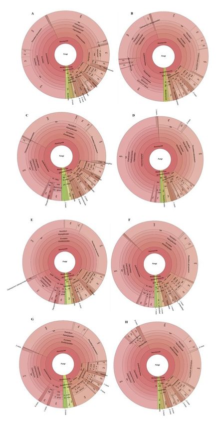
Krona charts of (A) total fungal diversity across all sampling sites, (B) King George Island, Ajax-
Stenhouse, (C) Greenwich Island, Fuerza Aérea, (D) Antarctic Peninsula, Sikorsky, (E) Antarctic Peninsula,
Leonardo-Blanchard, (F) Arctowski Peninsula, Rozier-Woodbury, (G) Livingston Island, Huron and (H)
Greenwich Island, Traub.
Page 17/18Supplementary Files
This is a list of supplementary files associated with this preprint. Click to download.
Suppl.Figure1.docx
Suppl.Figure2.docx
Suppl.Table1.docx
Suppl.Table2.docx
Suppl.Table3.docx
Page 18/18You can also read