Has the following disclosures to make: December 09, 2019 - Heartland National TB Center
←
→
Page content transcription
If your browser does not render page correctly, please read the page content below
8/17/2020

               TB and Chest Radiology
                    Megan Devine, MD
                    December 09, 2019

                Navigating Toward TB Free Pacific Islands
                         December 9‐10, 2019
                        Majuro, Marshall Islands

1

    Megan Devine, MD
    has the following disclosures to make:
    • No conflict of interests

    • No relevant financial relationships with any
      commercial companies pertaining to this
      educational activity

2

                                                                   1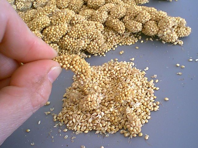
8/17/2020

                                 TB and Chest Radiology

                                         Megan Devine, MD
                                     Heartland National TB Center

                                    Assistant Professor of Medicine
                                    UT Health Science Center Tyler

                                                                      EXCELLENCE   EXPERTISE   INNOVATION

3

    Megan Devine has the following disclosures to
    make:

    • No conflict of interests
    • No relevant financial relationships with any commercial companies

4

                                                                                                                   2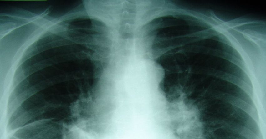
8/17/2020

                        Outline

    • Normal CXR
    • Images of Primary TB
    • Images of Post‐primary (Reactivation) TB
    • Images of pulmonary complications
    • Local images
    • Missed diagnosis

5

6

                                                        3
8/17/2020

7

    Normal CXR Child

8

                              4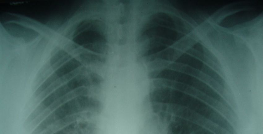
8/17/2020

                                                               Cellestis

9

                     Primary Tuberculosis

     • Most commonly in children and immune compromised
       patients
     • Opacities are seen the in middle and lower lungs
     • Commonly unilateral, bilateral 15%
     • Lymph node enlargement often occurs, and may cause
       bronchial compression
     • Hilar or paratracheal lymphadenopathy with or without
       infiltrates is characteristic.

10

                                                                                  5
8/17/2020

11

12

            6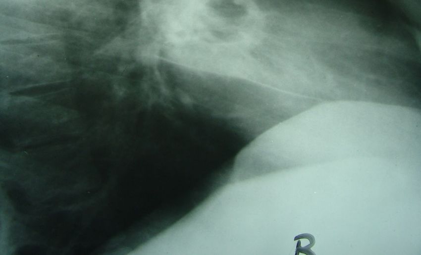
8/17/2020

     Courtesy: Dr. Santiago Restrepo

13

14

                                              7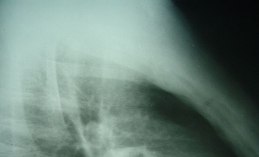
8/17/2020
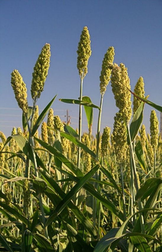
Millet Seeds
Slender plant, 1‐15 feet
Seeds ~ 2 mm in diameter
1/3 of grain for 3rd world
Africa and India
Producer: India
15
Miliary Tuberculosis
16
8
8/17/2020

17

       Post Primary, Reactivation Tuberculosis

     • Characterized by upper lobe predilection, cavitation
       and absence of lymphadenopathy.
     • Cavitation is the hallmark; can also see parenchymal
       disease (consolidation), hematogenous dissemination
       (miliary), bronchogenic spread (tree‐in‐bud) and
       pleural disease.
     • Fibrosis and calcification are seen after healing.

18

                                                                     9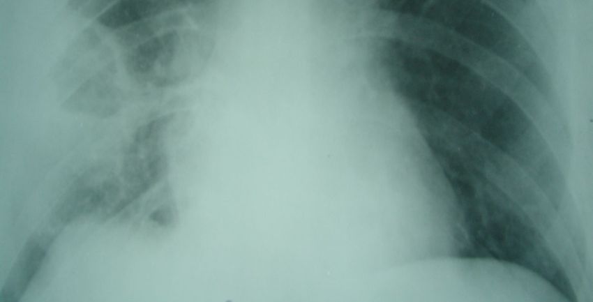
8/17/2020

19

20

           10
8/17/2020
21
22
118/17/2020
23
Reactivation TB Cavitary Consolidation
Courtesy: Dr. Santiago Restrepo
24
128/17/2020
Tree in Bud……
http://www.jellosalad.com/pic/favorites/index.html
25
26
138/17/2020
27
Pleural Effusions
• Primary TB (25%)
• Hypersensitivity reaction to TB proteins
• Organisms uncommonly isolated from fluid
• May not be associated with obvious parenchymal
disease on CXR
28
28
148/17/2020
29
30
158/17/2020
Pleural Effusions
• Post primary TB (20%)
• Caused by rupture of a tuberculous cavity into the
pleural space, causing empyema
• May cause bronchopleural fistula with air fluid levels
• Often results in irreversible pleural thickening and
calcification
31
31
32
168/17/2020
xxxxxx
33
Tracheobronchial TB
• TB is the most common cause of inflammatory
stricture of a bronchus
• 10% ‐ 20% of TB patients
• Circumferential wall thickening
• Luminal narrowing
• Long segment involvement
• Left > Right
Moon WK, et al. AJR 1997;169:649
34
178/17/2020
Courtesy: Dr. Santiago Restrepo
35
Complications of Pulmonary Tuberculosis
• Bronchiectasis
• Broncholithiasis
• Extensive pulmonary destruction
• Non‐tuberculous mycobacterial disease
• Chronic pulmonary aspergillosis
• Venous thromboembolism
36
188/17/2020
37
38
198/17/2020
39
Local Cases
40
208/17/2020
50yo woman
TST: 29mm
coughx3/day
Exposure (sister &
aunt)
No DM
MTB not detected
CXR: Aug 15, 2018
Copyrights apply
41
Copyrights apply
42
218/17/2020
Copyrights apply
43
28yo woman
TST: 16mm
No Sx/Sym
No Hx TB
No DM
MTB not detected
CXR: July 9, 2018
Copyrights apply
44
228/17/2020
Copyrights apply
45
Copyrights apply
46
238/17/2020
28yo man
TST: 0 MM
No Sx/Sym
No DM
MTB not detected
CXR: August 8, 2018
Copyrights apply
47
Copyrights apply
48
248/17/2020
Copyrights apply
49
Missed Diagnoses
50
258/17/2020
CXR in the ER
51
Missed Diagnoses
52
268/17/2020
Thank you
53
27You can also read