Jahf - EuroGUCH Meeting 2016 Harald Kaemmerer, Peter Ewert, Andreas Eicken (ed.) - Deutsches Herzzentrum München
←
→
Page content transcription
If your browser does not render page correctly, please read the page content below

Band 05 ⁄ April 2016
                                                      jahf
                                                         Journal für angeborene Herzfehler

EuroGUCH Meeting 2016
Harald Kaemmerer, Peter Ewert, Andreas Eicken (ed.)

                                                      Deutsches Herzzentrum München
                                                      des Freistaates Bayern
                                                      Klinik an der Technischen Universität München
Content
    Elements Required to Build a Strong ACHD Program                          6
    Gary Webb

    Echocardiographic evaluation of the tricuspid valve, the pulmonic valve   10
    and the right ventricle
    Jan Marek

    Advanced echocardiographic modalities in the assessment of
    right ventricular function                                                14
    Satoshi Yasukochi

    Assessment of RV function with pressure-volume loops –                    16
    impact on treatment indication for a volume-loaded RV
    Christian Apitz

    Cardiovascular Magnetic Resonance Evaluation of the tricuspid valve,      18
    the pulmonary valve and the right ventricle
    Christian Meierhofer

    Exercise physiology and pathology in right ventricular dysfunction        20
    Folkert Meijboom

    Indications for treatment in adults with tricuspid valve dysfunction      24
    due to a congenital cardiac anomaly
    Markus Schwerzmann

    Surgical techniques to improve tricuspid valve function                   26
    in congenital heart defects
    Victor Tsang, Henri Haapanen

    When Tricuspid Valvuloplasty fails … What is the best valve               28
    in tricuspid position?
    Alessandro Giamberti

    Which valve is best in right ventricular outflow tract dysfunction?       30
    Rüdiger Lange

    Indications for pulmonary valve replacement in adults with                32
    congenital heart disease
    Werner Budts

    Failing biological tricuspid valve – percutaneous tricuspid               36
    valve replacement
    Peter Ewert

    Transcatheter Percutaneous Pulmonary Valve-in-Valve Implantation          38
    Chessa Massimo

    When treatment fails – indication for and results of transplantation      40
    for failing hearts in Congenital Heart Disease (CHD)
    Harald Gabriel

    The John Hess Lecture – How to optimize ACHD patient care and             42
    future directions in physician education
    Gary Webb

2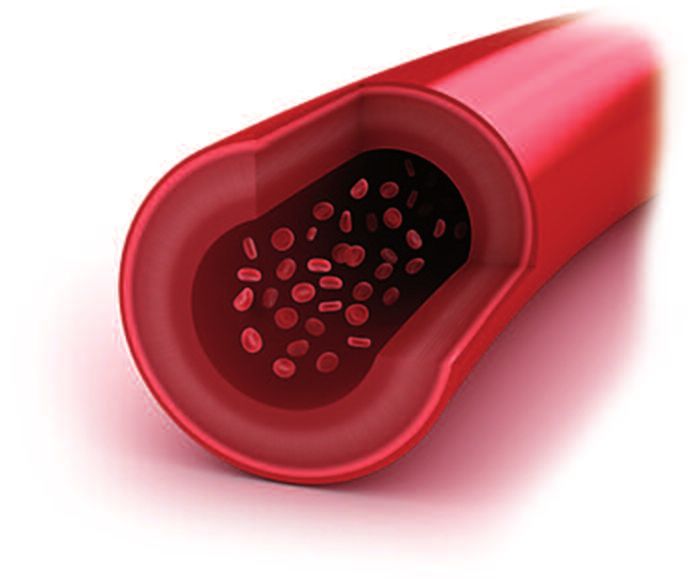
Prognostic Value of Brain Natriuretic Peptides in ACHD 43
Olga Hajnalka Balint
Heart failure in ACHD: What is different from acquired heart disease? 47
Pedro Trigo Trindade
Hyponatremia in ACHD: From prognosis to treatment 50
Oktay Tutarel
Can we prevent the development of heart failure in ACHD? 52
Helmut Baumgartner
Endstage heart failure in ACHD: Medical and surgical treatment concepts 54
Felix Berger
Aortic root dilatation in adults with congenital heart disease and
effects of pharmacotherapy on aortic growth 56
Julie De Backer
Oral Anticoagulants in Adult Congenital Heart Disease 62
Harald Kaemmerer, Claudia Pujol, Anne-Charlotte Niesert, David Pittrow, Peter Ewert
Morbus Fabry: Too often overlooked? 64
Tomas Palecek, Ales Linhart
Hypertrophic Cardiomyopathy: the renewed ESC-guidelines 67
Hubert Seggewiss
Modern survival analysis in Eisenmenger-Syndrome 68
Gerhard-Paul Diller
Pulmonary vasoactive medication in adults with Fontan circulation 70
Jamil Aboulhosn
Diagnosis and management of iron deficiency and 73
hyperuricemia in cyanotic ACHD
Koichiro Niwa
Treat and repair strategies for adults with septal defects and 74
pulmonary arterial hypertension
Dietmar Schranz
The future of PAH treatment on the horizon 76
Stephan Rosenkranz
Electrophysiologic considerations in ACHD with heart failure 80
Karl-Heinz Kuck
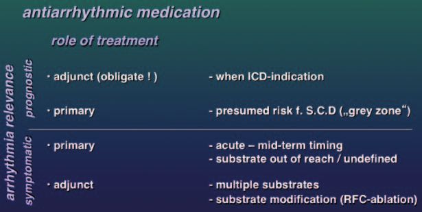
Efficacy of Antiarrhythmic Pharmacotherapy in ACHD 82
Joachim Hebe
Sudden death in ACHD: Risk stratification 88
Johan Holm
Device Therapy: Indications and Future Aspects in Primary Prevention 90
of Cardiac Death
Christof Kolb
Imprint 92
3
Chairs and Speakers

Jamir ABOULHOSN (Chair)        Bremen, Germany              Stefan ROSENKRANZ
Los Angeles, United States                                  Köln, Germany
                               Johan HOLM
Christian APITZ                Lund, Sweden                 Hubert SEGGEWIß
Ulm, Germany                                                Schweinfurt, Germany
                               Harald KAEMMERER
Julie DE BACKER                München, Germany             Dietmar SCHRANZ
Gent, Belgium                                               Gießen, Germany
                               Christoph KOLB
Olga Hajnalka BALINT           München, Germany             Markus SCHWERZMANN (Chair)
Budapest, Hungary                                           Bern, Switzerland
                               Karl Heinz KUCK
Helmut BAUMGARTNER (Chair)     Hamburg, Germany             Jane SOMERVILLE
Münster, Germany                                            London, United Kingdom
                               Rüdiger LANGE (Chair)
Felix BERGER                   München, Germany             Heiko STERN
Berlin, Germany                                             München, Germany
                               Ivan MALCIC (Chair)
Werner BUDTS (Chair)           Zagreb, Croatia              Öztekin OTO (Chair)
Leuwen, Belgium                                             Izmir, Turkey
                               Jan MAREK
Massimo CHESSA                 Londo n, United Kingdom      Pedro TRIGO-TRINDADE (Chair)
Mailand, Italy                                              Geneva, Switzerland
                               Christian MEIERHOFER
Gerhard DILLER                 München, Germany             Viktor TSANG
Münster, Germany                                            London, United Kingdom
                               Folkert MEIJBOOM (Chair)
Andreas EICKEN (Chair)         Utrecht, The Netherlands     Oktay TUTAREL
München, Germany                                            Hannover, Germany
                               Ina MICHEL-BEHNKE (Chair)
Peter EWERT                    Vienna, Austria              Gary WEBB
München, Germany                                            Cincinnati, Unite States
                               Koichiro NIWA (Chair)
Harald GABRIEL                 Tokyo, Japan                 Satoshi YASUKOCHI
Wien, Austria                                               Nagano, Japan
                               Tomas PALECEK
Alessandro GIAMBERTI (Chair)   Prag, Czech Republic
Mailand, Italy
                               Jolien ROOS-HESSELINK
Joachim HEBE                   Rotterdam, The Netherlands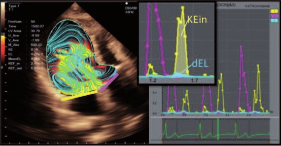
Dear colleagues and friends,

PD Dr. med. Andreas Eicken     Prof. Dr. Dr. Harald Kaemmerer    Prof. Dr. med. Peter Ewert

it is our great honour and pleasure to welcome you at the           day is focuses on different clinically relevant topics for this
occasion of the 7th annual scientific meeting of the ESC-           group of patients.
working group on adults with Congenital Heart Disease.              We believe the meeting will be attractive not only for adult –
It is worth mentioning, that this year the DACH-Symposium,          or pediatric cardiologists and surgeons, but also for cardiac
a GUCH-meeting of the German-speaking Central European              anesthesiologists, intensivists, nurse specialists and other
countries, has been integrated into this congress.                  health professional caring for GUCH patients.

The combined effort – cardiological and surgical – to treat         We hope that, besides an excellent scientific meeting, you
adults with congenital heart disease is a story of success.         will enjoy Bavarian hospitality and a stimulating atmosphere
Today an increasing number of patients, even with very              here in Munich.
complex congenital heart disease, survive to adulthood              It is an opportunity not only to share scientific thoughts but
with a good quality of life.                                        to promote personal relationships.
However, since true „correction“ of a congenital cardiac
lesion is rarely achievable, these patients need lifelong           Last but not least, we would like to sincerely thank
expert medical surveillance and close collaboration of              “Actelion Pharmaceuticals Germany GmbH”. Without
health professionals in many medical sub-specialities.              their support it would not have been possible to provide
                                                                    you with the companion volume of this meeting.
The first day of the current meeting is concentrating on            In addition, a printable pdf version of the booklet is
the right ventricle in patients with congenital heart disease.      also available (see: www.dhm.mhn.de/de/
Diagnostic modalities, indication for treatment and treat-          kliniken_und_institute/klink_fuer_kinderkardiologie_u.cfm).
ment options are presented and discussed. The second

PD Dr. med. Andreas Eicken     Prof. Dr. Dr. Harald Kaemmerer     Prof. Dr. med. Peter Ewert

                                                                                                                                 5
Elements Required to Build a Strong ACHD Program
Gary Webb, M.D.
Cincinnati Adolescent and Adult Congenital Heart Center
Cincinnati Children’s Hospital
The Heart Center
Cincinnati, USA

Over the last couple of generations, the number of adult        Another issue has to do with an agreed age to transfer from
patients with congenital heart defects has greatly increased,   pediatric to adult care. In Canada, this occurs at age 18.
and numerous ACHD clinics have opened. Perhaps you              Most Western European countries have an agreed age of
are thinking of starting one. Let’s have a look at the things   transfer. To me, this is a critical factor in growing successful
you should be thinking about and planning for in order to       ACHD programs. In the USA, there is no agreement on any
ensure that your clinic is successful, and that your patients   age of transfer, and this is a key factor in weakening ACHD
get excellent care.                                             care in the USA. A notable exception is at Emory University
The first consideration has to do with understanding where      in Atlanta, where patients are routinely transferred at age
your patients are going to come from. Will pediatric cardio-    21 to the adult facility.
logists send a lot of patients to you? Are you depending        Another process that needs to be monitored virtually from
on adult cardiologists doing so? Primary care physicians?       birth and the diagnosis of congenital heart defects is the
Patients and families finding you from their communities?       issue of attrition or loss to care of patients with moderate
What will be the trajectory of new graduates from pediatric     and complex congenital heart conditions (1). Attrition of
cardiology practices? If the pediatric cardiologists are wil-   patients who would benefit from ongoing lifelong care is a
ling to refer their graduates to your ACHD program, that is     widespread problem that will result in avoidable morbidity
welcome news. If not, that spells trouble.                      and mortality for some of these patients. Accordingly, a
How many patients are needed to build a strong ACHD             determined effort to identify these patients and keep them
program? In order to build a strong multidisciplinary ACHD      in care in the pediatric institutions or practices would be
program, I believe if you need approximately 250 new re-        very valuable for all concerned. If appointments are missed,
ferrals a year or a base of at least 1500 patients in order     the family/patient should be contacted and a follow-up
to develop and maintain team skills, to train fellows, and      made unless it is known that patients are receiving appro-
to provide adequate volumes for both diagnostic and inter-      priate care elsewhere.
ventional services.                                             For those patients who remain in pediatric cardiac care

6

after age 11 or 12, a consistent progressive patient and        complex patients include Fontan patients, severe pulmo-
family education program is in order. This is often called      nary hypertensive patients, Mustard/Senning patients,
“transition”, but I don’t like the word. At least in the USA,   patients with conduits, truncus arteriosus repairs, and
pediatric cardiologists hear the word “transition” and as-      cyanotic patients. It is critically important that such pa-
sume that we are going to try to steal their patients. Unfor-   tients remain within expert care whether in the pediatric
tunately, I haven’t found a suitable replacement. In dealing    clinic or, after a certain age, in an ACHD clinic. These
with adolescents and young adults, we need to remember          patients need to know not only the lifelong care message,
that our brains don’t mature until approximately age 25.        but how to identify expert care and where to find it (4).
A transition program should include teenagers and young         Part of the education process includes teaching the patient
adults and should be aimed at encouraging young people          and family the basics. What is the name of the condition
to take appropriate responsibility for their own health.        the patient has? What treatment has he or she received?
Healthy attitudes and behaviors are encouraged. The             What are future expectations? Are there any threats that
lifelong care message is emphasized for most patients.          can be anticipated, or any precautions that should be
Education should include career planning, lifestyle choices,    taken? What symptoms should be watched for? How fre-
                                                                quently should the patient be seen, and by whom and
                                                                where?
                                                                A multidisciplinary ACHD clinic needs to be based in an
                                                                institution or at least a large multidisciplinary ambulatory
                                                                practice. Your ACHD clinic will need the support of the insti-
                                                                tution or practice group. Is there a long-term commitment
                                                                to build and maintain the program, and to provide excellent
                                                                care for ACHD patients? There needs to be an agreement
                                                                on goals, objectives, financial considerations, and other
                                                                matters.
                                                                If you have a choice, should the program be located in an
                                                                adult hospital or a children’s hospital? There is no best
                                                                answer to the question. The ideal situation would be one in
Figure 1: Key members of a multidisciplinary ACHD team          which both adult and pediatric services are provided in the
                                                                same institution. There is evidence that successful transfer
                                                                to adult care is more effective and successful when the pa-
                                                                tient can be transferred within the same facility (5, 6). The
insurance planning, and reproductive counseling. Young          availability of both adult and pediatric subspecialty provi-
people who make good lifestyle choices are more likely          ders improves your potential flexibility in identifying the
to take responsibility for their own health and lives, so       right resources for your patient. In my experience having an
healthy lifestyles should be strongly encouraged.               ACHD clinic in an adult hospital means that you may lose
Some patients are expected to be at low risk of adverse         the opportunity to keep complex CHD children and adoles-
outcomes over the course of their lifetime. Such patients,      cents in care unless you have remarkable support from your
such as those with closed secundum ASDs at a young age,         pediatric cardiology colleagues. Having an ACHD clinic in an
small VSDs, and repaired pulmonary stenosis, need to            adult hospital means you may not be able to negotiate the
learn of our favorable expectations for them at a young age     smooth transfer of complex CHD patients to adult care,
so they can lead a normal life and not think of themselves      especially in the USA. On the other hand, having an ACHD
as “heart patients”. The same facts need to be conveyed         clinic in a children’s hospital may help you to avoid the loss
to their parents. Transfer of such patients from pediatric      to care of complex CHD children and adolescents and help
care is usually easy since good primary care physicians         effect a smooth transfer to adult care.
and cardiologists can usually provide appropriate care.         Caring for ACHD outpatients is fairly straightforward and
The medium to high risk patients need expert lifelong sur-      can occur in either a pediatric or adult clinic location. On
veillance. Expert care has been shown to have a survival        the other hand, the care of ACHD inpatients is a much more
advantage for these patients (2). Group life expectancy is      difficult challenge. Adult inpatients are not welcome in
clearly reduced in a lesion -dependent fashion (3). Many of     children’s hospitals. ACHD inpatients in adult hospitals
these patients will have limitations of their exercise capa-    may be cared for by teams not familiar with their conditions
city and lifestyle. Examples of moderately complex patients     or how best to treat them.
are arterial switch patients, repaired tetralogy, moderate to   There are important cultural differences between adult
severe pulmonary valve lesions, repaired AV septal defects,     hospitals and children’s hospitals. Team members with an
aortic coarctation, and Ebstein anomaly. Examples of very       adult background will align most easily with an adult envi-

                                                                                                                            7
ronment and team members with a pediatric background             for adult patients needs to be provided. Excellent nursing
will align best with a pediatric environment. The reverse        support adds value to the team. Whether nursing team
also applies, and alignment may be painful.                      members should come from an adult or pediatric back-
A related issue has to do with the need for separate and         ground can be debated. A long-term commitment to ACHD
distinct protocols for adult patients in any environment as      care will improve their knowledge and abilities.
regards echocardiography, exercise testing, cardiac cathe-       Access to excellent congenital heart surgery is essential.
terization, cardiac surgery, and other diagnostic procedures     This does not need to be in your own institution, but it cer-
and therapies. Extension of pediatric protocols to adult         tainly needs to be accessible. This includes the entire surgi-
patients is not appropriate, even though it is widely done.      cal team and intensive care personnel. The most common
In countries with national healthcare, the importance of en-     ACHD surgical procedure is pulmonary valve replacement.
gaging the governmental health department’s interest and         In my opinion, these are best handled by congenital heart
support cannot be underestimated in helping to establish         surgeons. Indeed, there is evidence that there is a survival
and maintain a well-organized service.                           advantage for ACHD patients being operated on by congeni-
You must commit yourself and your program to excellent           tal rather than adult cardiac surgeons (7). There are other
patient care. This will reduce the number of times you           patients that can be handled by adult cardiovascular sur-
do things you shouldn’t because of pride or self-interest.       geons skilled at aortic, mitral, aortic valve surgery, coronary
Honoring this principle will help you build and maintain         artery bypass, etc. If your ACHD program is going to include
your credibility.                                                patients with Marfan syndrome, there needs to be 24/7
You and your program should aim to be able to address the        availability of emergency surgical services to deal with
needs of any patient that comes through your door. You may       anticipated aortic dissections. If your program is going to
not be able to do everything in your own institution or com-     include patients with severe pulmonary hypertension,
munity. If not, you need to make sure that services of high      special resources also need to be available to manage
quality are available to your patients, and you should refer     these contingencies. If your program is going to include
them to the best available resources.                            cardiac patients who are pregnant, appropriate resources
The members of a multidisciplinary ACHD care team are            must be available 24/7.
many. Moreover, you don’t just want to have one person in        Access to excellent diagnostic and interventional catheteri-
each of your key categories. You must at least duplicate key     zation services is also imperative. Again, they don’t need to
skills.                                                          be in the same building, but they need to be accessible.
You need at least two ACHD cardiologists. These can come         The cardiologists doing these procedures usually come
either from an adult cardiology or a pediatric cardiology        from a pediatric cardiology background, although adult car-
background. If from pediatric cardiology, that person needs      diologists with training and experience in structural heart
to practice differently when dealing with adults than when       disease a well be capable of managing many of these ca-
dealing with children and their parents. The script used         theterization cases. In either case, the interests of the pa-
needs to be fundamentally different. Life is theater, and        tient should be primary. The procedure should be done by
this is a good example of where changing roles and respon-       someone with appropriate expertise and experience and
sibilities and customers are needed. Ideally, both types of      whose equipment and resources offer the patient the best
cardiologists will have received specific training in ACHD. If   possible chance of an excellent outcome. Is a biplane faci-
not, there is at least the need for a clear career commitment    lity required? Does the lab have enough adult-sized cathe-
to become an excellent ACHD cardiologist and to grow and         ters and devices to manage contingencies?
maintain one’s skills.                                           Access to excellent electrophysiology services are also very
As a rule, we believe it’s important to have both adult-trai-    important. EP issues are very prevalent amongst ACHD
ned and pediatric-trained cardiologists in an ACHD clinic.       patients. Pediatric EP cardiologists do very different work
Each brings hopefully offsetting strengths and weaknesses        from adult EP cardiologists, so selecting a consultant may
to the table.                                                    depend on the needs of the individual patient.
As you begin building your program, you’ll need to identify      Heart failure and transplant services will also need to be
potential consulting team members. Once done, you will           available. Heart failure is becoming an increasing issue in
need to develop and improve the capabilities of your con-        ACHD programs. Access to pulmonary hypertension resour-
sulting team in a constructive ongoing way.                      ces will also be needed. Many ACHD programs now include
You definitely need an excellent echocardiography service.       team members with pulmonary hypertension expertise.
This includes both physicians and sonographers. High-            Reproductive services also need to be available locally. This
quality echo work is critically important to the success of      begins with the ability to counsel women of childbearing
an ACHD clinic. Adult echoes and pediatric echoes are very       age regarding contraceptive options and their pregnancy
different and are reported differently. Adult approaches         risks. Some women should not use an estrogen-containing
are needed for adult patients. A full range of echo services     preparation. Risk factors for pregnancy should be evaluated

8
according to several standard scales. One or more interes-      and to meet whatever health needs they may develop. You
ted gynecologists may well make a valuable contribution         may need to decide in what type of institution to locate your
to the family planning aspects of your ACHD clinic. A good      program. Most of your care will be ambulatory, and you will
primary care physician might provide many of the same           need to establish suitable and different diagnostic and
services. The availability of maternal fetal medicine ser-      management protocols for the ACHD patients regardless of
vices is also vital. The ACHD program should provide the        whether you are in a pediatric institution or an adult insti-
cardiac component of surveillance and treatment planning        tution. If you are based in a children’s hospital, the admis-
in collaboration with one or more obstetrical teams.            sion of adult patients will be trying until you develop an ap-
Imaging services beyond echocardiography are also essential.    propriately adult trained care team to take the load off the
Excellence in MRI is particularly vital, and CT angiography     pediatric practitioners. You will need to keep the interests
will need to be available. Specific training and experience     of the patient uppermost in your planning and will need to
in congenital heart disease and/or ACHD is essential to         be clear what services you will be able to provide locally,
maximize value and minimize errors.                             and which services will need to be accessed elsewhere.
A number of other people might be important from time           You will be challenged to develop your multidisciplinary
to time. If you have Fontan patients, you should have a         care team and this will take both persistence and patience.
knowledgeable hepatologist available. Many ACHD patients        Collaboration of at least one pediatric trained cardiologist
have chronic kidney issues, so nephrology support and           with at least one adult trained cardiologist is a good foun-
involvement would be important. The availability of a gene-     dation for a program. An excellent echocardiography service
ticist and a genetics counselor may also be very important      is essential, and will need to be different from both pedia-
to the assessment and management of some patients.              tric echo services and adult acquired heart disease echo
Hematology involvement may be required at times. Mental         services. Success will not be easy, but there are an ever
health resources are an important part of a comprehensive       increasing number of patients who are depending on you
service.                                                        to get the job done, and to get it done well.
In starting a new ACHD program, you need to have the
essential elements in place before you open the doors.
Once you do open the door and patients come in in suffi-
cient numbers, your team will gain additional clinical expe-    References
rience and will become even more talented than they were
at the outset. The team would be well advised to focus on       1. Mackie, A.S., et al., Children and adults with congenital heart disease
quality outcome metrics in order to ensure themselves that          lost to follow-up: who and when? Circulation, 2009. 120(4): p. 302–9.
                                                                2. Mylotte, D., et al., Specialized adult congenital heart disease care: the
they are maintaining standards and following published              impact of policy on mortality. Circulation, 2014. 129(18): p. 1804–12.
guidelines to ensure the best care of their patients (8–10).    3. Diller, G.P., et al., Survival Prospects and Circumstances of Death in
The Adult Congenital Heart Association in the USA has crea-         Contemporary Adult Congenital Heart Disease Patients Under Follow-Up
                                                                    at a Large Tertiary Centre. Circulation, 2015. 132(22): p. 2118–25.
ted accreditation metrics for American ACHD clinics. This       4. Reid, G.J., et al., Prevalence and correlates of successful transfer from
information should, in my opinion, be accessed from all ju-         pediatric to adult health care among a cohort of young adults with com-
risdictions since it provides important reminders regarding         plex congenital heart defects. Pediatrics, 2004. 113(3 Pt 1): p. e197–205.
the many elements of high-quality ACHD patient care.            5. Goossens, E., et al., Transfer of adolescents with congenital heart
                                                                    disease from pediatric cardiology to adult health care: an analysis of
ACHD services need to be designed separately from pedia-            transfer destinations. J Am Coll Cardiol, 2011. 57(23): p. 2368–74.
tric cardiology services and from adult cardiology services.    6. Norris, M.D., et al., Prevalence and patterns of retention in cardiac care
This applies to a wide range of activities including echocar-       in young adults with congenital heart disease. J Pediatr, 2013. 163(3): p.
                                                                    902–904 e1.
diography, exercise testing, heart catheterization, and         7. Karamlou, T., et al., National practice patterns for management of adult
inpatient care. This is a difficult process, but extending          congenital heart disease: operation by pediatric heart surgeons de-
pediatric services into the adult age range is not a formula        creases in-hospital death. Circulation, 2008. 118(23): p. 2345–52.
for success, nor is the extension of acquired heart disease     8. Warnes, C.A., et al., ACC/AHA 2008 Guidelines for the Management of
                                                                    Adults with Congenital Heart Disease: Executive Summary: a report of
services into the congenital heart population likely to be          the American College of Cardiology/American Heart Association Task
successful. Parallel systems in all these dimensions should         Force on Practice Guidelines (writing committee to develop guidelines
be created.                                                         for the management of adults with congenital heart disease). Circula-
                                                                    tion, 2008. 118(23): p. 2395–451.
If you want to build a strong ACHD program, you will need:      9. Silversides, C.K., et al., Canadian Cardiovascular Society 2009 Consen-
lots of suitable patients; a supportive institution; suppor-        sus Conference on the management of adults with congenital heart
tive colleagues; a talented team; a commitment to excellent         disease: shunt lesions. Can J Cardiol, 2010. 26(3): p. e70–9.
                                                                10. Baumgartner, H., et al., ESC Guidelines for the management of grown-
patient care; educated patients and families; an effective
                                                                    up congenital heart disease (new version 2010). Eur Heart J, 2010.
transition/education process; and a clear transfer/gradua-          31(23): p. 2915–57.
tion policy. You will need to focus mainly on medium risk to
high-risk ACHD patients, and will work to keep them in care

                                                                                                                                            9
Echocardiographic evaluation of the tricuspid valve,
the pulmonic valve and the right ventricle
Jan Marek
Great Ormond Street Hospital for Children
London, UK

KEY POINTS                                                    S/D ratio, change of the pressure over the time (+dP/dt
                                                              max/mmHg-s), tricuspid annular plane systolic excursion
Significant improvement has been achieved in assessing        (TAPSE/mm), or based on Tissue Deformation Imaging tech-
RV function using conventional and advanced ECHO              niques (tissue Doppler velocity and colour coded and 2D
techniques. Despite high sensitivity to loading conditi-      Strain imaging measures) for assessing myocardial veloci-
ons, many ECHO measures and indices can be applied to         ties (S`, E`, A`), measuring myocardial performance index
clinical practice for their high predicting value, particu-   (MPI), Strain (S/%) or Strain rate (SR/-1), or isovolemic
larly in conditions where RV is exposed to high afterload.    acceleration index (IVA), perhaps least load dependent but
                                                              strongly heart-rate dependent parameter. Real-time 3D
                                                              ECHO appeared to be alternative to MRI in assessing RV
Echocardiographic (ECHO) assessment of RV function is         function. Twenty-three studies including 807 subjects how-
complex and no single functional parameter is generally       ever showed that 3D ECHO significantly underestimates
accepted in clinical practice. Several cross-sectional ima-   RV enddiastolic volumes as well as RV ejection function (5).
ging parameters have been tested and validated and many
article published suggesting advantages and limitations.      Right ventricle exposed to high preload (atrial septal de-
Major limitation of all of them is more or less load depen-   fect – ASD, tetralogy of Fallot with postoperative pulmonary
dency; however, many of them are giving insight in under-     regurgitation – TOF/PR, Ebstein anomaly – EA): In ASD
standing of mechanism of RV failure and should be routi-      patients, longitudinal deformation measured conventional
nely used in clinical practise (1.–4.). Parameters used for   and TDI showed in general normal or supra-normal functio-
assessment of RV function are based on conventional ECHO      nal parameters. Significant increase was found in the api-
methods (fractional area change - FAC/%, M-mode derived       cal segment by using 2D Strain technique (6), apical strain
fractional shortening – FS/%), Doppler derived E/A ratio,     correlated with shunt-ratio, RV end-diastolic area, and

10
Figure 1. Clinical information (A) and RV
echocardiographic indices (B.-D.) before
and after cone operation for Ebstein anomaly.
NYHA = New York Heart Association,
TAPSE = tricuspid annular plane systolic
excursion, RV FAC = right ventricular fractioning
area change
Figure 2. Conventional LV echocardiographic indices
(4.B.), 2D Strain (C.) and dyssynchrony assessment
(D) before and after cone operation for Ebstein
anomaly. LVEF = left ventricular ejection fraction,
TTP = time-to-peak, HR = heart rate
RV stroke volume (7). Reduced functional parameters after prolongation (13, 14). Early septal activation leads to early
surgical closure as compared to preoperative finding and pre-stretch and late contraction of the RV basal lateral
post-catheter closure are related to reduced preload, car- segments that are hallmarks of electromechanical dys-
diopulmonary bypass procedure and pericardial constrain. synchrony similar to LV dysfunction and LBBB resulting in
In TOF/PR, functional parameters are compounded by seve- mechanical inefficiency. Diastolic dysfunction is also well
ral factors such as type and time of initial surgical inter- described in these patients, potentially preceding systolic
ventions, degree of residual pulmonary regurgitation and functional impairment, concept of Doppler derived presys-
stenosis and QRS time. Several studies (8, 9, 10) confirm tolic atrial antegrade flow in pulmonary artery suggestive
reduced overall longitudinal deformation but more signifi- of restrictive RV physiology (15) has been routinely used
cantly in the more apical segments of the RV free wall and in clinical practise. Our study however suggested that RV
the interventricular septum but predictive value of these physiology is influenced by degree of PR and this Doppler
changes is still not clearly identified. Higher pulmonary re- flow pattern can resolve once PR is eliminated (16).
gurgitation volume and larger RV end-diastolic volume can
also be associated with decreased LV radial strain (11) due In EA, the RV is chronically exposed to high preload (signifi-
to apical RV dilatation. In our study (12) we confirmed that cant TR±ASD) and low afterload (low PVR). Despite the TR
the increase in early LV diastolic filling post percutaneous and large atrialised portion of the RV together with under-
pulmonary valve implantation correlated with the reduction filled LV, this physiology can be tolerated well even for
in RV to LV mechanical delay and change in septal curva- years. Successful cone operation eliminates TR and incor-
ture. Abnormally delayed septal contraction can be often porates atrial RV into functional RV. This acute change leads
explained by electromechanical dyssynchrony due to QRS to rather dramatic RV dysfunction documented by ECHO
11and MRI indices (17, 18) but in the other hand, improved MRI or catheter derived data (21). Simple measure of RV
clinical condition is explained by increased cardiac index longitudinal function TAPSE is associated with survival in
due to increased net forward flow from the right ventricle. patients with PAH (22), however this parameter analyse
Our recent unpublished data on assessment of the RV very small part of the RV. Other ECHO predictors of progno-
(Figure 1.) and LV function (Figure 2.) did not show signifi- sis include pericardial effusion, indexed right atrial area,
cant change in LV contractility or mechanical synchrony the degree of septal shift toward the left ventricle in dias-
(marginally better) before and after cone operation sugges- tole, pulmonary vascular capacitance, and RV myocardial
ting that obvious septal “dyskinesia” might be visual im- performance index (MPI, Tei index) (23, 24). Relatively
pression caused by competitive pressure/volume dynamics simple methods such as Doppler derived systolic/diastolic
between atrialised RV and functional LV rather than intrin- index (25) might be used effectively as well as rather so-
sic myocardial impairment and this seems to be in agree- phisticated RV myocardial strain (26).
ment with recent MRI study (19).
In patients with ccTGA, in TGA after intra-atrial baffling,
Right ventricle exposed to high afterload (pulmonary arte- and in HLHS, the morphological RV is the systemic ventri-
rial hypertension – PAH, congenitally corrected transpo- cle. Several studies in patients with ccTGA and TGA after
sition of the great arteries (ccTGA), transposition of the Mustard or Senning operation showed that quantitative
great arteries (TGA) operated via intra-atrial baffling, and echocardiographic assessment of global systemic RV
in hypoplastic left heart - HLHS (before or after staged function such as MPI highly correlates with RVEF obtained
Fontan palliation), by CMR and can be used in clinical practice (27). Speckle
tracking derived global longitudinal strain is lower especi-
In PAH patients, echocardiography provides several varia- ally in the apical segment, and tended to be lower in TGA-
bles which correlate with right heart haemodynamics and Mustard than ccTGA patients (28) and is related to adverse
should always be performed in the case of suspected PAH clinical outcome (29).
(20). In contrast to volume overloaded RV, conventional and
TDI ECHO values and indices in pressure overloaded RV are
more accurate and correlating better when compared to
12References
14. Mueller M, Rentzsch A, Hoetzer K, Raedle-Hurst T, Boettler P, Stiller B,
1. Rudski LG, Lai WW, Afilalo J, Hua L, Handschumacher MD, Chandrase- Lemmer J, Sarikouch S, Beerbaum P, Peters B, Vogt M, Vogel M, Abdul-
karan K, Solomon SD, Louie EK, Schiller NB. Guidelines for the Echocar- Khaliq H. Assessment of interventricular and right-intraventricular dys-
diographic Assessment of the Right Heart in Adults: A Report from the synchrony in patients with surgically repaired tetralogy of Fallot by two-
American Society of Echocardiography Endorsed by the European Asso- dimensional speckle tracking. Eur J Echocardiogr. 2010; 11: 786–92.
ciation of Echocardiography, a registered branch of the European So- 15. Redington AN, Oldershaw PJ, Shinebourne EA, et al. A new technique
ciety of Cardiology, and the Canadian Society of Echocardiography. J Am for the assessment of pulmonary regurgitation and its application to
Soc Echocardiogr 2010; 23: 685–713. the assessment of right ventricular function before and after repair of
2. Lang RM, Badano LP, Mor-Avi V, Afilalo J, Armstrong A, Ernande L, tetralogy of Fallot. Br Heart J 1988; 60: 57e65.
Flachskampf FA, Foster E, Goldstein SA, Kuznetsova T, Lancellotti P, Mu- 16. Frigiola A, Giardini A, Taylor A, Tsang V, Derrick G, Khambadkone S, Wal-
raru D, Picard MH, Rietzschel ER, Rudski L, Spencer KT, Tsang W, Voigt ker F, Cullen S, Bonhoeffer P, and Jan Marek. Echocardiographic assess-
JU. Recommendations for cardiac chamber quantification by echocar- ment of diastolic biventricular properties in patients operated for se-
diography in adults: an update from the American Society of Echocar- vere pulmonary regurgitation and association with exercise capacity.
diography and the European Association of Cardiovascular Imaging. European Heart Journal Cardiovascular Imaging (2012) 13, 697–702.
Eur Heart J Cardiovasc Imaging. 2015; 16: 233–70. 17. Ibrahim M, Tsang VT, Caruana M, Hughes ML, Jenkyns S, Perdreau E,
3. Lopez L, Colan SD, Frommelt PC, Ensing GJ, Kendall K, Younoszai AK, Lai Giardini A, Marek J. Cone reconstruction for Ebstein's anomaly: Patient
WW, Geva T. Recommendations for quantification methods during the outcomes, biventricular function, and cardiopulmonary exercise capa-
performance of a pediatric echocardiogram: a report from the Pediatric city. J Thorac Cardiovasc Surg. 2015; 149: 1144–50.
Measurements writing Group of the American Society of echocardiogra- 18. Lange R, Burri M, Eschenbach LK, Badiu CC, da Silva JP, Nagdyman N,
phy Pediatric and Congenital Heart Disease Council. J. Am. Soc. Echo- Fratz S, Hörer J, Kühn A, Schreiber C, Vogt MO. Da Silva cone repair for
cardiogr. 2010; 23, 465–495. Ebsteon;s anomaly: effect on right ventricular size and function. Eur J
4. Abman SH, Hansmann G, Archer SL, Ivy DD, Adatia I, Chung WK, Hanna Cardiothorac Surg. 2015; 48: 316–20; discussion 320–1.
BD, Rosenzweig EB, Raj JU, Cornfield D, Stenmark KR, Steinhorn R, Thé- 19. Goleski PJ, Sheehan FH, Chen SSM, Kilner PJ, Gatyoulis MA. The shape
baud B, Fineman JR, Kuehne T, Feinstein JA, Friedberg MK, Earing M, and function of the left ventricle in Ebstein's anomaly. Int J Cardiol.
Barst RJ, Keller RL, Kinsella JP, Mullen M, Deterding R, Kulik T, Mallory 2014 15; 171: 404–12.
G, Humpl T, Wessel DL. American Heart Association Council on cardio- 20.Authors/Task Force Members: Galie`N (Chairperson). Guidelines for the
pulmonary, critical care, perioperative and resuscitation; Council on diagnosis and treatment of pulmonary hypertension. The Task Force for
Clinical Cardiology; Council on Cardiovascular Disease in the Young; the Diagnosis and Treatment of Pulmonary Hypertension of the Euro-
Council on Cardiovascular Radiology and Intervention; Council on Car- pean Society of Cardiology (ESC) and the European Respiratory Society
diovascular Surgery and Anesthesia; and the American Thoracic So- (ERS), endorsed by the International Society of Heart and Lung Trans-
ciety. Pediatric Pulmonary Hypertension: Guidelines From the American plantation (ISHLT). European Heart Journal 2009; 30, 2493–2537.
Heart Association and American Thoracic Society. Circulation. 2015; 24: 21. Lammers AE, Haworth SG, Riley G, Maslin K, Diller GP, Marek J. Value of
132: 2037–99. tissue Doppler echocardiography in children with pulmonary hyperten-
5. Shimada YJ, Shiota M, Siegel RJ, Shiota T. Accuracy of right ventricular sion. J Am Soc Echocardiogr. 2012; 25: 504–10.
volumes and function determined by three-dimensional echocardiogra- 22.López-Candales A, Rajagopalan N, Saxena N, Gulyasy B, Edelman K,
phy in comparison with magnetic resonance imaging: a meta-analysis Bazaz R. Right ventricular systolic function is not the sole determinant
study. J Am Soc Echocardiogr. 2010; 23: 943–53. of tricuspid annular motion. Am. J. Cardiol. 2006; 98: 973–977.
6. Dragulescu A, Grosse-Wortmann L, Redington A, Friedberg MK, Mertens 23. Bossone E, D'Andrea A, D'Alto M, Citro R, Argiento P, Ferrara F, Cittadini A,
L. Differential effect of right ventricular dilatation on myocardial defor- Rubenfire M, Naeije R. Echocardiography in pulmonary arterial hyperten-
mation in patients with atrial septal defects and patients after tetra- sion: from diagnosis to prognosis. J Am Soc Echocardiogr. 2013; 26: 1–14.
logy of Fallot repair. Int J Cardiol. 2013, 30; 168: 803–10. 24.Zimbarra Cabrita I, Ruisanchez C, Dawson D, Grapsa J, North B, Howard
7. Van De Bruaene A, Buys R, Vanhees L, Delcroix M, Voigt JU, Budts W. Regio- LS, Pinto FJ, Nihoyannopoulos P, Gibbs JS. Right ventricular function in
nal right ventricular deformation in patients with open and closed atrial patients with pulmonary hypertension; the value of myocardial perfor-
septal defect. European Journal of Echocardiography 2011; 12, 206–213. mance index measured by tissue Doppler imaging. Eur J Echocardiogr.
8. Kowalik E, Kowalski M, Ró a ski J, Ku mierczyk M, Hoffman P. The impact 2010; 11: 719–24.
of pulmonary regurgitation on right ventricular regional myocardial 25. Alkon J, Humpl T, Manlhiot C, McCrindle BW, Reyes JT, Friedberg MK.
function: an echocardiographic study in adults after total repair of Usefulness of the right ventricular systolic to diastolic duration ratio
tetralogy of Fallot. J Am Soc Echocardiogr 2011; 24: 1199–204. to predict functional capacity and survival in children with pulmonary
9. Sabate Rotes A, Bonnichsen CR, Reece CL, Connolly HM, Burkhart HM, arterial hypertension. Am J Cardiol. 2010; 106: 430–6.
Dearani JA, Eidem BW. Long-term follow-up in repaired tetralogy of fal- 26.Okumura K, Humpl T, Dragulescu A, Mertens L, Friedberg MK. Longitu-
lot: can deformation imaging help identify optimal timing of pulmonary dinal assessment of right ventricular myocardial strain in relation to
valve replacement? J Am Soc Echocardiogr. 2014. transplant-free survival in children with idiopathic pulmonary hyper-
10. Kempny A, Diller GP, Orwat S, Kaleschke G, Kerckhoff G, Bunck ACh, tension. J Am Soc Echocardiogr. 2014; 27: 1344–51.
Maintz D, Baumgartner H. Right ventricular-left ventricular interaction 27. Salehian O, Schwerzmann M, Merchant N, Webb GD, Siu SC, Therrien J.
in adults with Tetralogy of Fallot: a combined cardiac magnetic reso- Assessment of systemic right ventricular function in patients with
nance and echocardiographic speckle tracking study. Int J Cardiol. transposition of the great arteries using the Myocardial Performance
2012; 154: 259–64. Index. Comparison with cardiac Magnetic Resonance Imaging. Circula-
11. Menting ME, van den Bosch AE, McGhie JS, Eindhoven JA, Cuypers JA, tion. 2004; 110: 3229–3233.
Witsenburg M, Geleijnse ML, Helbing WA, Roos-Hesselink JW. Assess- 28.Eindhoven JA, Menting ME, van den Bosch EA, McGhie JS, Witsenburg
ment of ventricular function in adults with repaired Tetralogy of Fallot M, Cuypers JAAE, Boersma E, Roos-Hesselink JW. Quantitative assess-
using myocardial deformation imaging. Eur Heart J Cardiovasc Imaging. ment of systolic right ventricular function using myocardial deformation
2015; 16: 1347–57. in patients with a systemic right ventricle. Eur Heart J Cardiovasc Ima-
12. Lurz P, Puranik R, Nordmeyer J, Muthurangu V, Hansen MS, Schievano ging. 2015; 16: 380–8.
S, Marek J, Bonhoeffer P, Taylor AM. Improvement in left ventricular fil- 29.Diller GP, Radojevic J, Kempny A, Alonso-Gonzalez R, Emmanouil L,
ling properties after relief of right ventricle to pulmonary artery conduit Orwat S, Swan L, Uebing A, Li W, Dimopoulos K, Gatzoulis MA, Baum-
obstruction: contribution of septal motion and interventricular mecha- gartner H. Systemic right ventricular longitudinal strain is reduced in
nical delay. European Heart Journal 2009, 30; 2266–2274. adults with transposition of the great arteries, relates to subpulmonary
13. Hui W, Slorach C, Dragulescu A, Mertens L, Bijnens B, Friedberg MK ventricular function, and predicts adverse clinical outcome. Am Heart J.
Mechanisms of right ventricular electromechanical dyssynchrony and 2012; 163: 859–66.
mechanical inefficiency in children after repair of tetralogy of fallot.
Circ Cardiovasc Imaging. 2014; 7: 610–8.
13Advanced echocardiographic modalities in the assessment
of right ventricular function
Satoshi Yasukochi, MD
Heart Center, Nagano Children’s Hospital
Toyoshina, Azumino, Nagano, Japan
The newer echocardiographic techno-
logy provides more information to dis-
KEY POINTS The right ventricular (RV) structure sect out the detailed contractile mode
and function have been found to be an and intra-cardiac flow dynamics of RV,
New imaging modalities of echocar- important determinant of prognosis besides the complexity of its geometri-
diography could demonstrate the de- and the outcome of the treatment in cal shape and structure.
tails of RV structure and mechanics congenital heart disease. Currently, The contractile mode and morphology
based on its fiber orientation (strain echocardiography and cardiac magnetic of anatomical RV is different in case
analysis) and enables the blood flow resonance are the two major imaging of pulmonic ventricle from that of
kinetic energy for assessing further modalities to visualize the structure systemic ventricle.
integrated cardiac performance. and functions of the RV. The 3D morphology of pulmonic RV is
14Figure 1
lower 3D peak strain and longer time-
to-peak strain, compared to those in
normal LV. Recently, the intra-blood
flow kinetics could be analyzed by
novel imaging modality using color
Figure 2 Doppler to calculate “blood flow
kinetic energy loss” (EL, mW) from the
reconstructed velocity vector compo-
nents transformed into Cartesian coor-
dinate system as previously reported
by K. Itatani et al., 2013.
The EL data were indexed by measu-
ring a ratio of EL to the inflow kinetic
energy (KEin) through systemic atrio-
ventricular valve in diastole. EL/KEin in
diastole of HLHS is higher than that of
normal LV. This means systemic RV of
HLHS may lose intracardiac blood flow
kinetic energy besides impairment of
chamber and wall kinetics. (Figure 2)
References
crescent while that of systemic RV is ventricle which means more circum-
ellipsoid. (Figure 1) ferential and longitudinal strain.
1. Keiichi Itatani, Takashi Okada, Tokuhisa Ue-
The contractile mode of pulmonic RV However, both pulmonic and systemic jima, Tomohiko Tanaka, Minoru Ono, Kagami
shows peristaltic from inlet to outlet, RV DO NOT have a torsion or twist Miyaji and Katsu Takenaka. Intraventricular
which means dominant longitudinal which a normal systemic LV does have. Flow Velocity Vector Visualization Based on
the Continuity Equation and Measurements
strain with less circumferential strain. (Figure 1) of Vorticity and Wall Shear Stress. Japanese
On the other hand, the systemic RV The systemic RV in hypoplastic left Journal of Applied Physics, Volume 52, Num-
contracts more like a systemic left heart syndrome (HLHS) demonstrates ber 7S. Published 22 July 2013
15Assessment of RV function with pressure-volume loops –
impact on treatment indication for a volume-loaded RV
Prof. Dr. Christian Apitz
Division of Pediatric Cardiology
University Children’s Hospital Ulm
Ulm, Germany
The assessment of right ventricular function is aggravated By recording a family of pressure-volume loops during
by the complex anatomy of the right ventricle and the reduction of preload, preferably achieved by temporary
variable effects of abnormal loading conditions. balloon occlusion of the inferior caval vein, the systolic
Pressure-volume loop analysis by conductance catheters ventricular function could be calculated by the slope of
is extensively used in experimental studies especially in the endsystolic pressure-volume relation (Figure).
models of acute and chronic right ventricular pressure or
volume overload and is generally considered the most reliable Main drawback for the routine use of pressure-volume loop
way to quantify right ventricular contractile function. (1–6) analysis in clinical everyday practice is its invasive nature.
Nevertheless, in individual cases it might be a helpful
A conductance catheter is a specialised multi-electrode tool to support decision-making for change in therapy,
catheter which allows accurate measurement of ventricular RVOT intervention or reoperation, as well as monitor
volume and pressure continuously throughout the cardiac changes after treatment and as a predictor for outcome
cycle. A variety of physiological parameters can be derived of patients with congenital heart disease and adverse RV
from pressure-volume loops. loading condition.
16References
1. Leeuwenburgh BP, Helbing WA, Steendijk P, Schoof PH, Baan J. Biventri-
cular systolic function in young lambs subject to chronic systemic right
ventricular pressure overload. Am J Physiol Heart Circ Physiol. 2001
Dec; 281(6): H2697–704.
2. Redington AN, Oldershaw PJ, Shinebourne EA, Rigby ML. A new techni-
que for measuring pulmonary regurgitation: Application to the assess-
ment of right ventricular function before and after repair of tetralogy of
Fallot. Br Heart J 1988; 60: 57–65.
3. Chaturvedi RR, Kilner PJ, White PA, Bishop AJ, Szwarc R, Redington AN.
Increased airway pressure and simulated branch pulmonary artery ste-
nosis increase pulmonary regurgitation after repair of tetralogy of Fal-
lot. Real-time analysis with a conductance catheter technique. Circula-
tion 1997; 95: 643–649.
4. Apitz C, Sieverding L, Latus H, Uebing A, Schoof S, Hofbeck M. Right
ventricular dysfunction and B-type natriuretic peptide in asymptomatic
patients after repair of tetralogy of Fallot. Pediatr Cardiol 2009; 30:
898–904.
5. Apitz C, Latus H, Binder W, et al. Impact of restrictive physiology on in-
trinsic diastolic right ventricular function and lusitropy in children and
adolescents after repair of tetralogy of Fallot. Heart 2010; 96: 1837–41.
6. Latus H, Binder W, Kerst G, Hofbeck M, Sieverding L, Apitz C. Right ven-
Figure: Family of pressure-volume loops during reduction of preload tricular-pulmonary arterial coupling in patients after repair of tetralogy
achieved by temporary balloon occlusion of the inferior caval vein. of Fallot. J Thorac Cardiovasc Surg. 2013 Dec; 146(6): 1366–72.
17Cardiovascular Magnetic Resonance Evaluation of
the tricuspid valve, the pulmonary valve and the right ventricle
Dr. Dr. med. Christian Meierhofer
Pediatric Cardiology and Congenital Heart Disease
Deutsches Herzzentrum München
Technical University Munich
Munich, Germany
KEY POINTS ons after repair of tetralogy of Fallot for evaluation of right
ventricular volumes, function and assessment of pulmo-
The right ventricle can easily be assessed by cardiovascu- nary regurgitation.
lar magnetic resonance. Right ventricular volumes, flow In patients after atrial switch operation for transposition
in the pulmonary artery and even through the tricuspid of the great arteries failing of the systemic right ventricle
valve are measured and provide valuable data on ventri- is the main focus of the assessment in CMR. Additionally,
cular function and information about tricuspid and pul- tricuspid valve regurgitation can be evaluated. Evaluation
monary valve regurgitation. of the ventricular myocardium to detect myocardial scars
may be useful, but this issue is controversially discussed
in patients with systemic right ventricle. (2, 3) Therefore
we do not perform routinely late gadolinium enhancement
Cardiovascular Magnetic Resonance (CMR) has become a for detection of myocardial scars in follow-up assessment
valuable tool for assessment of right ventricular structures of patients with systemic right ventricles.
in routine follow-up of right ventricular disease. Anatomic The assessment of the systemic right ventricular myocar-
reasons may limit assessment of the right ventricle by dium for scientific purpose includes e. g. T1 mapping. Most
echocardiography. of the right ventricular disorders can be evaluated by CMR
Is has been shown that right ventricular volumes and with scan times less than one hour.
function can be assessed very precisely by CMR, since In patients after arterial switch the focus of the examina-
other methods tend to over- or underestimate right ventri- tion lies on the anteriorly positioned pulmonary artery that
cular volumes. (1) may be become stenotic over time. The right and left pul-
The main conditions for patients grown-up with congenital monary artery may also get stenotic mainly due to anatomic
heart disease to be referred to CMR assessment are situati- reasons and due to tension on the pulmonary arteries due
18to relocation of the pulmonary artery anterior to the aorta. also largely widened ECG complexes may cause problems.
Further congenital cardiac conditions that are routinely eva- Most CMR scans need a stable ECG signal for averaging
luated in adulthood by CMR to evaluate the right ventricular image information that has been measured over several
situation are congenital corrected transposition of the heart beats. Bad ECG trigger may impair the quantitative
great arteries, complex DORV situations, single ventricle analysis, but it may still be possible to qualitatively assess
performance and Ebstein’s anomaly. ventricular volumes and function in such conditions. Vo-
In Ebstein’s anomaly right ventricular function and tricus- lume overloaded ventricles may also be detected and end-
pid regurgitation play a target role. diastolic volume indices can be used, but with precaution.
Figure 1 shows a 68 years old woman with Ebstein’s ano- In such situations volume indices, structural assessment,
maly with severe tricuspid regurgitation. Figure 2 shows size and function of the right ventricle by eyeballing and
the same patient after cone repair of the tricuspid valve. relation of size and function of the right ventricle to left
Generally, regurgitation of the tricuspid valve can be ventricle parameters may be used.
Figure 1: 68 years old woman with Ebstein’s anomaly. Figure 2: Same patient after cone repair of the tricuspid valve.
(a) transversal view, arrows show position of the tricuspid valve;
(b) parasagittal view; (c) direct view on opening area of the tricuspid
valve, anatomic information; (d) direct view on opening area of the
evaluated by two methods in CMR. It can be assessed by tricuspid valve, phase contrast image with flow information;
measuring directly the flow through the tricuspid valve. (e) flow profile through the tricuspid valve.
This measurement implements some technical problems
since the tricuspid valve area is moving during the cardiac
circle and therefore during flow measurement. The second References
method to assess tricuspid regurgitation is based on mea-
suring the stroke volume of the right ventricle by evaluation 1. Sugeng L, Mor-Avi V, Weinert L, Niel J, Ebner C, Steringer-Mascherbauer
of the end-diastolic and the end-systolic volume. The stroke R, et al. Multimodality comparison of quantitative volumetric analysis
volume is calculated and the flow in the main pulmonary of the right ventricle. JACC Cardiovasc Imaging. 2010; 3: 10–8.
2. Babu-Narayan SV, Goktekin O, Moon JC, Broberg CS, Pantely GA, Pen-
artery is measured. Further the part of the stroke volume nell DJ, et al. Late gadolinium enhancement cardiovascular magnetic
that is not running through the pulmonary artery is assu- resonance of the systemic right ventricle in adults with previous atrial
med to be regurgitated through the tricuspid valve. (Figure 3) redirection surgery for transposition of the great arteries. Circulation.
2005; 111: 2091–8.
Major limitations for volume and flow assessment by CMR 3. Fratz S, Hauser M, Bengel FM, Hager A, Kaemmerer H, Schwaiger M,
are rhythm abnormalities that do not allow sufficient trigge- et al. Myocardial scars determined by delayed-enhancement magnetic
ring of the ECG signal. Therefore, atrial fibrillation, atrial resonance imaging and positron emission tomography are not common
flutter, many kinds of ectopic beats during the scan and in right ventricles with systemic function in long-term follow up. Heart.
2006; 92: 1673–7.
(stroke volume right ventricle [ml] – forward flow main pulmonary artery [ml])
Tricuspid regurgitation (%) = ~ 100
(stroke volume right ventricle [ml] – pulmonary regurgitation [ml])
Figure 3: This formula is used to calculate tricuspid regurgitation
19Exercise physiology and pathology in right ventricular dysfunction
Folkert Meijboom
Centre for Congenital Heart Disease Utrecht
Dept Cardiology
University Medical Centre Utrecht
Utrecht, The Netherlands
At the start of exercise, pulmonary vascular resistance
drops, allowing the right ventricle to increase pulmonary
KEY POINTS perfusion with only limited rise of right ventricular pres-
sure. There will be more pulmonary venous return to the
• The right ventricle is inseparably connected with the left atrium and thus the left ventricle. In turn, the right
venous system, that determines preload, and the ventricle can only increase pulmonary blood flow when
pulmonary vascular bed, that determines afterload. provided with more systemic venous return. This is achie-
• Right ventricular function can only be judged when ved by increase of the vascular tone in the capacitance
preload and afterload are taken into account. vessels of the venous system, for which in increase of
• Understanding of both basics of the exercise physio- sympatic nerve activity is responsible (1).
logy and the limitations in the non-invasive assess-
ment of right ventricular function, helps in the assess- At rest, a large part of the circulating volume is in the
ment of right ventricular function in clinical practice. venous compartment. The thin-walled and very compliant
venous system holds approximately 10 times the volume
that the stiffer and thicker-walled arterial system holds (2).
Physiology of the circulation during exercise In other words, the total systemic vascular capacitance
(or blood-holding capacity) is predominantly dependent on
During exercise, the metabolic demands of the body in- the venous system. In this system, the biggest reservoirs
crease instantaneously. These increased demands should are in the splanchnic system, in the spleen and in the liver.
be met by an increase in cardiac output. This is achieved A small increase in vascular tone will directly lead to an
by the left ventricle, by increasing stroke volume and increase in venous return towards the right atrium. A prere-
heart rate. The left ventricle can only do so, if supplied quisite is that right atrial pressures remain low at exercise.
adequately by the right ventricle. Only then, a higher systemic venous pressure will lead to a
20You can also read