CHEST X-RAYS: Case Studies from the Field
←
→
Page content transcription
If your browser does not render page correctly, please read the page content below
2/28/2021

        CHEST X-RAYS:
 Case Studies from the Field

                Michael Zacharisen, M.D

                                                               THANK YOU
    Amy Saltzman                      Marcus Paske, Pharm.D., AE‐C
    BJ Biskupiak                      Marcy Ballman, Ph.D.
    Dewey Halbohm, PA, AE‐C           Michael Zacharisen, MD
    Greg Holzman, MD                  Trista Gilmore, LPN
    Jessie Fernandes, MPH             Vicki Ann Lint, RN, LPN
    Kendra Procacci, Pharm.D., AE‐C

                          DISCLOSURES & CONFLICT OF
                                           INTEREST
• 1. FYI, I’m an allergist, not a radiologist!
• 2. In medical school, I nearly chose radiology as a specialty.

                                                                                  1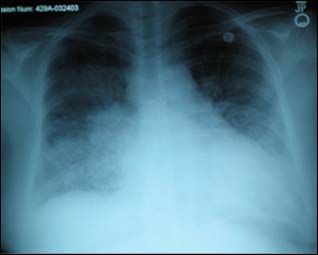
2/28/2021

                                                      OUTLINE
Chest x-rays:
• History
• Terminology
• Normal vs abnormal
• Case studies to highlight cardio-respiratory diseases

                                     HISTORY OF X-RAYS
• Germany: 1895
• Dr. Rector Wilhelm Conrad Roetgen
• “Discovery of a new form of photography, which
  revealed hidden solids, penetrated wood, paper,
  and flesh, and exposed the bones of the human
  frame.” from: Early History of X Rays by Alexi Assmus.
• First x-ray: his wife’s hand with her ring.
• First X-ray made in public. Hand of the famed anatomist,
• Albert von Kölliker on January 1896.

                                               CHEST X-RAY
• 1896: Chest x-ray: 2 dimensional

                                                                       2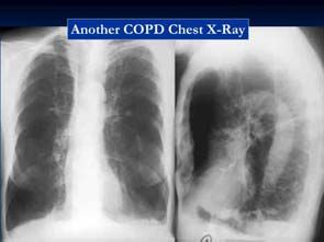
2/28/2021

                          CHEST X-RAY: INDICATIONS
• Respiratory disease                      • Trauma
   • Pneumonia                             • Suspected cancer metastasis
   • Pneumothorax
   • Chronic dyspnea                       • Pneumoperitoneum
   • Hemoptysis                            • Check position of nasogastric
   • Pulmonary embolism                      tubes, endotracheal tubes, etc.
   • Investigation of TB
                                           • Radiopaque foreign bodies
• Heart disease
                                           • Post-operative imaging
                                           • Pre-employment screening

               POSITIONING: PA AND LATERAL

                                                      WHERE DO I START?

                 This Photo by Unknown Author is licensed under CC BY-SA

                                                                                      3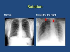
2/28/2021

                      USE A SYSTEMATIC APPROACH
• RIP
  Rotation: square on – if rotated, then distortion occurs (clavicles)
  Inspiration: 7 to 9 ribs visible (9 ribs = asthma, COPD)
  Penetration: over or under-exposed (makes it too light or dark)
• ABCDE
  A – airway (trachea, bronchi, etc)
  B – bones (and soft tissues)
  C – cardiac silhouette
  D – diaphragm
  E – everything else (hardware) and THEN the LUNGS!

                                               CXR: ROTATION

                                            CXR: INSPIRATION

                                                 Take a deep
                                                 breathe and hold it

                                                                                4
2/28/2021
CXR: PENETRATION
NORMAL LANDMARKS
A – airway (trachea, bronchi)
B – bones (and soft tissues)
C – cardiac silhouette
D – diaphragm
E – everything else
COMMON CXR TERMS
• Opacities
○ Appears radio-opaque (white) compared to normal lung
○ Alveolar opacity vs Interstitial opacity
• Mass/Nodule
○ Discrete appearance with borders. Nodule < 3 cm. Pleural- or parenchymal
• Consolidation
○ Focal confluence of alveolar opacities.
• Atelectasis vs. Effusion
○ Discrete lines or lobar distribution for atelectasis (small airways collapse)
○ Effusions (liquid) are usually dependent (starts at bases and moves upwards)
• Edema: swelling in Alveolar vs. Interstitial patterns
• Fibrosis: Septal thickening vs Honeycombing
52/28/2021
CASE 1
• 20 y/o with cough, wheeze and
shortness of breath at night, with
colds and with exercise.
• Exam: wheezing on expiration.
• Your diagnosis?
• COPD
• Asthma
• Bronchitis
• Foreign body aspiration
ASTHMA
• Hyper-inflated
• Flat diaphragms
• Heart appears small
• Often normal: used to rule
out other diagnoses.
• Note: endotracheal tube
CASE 2
• 60 y/o with shortness of breath with
exertion and swollen ankles after a
viral infection.
• PMH: high blood pressure, diabetes,
thyroid disease and alcoholism
• Family hx: coronary artery disease
• Exam: obese, decreased lung sounds
62/28/2021
CXR
• CXR features:
• Increased width of vascular pedicle
• Perihilar haze: excess fluid
• Large cardiac silhouette
HEART FAILURE FROM
CARDIOMYOPATHY
Cardiomegaly – width of the
silhouette is greater than 1⁄2 the
thoracic cage width.
CASE 3
• 25 year old.
A. Normal CXR but placed backward?
B. Dextrocardia?
72/28/2021
DEXTROCARDIA
• Dextrocardia: abnormal
congenital condition where the
heart points to right side of chest
instead of left.
• Dextro: right and Levo: left
• 1 in 12,000 pregnancies.
• Exam: heart sounds louder on
the right side.
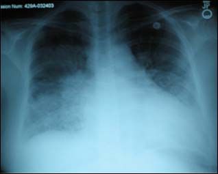
CASE 4
• 50 y/o with fever, muscle aches,
shortness of breath and
dysgeusia
• Exam: rales (“Velcro” sound)
• CXR Features:
• “Ground glass” opacity: hazy
opacity
COVID-19 PNEUMONIA
• 10 days later
• Consolidation: middle and
upper lobes
82/28/2021
CASE 5
• 60 y/o with 50 pack yr
of smoking
• Cough and short of
breath.
• Exam: barrel chest
• CXR Features:
Hyperinflated
“Floating heart” sign
COPD
CXR Features:
Hyperinflated:
Flat diaphragms
“Small” heart
Retrosternal air
“Floating heart” sign
Bullae (blebs)
Washed out bones
(steroids)
CASE 6
1 y/o boy with wheezing x 2 months
Exam: wheezing on right side
CXR features:
Hyper-lucency of right
Hyper-expansion of right
92/28/2021
INHALED FOREIGN BODY
1 y/o boy with wheezing x 2 months
Exam: wheezing on right side
CXR features:
Hyperlucency of right
Hyperexpansion of right
Bronchoscopy: kernel of corn
removed
500-2000 deaths occurring each year from foreign body
aspiration.
• 70 yr old with 4 months of
CASE 7
• Shortness of breath
• Cough
• Bone pain
• Weight loss
• Fatigue
• CXR features:
• Opacity near hilum
LUNG CANCER
102/28/2021
CASE 8
• 40 yr old doctor (nice guy)
• Sudden onset chest pain,
fever, shaking chills, cough
with thick green sputum
• CXR features:
• Infiltrate with opacification
BACTERIAL PNEUMONIA
• 40 yr old doctor (nice guy)
• Chest pain, fever, chills,
cough with green sputum
• Sputum culture: Strep
pneumonia and H influenza
• CXR features:
• Infiltrate with opacification
CONCLUSIONS
Chest X-rays
• Important tool to rule out processes and confirm diagnoses
• Quick, easy to obtain
• Cost: $200 to $400
• Look at the lungs last!
112/28/2021
Thank you for attending!
12You can also read