Hepatic Injury Induced by Dietary Energy Level via Lipid Accumulation and Changed Metabolites in Growing Semi-Fine Wool Sheep
←
→
Page content transcription
If your browser does not render page correctly, please read the page content below
ORIGINAL RESEARCH
                                                                                                                                     published: 23 September 2021
                                                                                                                                    doi: 10.3389/fvets.2021.745078

                                             Hepatic Injury Induced by Dietary
                                             Energy Level via Lipid Accumulation
                                             and Changed Metabolites in Growing
                                             Semi-Fine Wool Sheep
                                             Benchu Xue 1 , Qionghua Hong 2 , Xiang Li 1 , Mingli Lu 1 , Jia Zhou 1 , Shuangming Yue 3 ,
                                             Zhisheng Wang 1 , Lizhi Wang 1 , Quanhui Peng 1 and Bai Xue 1*
                                             1
                                               Animal Nutrition Institute, Sichuan Agricultural University, Chengdu, China, 2 Yunna Academy of Animal Science and
                                             Vetarinary Medicine, Kunming, China, 3 Department of Bioengineering, Sichuan Water Conservancy College, Chengdu, China

                                             Liver injury threatens the overall health of an organism, as it is the core organ of the
                                             animal body. Liver metabolism is affected by numerous factors, with dietary energy
                                             level being a crucial one. Therefore, the present study aimed to evaluate hepatic injury
                                             and to describe its metabolic mechanism in ruminants fed diets with different dietary
                                             energy levels. A total of 25 Yunnan semi-fine wool sheep were fed diets with five dietary
                                             metabolic energy levels and were randomly assigned to five groups as follows: low energy
                         Edited by:
                                             (LE), medium–low energy (MLE), medium energy (ME), medium–high energy (MHE), and
                     Jianguo Wang,           high energy (HE). The results revealed that the average optical density (AOD) of lipid
      Northwest A&F University, China
                                             droplets in the LE, MLE, and HE groups was higher than that in the ME and MHE
                       Reviewed by:
                                             groups. The enzyme activity of alanine aminotransferase (ALT) was the lowest in the
                                Lei Liu,
  Hunan Agricultural University, China       ME group. An increase in dietary energy level promoted the superoxide dismutase (SOD)
                                  Yu Li,     and glutathione peroxidase (GSH-Px) activity and altered the malondialdehyde (MDA) and
   Anhui Agricultural University, China
                                             protein carbonyl (PCO) concentration quadratically. In addition, both high and low dietary
                  *Correspondence:
                            Bai Xue
                                             energy levels upregulated the mRNA abundance of proinflammatory cytokine interleukin
                 13592@sicau.edu.cn          (IL)-1β, nuclear factor-kappa B (NF-κB), and tumor necrosis factor (TNF)-α. Metabonomic
                                             analysis revealed that 142, 77, 65, and 108 differential metabolites were detected in the
                    Specialty section:
         This article was submitted to
                                             LE, MLE, MHE, and HE groups, compared with ME group respectively. These metabolites
     Animal Nutrition and Metabolism,        were involved in various biochemical pathways, such as glycolipid, bile acid, and lipid
                a section of the journal     metabolism. In conclusion, both high and low dietary energy levels caused hepatic injury.
        Frontiers in Veterinary Science
                                             Section staining and metabonomic results revealed that hepatic injury might be caused
             Received: 21 July 2021
         Accepted: 17 August 2021            by altered metabolism and lipid accumulation induced by lipid mobilization.
      Published: 23 September 2021
                                             Keywords: liver, injury, metabonomics, energy level, oxidative stress
                              Citation:
   Xue B, Hong Q, Li X, Lu M, Zhou J,
 Yue S, Wang Z, Wang L, Peng Q and           INTRODUCTION
  Xue B (2021) Hepatic Injury Induced
      by Dietary Energy Level via Lipid
                                             Yunnan semi-fine wool sheep, characterized by optimal reproductive performance, stable genetic
          Accumulation and Changed
    Metabolites in Growing Semi-Fine
                                             performance, strong adaptability, and excellent meat performance and shearing capacity, is an
                         Wool Sheep.         important breed in the development of China’s livestock industry (1). In sheep breeding, the focus
            Front. Vet. Sci. 8:745078.       is often on growth performance, and hence, the health of the heart, lungs, kidneys, spleen, liver,
     doi: 10.3389/fvets.2021.745078          and other organs is usually ignored. Organ health is an important factor that not only determines

Frontiers in Veterinary Science | www.frontiersin.org                             1                                     September 2021 | Volume 8 | Article 745078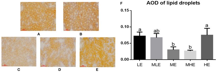
Xue et al.                                                                                                       Hepatic Injury Induced by Energy

whether the lamb can develop quickly but also reflects its overall        MS (LC-MS/MS) in combination with multivariate statistical
health (2). The liver is the most important metabolic organ,              analysis. The objective of this study was to evaluate the injury
with functions such as bile secretion, glycogen storage, and the          caused by dietary energy levels, identify an energy level that is
regulation of protein, fat, and carbohydrate metabolism.                  the most beneficial for the hepatic health of Yunnan semi-fine
    Several studies have reported that dietary energy                     hair sheep, and investigate its metabolic mechanism.
concentration exerts an important effect on the development
of ruminant organs, with the liver being the most significantly
affected (3). However, with an increase in energy intake, the liver       MATERIALS AND METHODS
may also be affected by simultaneous damage, including liver              Experimental Design, Animals, and Sample
function impairment and oxidative damage (4, 5), which may
                                                                          Collection
subsequently aggravate hepatocyte apoptosis and inflammatory
                                                                          All experimental protocols were approved by the Animal Ethical
response. Researchers have reported that fatty liver causes
                                                                          and Welfare Committee (AEWC) of the Sichuan Agricultural
hepatic injury (6, 7). Fatty liver is a reversible disease, with
                                                                          University Academy of Sciences (approval no. 20180601). This
large amounts of triglycerides or lipid droplets accumulating
                                                                          study was conducted in accordance with the Chinese Guidelines
in hepatocytes through steatosis, which adversely affects the
                                                                          for Animal Welfare. This study was conducted at the farm of
development, health, and reproduction of cows (8, 9). Diets
                                                                  ′       the Sichuan Agricultural University (Ya’an, Sichuan Province,
with high energy levels downregulated the expression of 5
                                                                          China). In this study, a total of 25 Yunnan semi-fine hair
adenosine monophosphate-activated protein kinase (AMPK)
                                                                          wether sheep, with similar BW (33.30 ± 1.77 kg), were randomly
signaling pathways, thereby enhancing the expression of lipid
                                                                          assigned to five groups. Each group of sheep was reared in
synthesis-related genes, promoting lipid synthesis in the liver
                                                                          a pen. The sheep in the five groups were fed diets with five
cells, reducing lipid oxidation, and increasing the triglyceride
                                                                          metabolic energy levels as follows: 8.0, 8.6, 9.2, 9.8, and 10.4
concentration (10, 11). However, hepatic lipid accumulation in
                                                                          MJ/kg. According to the standard energy requirement of Yunnan
animals fed diets with low energy levels has not been reported
                                                                          semi-fine hair sheep (BW: 33.3 kg and ADG: 80 g/day) (18), these
yet. Studies on fatty liver disease in dairy cows have reported
                                                                          five groups were distinguished as follows: low energy (LE; 86%
a similar insufficient energy intake condition, which suggests
                                                                          energy requirement), medium–low energy (MLE; 93% energy
that lipid accumulation is frequently observed in the liver of
                                                                          requirement), medium energy (ME; 100% energy requirement),
animals fed diets with low energy levels (12). Studies on dietary
                                                                          medium–high energy (MHE; 107% energy requirement), and
energy levels that affect hepatic health are now mostly focused
                                                                          high energy (HE; 114% energy requirement). The ingredients and
on poultry (13, 14). The systematic evaluation of hepatic health
                                                                          chemical compositions of each diet are provided in Table 1.
is overlooked when ruminants are fed diets with different dietary
                                                                              The trial lasted 45 days, with the first 15 days being the
energy levels. Therefore, it is necessary to systematically assess
                                                                          preliminary period for the sheep to adapt to the diets and 30
the effects of dietary energy levels on hepatic health in Yunnan
                                                                          days being the formal trial period. The sheep were fed twice
semi-fine wool sheep to understand their metabolism.
                                                                          daily at 8:00 AM and 6:00 PM and had free access to water. The
    Metabolomics displays all small-molecule metabolites
                                                                          criteria for euthanizing the sheep before the experiment included
produced by alterations in the nutritional status of an organism,
                                                                          anesthesia and neck bleeding.
thereby providing a more comprehensive and direct insight into
                                                                              On the 30th day of the formal trial, three sheep from each
the chemical processes and changes in nutritional status (15).
                                                                          group were selected to be weighed and anesthetized, which
Ippolito et al. used gas chromatography–mass spectrometry
                                                                          were eventually euthanized. Subsequently, the whole liver was
(GC-MS) to analyze changes in the plasma nutrient metabolome
                                                                          separated and weighed. The left liver was collected and stored
of rats under heat stress and obtained 28 heat stress markers,
                                                                          in 4% paraformaldehyde and PBS for liver section staining. The
involving those for apoptosis or catabolism, altered energy
                                                                          right liver was collected in a 30-ml Eppendorf (EP) tube for
balance, and cholesterol and nitric oxide metabolism (16). Zhang
                                                                          later measurement.
et al. employed nuclear magnetic resonance (NMR) for analyzing
the plasma of cows exhibiting postpartum estrus, which showed
that the levels of seven plasma metabolites were significantly            Weight of the Liver and Liver Index
lower in the estrus period than those in the normal period,               The data for the final BW and liver weight (LW) of sheep
demonstrating that estrus is accompanied by altered amino acid,           were recorded, and the liver index (LI) was calculated as LI =
glucose, and lipid metabolism (17).                                       LW/BW (%).
    Various studies have been reported on the energy
requirements of the sheep. However, only a few studies                    Determination of Hepatic Lipid
have focused on the local breed of Yunnan semi-fine hair                  Accumulation
sheep. Li reported that the metabolic energy requirement of               Liver tissues were fixed with 10% formalin; they were embedded,
the sheep was 0.4359 MJ/kg of body weight (BW) 0.75/day +                 sliced, and stained with Oil Red O. A microscopic imaging system
0.0387 average daily gain (ADG) (18). Based on her study, we              was used to capture pictures. A total of three pictures were
designed a trial to investigate hepatic health and metabolism             captured for each sample. Image-Pro Plus 6.0 software was used
by controlling the metabolic energy level of the diet using the           for analyzing the average optical density (AOD) of the positive
metabolomic technique liquid chromatography with tandem                   results of Oil Red O staining.

Frontiers in Veterinary Science | www.frontiersin.org                 2                                September 2021 | Volume 8 | Article 745078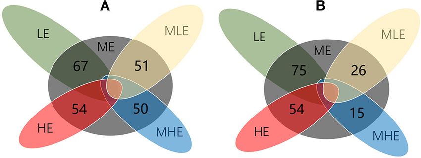
Xue et al.                                                                                                                             Hepatic Injury Induced by Energy

TABLE 1 | Composition and nutrient levels of experimental diets.                               melting phase set to 95◦ C for 15 s, 60◦ C for 1 min, and 95◦ C for
                                                                                               15 s. Glyceraldehyde 3-phosphate dehydrogenase (GAPDH) was
Items                                                    Groups
                                                                                               used as an internal reference gene, and its expression level was
                                    LEc        LME         ME         MHE          HE          analyzed using the 2−11Ct method.

Ingredients                                             Content (%)
Corn                               11.00       19.35      28.15       34.15      30.00
                                                                                               Untargeted Metabonomics and Its Analysis
                                                                                               Samples for metabolites were extracted from liver tissues
Wheat bran                         26.15       16.80       7.00       0.00        0.00
                                                                                               (20). LC-MS/MS analyses were performed using a Vanquish
Soybean meal                        6.00       7.00        8.00       9.00        9.00
                                                                                               UHPLC system (Thermo Fisher Scientific) coupled with an
Corn starch                         0.00       0.00        0.00       0.00        4.15
                                                                                               Orbitrap Q Exactive HF-X mass spectrometer (Thermo Fisher
NaCl                                0.50       0.50        0.50       0.50        0.50
                                                                                               Scientific) in both positive and negative modes. The raw
NaHCO3                              0.35       0.35        0.35       0.35        0.35
                                                                                               data files generated by UHPLC-MS/MS were processed using
Premixa                             1.00       1.00        1.00       1.00        1.00
                                                                                               the Compound Discoverer 3.1 (CD3.1, Thermo Fisher) to
Corn silage                        10.00       21.00      33.00       40.00      55.00
                                                                                               perform peak alignment, peak picking, and quantitation for
Wheat straw                        45.00       34.00      22.00       15.00       0.00
                                                                                               each metabolite.
Total                               100         100        100         100        100
                                                                                                   Principal component analysis (PCA) and partial least
Concentrate : roughage             45:55       45:55      45:55       45:55      45:55         squares discriminant analysis (PLS-DA) were performed at
nutrient levelsb
                                                                                               metaX (a flexible and comprehensive software for processing
ME (MJ/kg)                          8.00       8.60        9.20       9.80       10.40
                                                                                               metabolomics data). Variable importance in the projection (VIP)
Dry matter (%)                     92.62       92.19      91.88       91.47      91.20
                                                                                               is the importance of the variables to the model and describes the
CP (%)                             10.42       10.42      10.42       10.48      10.46
                                                                                               overall contribution of each variable to the differences between
NDF (%)                            53.80       46.71      39.10       34.24      28.10
                                                                                               groups. We applied univariate analysis (t-test) to calculate the
ADF (%)                            21.96       20.92      19.62       19.63      19.20
                                                                                               statistical significance (p-value). The metabolites with a VIP >1,
Ca (%)                              0.42       0.40        0.37       0.35        0.35
                                                                                               p-value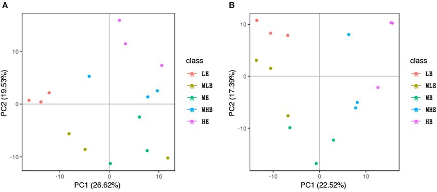
Xue et al.                                                                                                                              Hepatic Injury Induced by Energy

TABLE 2 | The effects of dietary energy level on liver weight and index of Yunnan semi-fine wool sheep.

Items                                                             Energy level                                    SEM                          P-value

                                 LE                MLE                    ME        MHE                HE                    Energy           Linear          Quadratic

Liver Weight (g)              471.93c            534.30b             603.87a       600.93a           608.97a      15.68       0.001            0.05; Figure 2B).
Assessment of Hepatic Injury                                                                        The assessment of oxidative injury in the liver is provided
The assessment of hepatic injury was based on transaminase                                      in Table 3. With an increase in dietary energy levels, the SOD
activity, oxidative damage, and inflammation.                                                   and GSH-Px enzyme activities increased linearly (p < 0.05).
   The assessment of transaminase activity in the liver is                                      MDA concentration in the ME and MHE groups was significantly
provided in Figure 2. The ALT enzyme activity in the liver                                      lower than that in the remaining three groups (p < 0.05). PCO
decreased and subsequently increased with an increase in dietary                                concentration in the ME group was significantly lower than that
energy levels (p < 0.05). The ALT enzyme activity of the ME                                     in the HE group (p < 0.05). CAT concentrations in the LE and

Frontiers in Veterinary Science | www.frontiersin.org                                       4                                September 2021 | Volume 8 | Article 745078
Xue et al. Hepatic Injury Induced by Energy
TABLE 3 | The effects of dietary energy level on oxidative injury of Yunnan semi-fine wool sheep.
Items Energy level SEM P-value
LE MLE ME MHE HE Energy Linear Quadratic
MDA (nmol/L) 11.57a 11.91a 9.10b 8.84b 12.67a 0.39Xue et al. Hepatic Injury Induced by Energy
FIGURE 4 | The PCA score plots based on the liver metabolic profiling. (A) Positive mode. (B) Negative mode.
DISCUSSION
Organ tissues only constitute 6%−10% of BW; however, their
energy consumption is as high as 50% of total energy in
ruminants (21). Therefore, organ development requires energy,
especially liver development (22). The organ weight and index
can reflect organ development to a certain extent. As shown in
Table 1, LW and LI were the highest in the ME group because
excess energy may not promote liver development once the
energy requirements have been fulfilled. However, an injury may
FIGURE 5 | The Venn diagram for illustrating the number of differential
metabolites among groups. (A) Positive mode. (B) Negative mode.
also be caused by increased energy levels.
We found that dietary energy levels induced significantly
different degrees of hepatic injury in semi-fine wool sheep. In
particular, the enzyme activity of ALT varied quadratically with
PLS-DA in the positive and negative modes are provided in an increase in dietary energy level and was the lowest in the ME
Supplementary Figures 2, 3, respectively. group. Because the enzyme activity of transaminase is an effective
indicator of hepatic injury (23), both high and low dietary energy
Identification of Different Metabolites intakes may cause hepatic injury. With regard to oxidative injury,
Compared with the ME group, 67, 51, 50, and 54 differential the effects of SOD and GSH-Px were similar, which increased
metabolites were detected in the LE, MLE, MHE, and HE groups, with an increase in dietary energy levels. However, MDA and
respectively, in the positive mode (Figure 5A). Similarly, 75, 26, PCO concentrations demonstrated a quadratic trend similar
15, and 54 differential metabolites were detected in the LE, MLE, to that of ALT, which is the peroxide product. These results
MHE, HE, and ME groups, respectively, in the negative mode indicated that the antioxidant function was increased with an
(Figure 5B). The metabolites related to hepatic metabolism and increase in dietary energy levels; however, the liver exhibited
health are listed in Table 4. more oxidative injury with high dietary energy levels. This may
be because excessive nutrient intake and absorption aggravate the
Integration of Key Different Metabolic metabolism of the body, which inevitably produces products such
Pathways as reactive oxygen radicals (24). Similar results were obtained
The metabolic pathway analysis by the KEGG database and in the expression of inflammation-related hepatic genes (TLR-
literature showed that these differential metabolites were 4 and TNF-α). Owing to TLR signaling, a large number of
involved in various biochemical pathways, such as amino inflammatory mediators such as IL-1β, IL-6, and TNF-α reacted
acid metabolism, glycolipid metabolism, bile acid metabolism, on the organism and produced a series of inflammatory responses
nucleotide metabolism, and energy metabolism. In order to (25), demonstrating that sheep in the LE and HE groups were
visualize the correlation between these metabolites, the results more likely to exhibit inflammation.
were finally combined, and a metabolic network diagram was The liver is a core organ in the regulation of lipid metabolism.
drawn (Figure 6). Limited production of very-low-density lipoproteins (VLDL)
Frontiers in Veterinary Science | www.frontiersin.org 6 September 2021 | Volume 8 | Article 745078Xue et al. Hepatic Injury Induced by Energy
TABLE 4 | Identification of different metabolites in the liver.
Metabolites LE vs. ME MLE vs. ME ME vs. MHE ME vs. HE
P VIP FC P VIP FC P VIP FC P VIP FC
6-Phosphogluconic acidXue et al. Hepatic Injury Induced by Energy
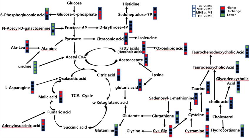
FIGURE 6 | Different metabolic pathways from the sheep of five groups. The red mark means that the concentration of the former group is higher than that of the latter
group. The black mark means the difference is not significant, and the green mark means the concentration of the former group is lower than that of the latter group.
oxidative injury. Therefore, hepatic injury may be induced by glycolysis (29), which was consistent with the assessment of
lipid accumulation. inflammation-related gene expression.
Because hepatic injury is confirmed, hepatic lipid
accumulation may be one of the causes. However, other Lipid Metabolism
factors may affect liver health as well. Therefore, we investigated The liver tissues of the LE, MLE, and HE groups exhibited more
differences in the global metabolic profiles of five dietary severe lipid deposition. Metabolomic results revealed that the
energy levels. concentration of hexanoic acid, as a free fatty acid (FFA), was
In this study, we employed LC-MS/MS to determine significantly higher in the LE group than in the ME group,
differences in the metabolic profiles of the liver. A huge number confirming that liver lipid deposition in the LE group may
of differential metabolites were detected, which were involved in be caused by the reesterification of FFA in the liver owing to
various biochemical pathways, such as amino acid metabolism, lipid mobilization, which results from insufficient energy intake.
glycolipid metabolism, bile acid metabolism, nucleotide The assessment of acetoacetic acid, one of the intermediate
metabolism, and energy metabolism. We have described below metabolites of fatty acid oxidation in the liver (30), and acetic
the involvement of these pathways in hepatic injury. acid yielded similar results, suggesting that they supplied energy
through fatty acid oxidation in the LE group. Owing to a
sufficient energy intake in the HE group, the level of fatty acid
Glucose Metabolism oxidation in the liver should be lower (31), and lipid synthesis
We found that the MHE and HE groups exhibited a metabolism should be enhanced (32). As a result, lipid deposition
significantly higher concentration of glucose 6-phosphate than was higher in the HE group than in the ME group. As amounts
that exhibited by the ME group. The concentration of N- of very low-density lipoproteins (VLDLs) were restricted, their
acetyl-D-galactosamine (a precursor of fructose 6-phosphate) ability to export triglycerides is extremely limited. Therefore,
was significantly lower in the LE group than in the ME group, sheep fed diets with both high and low energy levels are at a
which was consistent with previous studies, suggesting that the higher risk for developing fatty liver disease.
glycolysis pathway is enhanced in the presence of adequate
energy substrates (28). The concentration of glucose 6-phosphate Energy Metabolism
was also significantly lower in the MLE group than in the ME In this study, the three intermediate metabolites of the
group, which may be due to inflammation. The proinflammatory tricarboxylic acid cycle, namely, citric acid, malic acid, and
cytokines IL-6, TNF-α, and IL-1β were found to enhance adenylosuccinic acid (one of the precursors of ferredoxin),
Frontiers in Veterinary Science | www.frontiersin.org 8 September 2021 | Volume 8 | Article 745078Xue et al. Hepatic Injury Induced by Energy
exhibited significantly higher concentrations in the LE group is consistent with the assessment of inflammation-related
than that exhibited by the ME group, which is inconsistent with gene expression.
Wu’s study (30). Combined with lipid metabolism, this may be
due to the higher concentration of acetyl coenzyme generated by Other Metabolites
FFA β-oxidation in the LE and MLE groups. Owing to different dietary energy levels, in addition to the
Nicotinamide adenine dinucleotide (NAD+), guanosine above-mentioned liver metabolites, other differential metabolites
diphosphate (GDP), and adenosine diphosphate (ADP) are are related to hepatic health as well, including glutathione,
involved in energy metabolism (33). The concentrations of ergothioneine, p-cresol, betaine, and cortisol.
NAD+, GDP, and ADP were significantly higher in the HE Glutathione is a tripeptide of glutamic acid, cysteine, and
group than those in the ME group, which indicates that the glycine, which contains γ-amide bonds and sulfhydryl groups.
sheep in the HE group were more likely to exhibit metabolic It is involved in converting harmful toxic substances into
injury compared with those in the ME group owing to a high harmless substances (43, 44), thereby maintaining the normal
energy intake. High dietary energy levels significantly enhanced immune functions of an organism (45). In this study, glutathione
the levels of substrate energy metabolism and bio-oxidation (34, concentration was significantly lower in the MLE group than that
35). In addition, it has been demonstrated that some metabolic in the ME group, suggesting that the ME group exhibited better
intermediates produced during the tricarboxylic acid cycle, such immune function. The results were partially consistent with
as citric acid, are inflammatory signals (36), suggesting that there those of antioxidant analysis because glutathione is a component
is a greater possibility of inflammation in the LE group than of GSH-Px.
in the ME group, which is consistent with the assessment of Ergothioneine is a natural antioxidant with anti-inflammatory
inflammation-related gene expression. and cytoprotective effects (46, 47), which is distributed in
certain tissues and organs of mammals. Melville reported
Nucleotide Metabolism that ergothioneine is also found in cereal plants (48). In
Nucleotides are important components of cells; they are this study, ergothioneine concentration in the ME group
not only involved in the synthesis of genetic material but was significantly higher than that in the LE and MLE
also play a significant role in energy metabolism, function groups, which may be owing to differences in the intake of
regulation, and immunity (37). In addition, nucleotides can maize. However, compared with the ME group, ergothioneine
particularly affect the growth, structure, morphology, and concentration did not increase in the MHE and HE groups,
function of the liver. Researchers have demonstrated that suggesting that its uptake was limited. However, further
exogenous nucleotides or endogenous nucleosides promote investigation is required. Owing to the natural antioxidant
the growth of liver cells (38). Uridine, a component of function of ergothioneine, the antioxidant activity in the ME
uridine monophosphate (UMP), is naturally produced by group was better than that in the LE and MLE groups,
the liver. In this study, uridine concentration in the LE, which is partially consistent with the results of oxidative
MLE, HE, and MHE groups was significantly lower than stress analysis.
that in the ME group, indicating that the liver of the Oxoadipate is metabolized by lysine through the zymocin and
ME group exhibited a better growth potential, which is pipecolic acid pathways. If lysine, tryptophan, and hydroxylysine
consistent with the assessment of liver growth provided in are metabolized incorrectly, oxoadipate production is greatly
Table 1. Furthermore, uridine phosphorylase disrupts hepatic increased (49). In this study, oxoadipate concentration was
pyrimidine nucleotide metabolism by expressing or inhibiting significantly higher in the LE and HE groups than that in the
dihydroorotate dehydrogenase, leading to liver steatosis. LME and MHE groups, indicating that low or high dietary
Uridine supplementation can inhibit liver steatosis caused by energy levels may cause disorders in the lysine, tryptophan,
dihydroorotate dehydrogenase (39). The results were consistent and hydroxylysine metabolism. This is not conducive to
with the trend of hepatic steatosis scores provided in Table 2, liver health.
indicating that the degree of hepatic steatosis may be minimum
in the ME group. CONCLUSION
Bile Acid Metabolism In conclusion, based on apparent and molecular evidence,
Bile acid is an important component of bile, which is we confirmed that hepatic injury may be induced by lipid
important for digestion and lipid metabolism (40). In recent accumulation and other altered metabolites. In particular, both
years, researchers have demonstrated that bile acids affect and high and low dietary energy levels cause hepatic injury in Yunnan
regulate physiological processes such as glucolipid metabolism semi-fine wool sheep. Based on our research findings, the dietary
and inflammatory reaction by activating downstream signals metabolic energy requirement of Yunnan semi-fine wool sheep is
through their receptors (41). It has been reported that 9.2–9.8 MJ/kg (ME and MHE groups). This study also provides
primary and some secondary bile acids can inhibit the useful information regarding the effect of dietary energy level on
release of TNF, suggesting that bile acids exert an anti- the hepatic health of growing sheep at the metabolic level, thereby
inflammatory effect (42). Therefore, the LE and HE groups providing guidance for improving the production efficiency of
were more likely to exhibit an inflammatory reaction, which the sheep.
Frontiers in Veterinary Science | www.frontiersin.org 9 September 2021 | Volume 8 | Article 745078Xue et al. Hepatic Injury Induced by Energy
DATA AVAILABILITY STATEMENT FUNDING
The original contributions presented in the study are included This research was funded by the National Key Research
in the article/Supplementary Material, further inquiries can be and Development Program of China (2018YFD0502303)
directed to the corresponding author/s. and National Technical System of Wool Sheep Industry
(CARS39-08).
ETHICS STATEMENT
The animal study was reviewed and approved by the Animal ACKNOWLEDGMENTS
Ethical and Welfare Committee (AEWC) of the Sichuan
We would like to thank the staff at our laboratory for their
Agricultural University Academy of Sciences.
ongoing assistance.
AUTHOR CONTRIBUTIONS
SUPPLEMENTARY MATERIAL
BCX, QH, and XL designed the studies and prepared the
manuscript with comments from all authors. ML, BX, SY, and JZ The Supplementary Material for this article can be found
performed all the experiments and analyzed the data. LW, ZW, online at: https://www.frontiersin.org/articles/10.3389/fvets.
and QP revised the manuscript. 2021.745078/full#supplementary-material
REFERENCES endoplasmic reticulum stress. J Anim Physiol a Anim Nutr. (2015) 99:626–
45. doi: 10.1111/jpn.12263
1. Zhao Y, Hong Q, Xie P, Chen G, Wang W. Study on yunnan semi-wool sheep 13. Hermier D, Salichon MR, Guy G, Peresson R. Differential channelling of
production performance. Acta Ecologiae Animalis Domastici. (2011) 32:51–6. liver lipids in relation to susceptibility to hepatic steatosis in the goose—
doi: 10.3969/j.issn.1673-1182.2011.06.010 sciencedirect. Poultry Sci. (1999) 78:1398–406. doi: 10.1093/ps/78.10.1398
2. Zhang X, Xin W, Chen W, Zhang Y, Zhou Z. Growth performance 14. Baraona E, Lieber CS. Alcohol and lipids. Recent DevAlcohol. (1998) 14:97–
and development of internal organ, and gastrointestinal tract of calf 134. doi: 10.1007/0-306-47148-5_5
supplementation with calcium propionate at various stages of growth 15. Nicholson JK, Lindon JC, Holmes E. “Metabonomi cs” unders tanding
period. PLoS ONE. (2017) 12:e0179940. doi: 10.1371/journal.pone.0179 the metabolic responses of living systems to pathophysiological stimuli
940 viamultivariate statistical analysis of biological NMR spectroscopic data.
3. Shen Z, Syfert H, Loehrke B, Schneider F, Zitnan R, Chudy A. An energy- Xenobiotica. (1999) 29:1181–9. doi: 10.1080/004982599238047
rich diet caused rumen papillae proliferation associated with more IGF type 16. Ippolito DL, Lewis JA, Yu C, Leon LR, Stallings JD. Alteration in circulating
1 receptors and increased plasma IGF-1 concentration in young goats. J Nutr. metabolites during and after heat stress in the conscious rat: potential
(2004) 134:11–17 doi: 10.1093/jn/134.1.11 biomarkers of exposure and organ-specific injury. BMC Physiol. (2014)
4. Noziere P, Attaix D, Bocquier F, Doreau M. Effects of underfeeding and 14:14. doi: 10.1186/s12899-014-0014-0
refeeding on weight and cellularity of splanchnic organs in ewes. J Anim Sci. 17. Zhang J, Wang G, Zhao C. 1H NMR plasma metabolomic profiling of
(1999) 77:2279–90. doi: 10.2527/1999.7782279x ovarianquiescence in energy balanced postpartum dairy cows. Vet Quart.
5. Buhler S, Frahm J, Tienken R, Kersten S, Meyer U, Huber K, et al. (2018) 38:47–52. doi: 10.1080/01652176.2018.1473660
Effects of energy supply and nicotinic acid supplementation on serum anti- 18. Li X, Lu ML, Hong QH, Xue B, Wu YH, Hu AH, et al. Energy
oxidative capacity and on expression of oxidative stress-related genes in blood requirement of yunnan semi-fine-wool sheep during growing period
leucocytes of periparturient primi- and pluriparous dairy cows. J Anim Physiol by regression model method. Acta Zoonutr Sin. (2020) 32:447–54.
Anim Nutr. (2018) 102:e82–9. doi: 10.1111/jpn.12705 doi: 10.3969/j.issn.1006-267x.2020.01.052
6. Clark JM, Diehl AM. Nonalcoholic fatty liver disease: an 19. Fujiang W, Ruiyan L, Pengfei T, Jianping C, Kewu Z, Yong J. Total glycosides
underrecognized cause of cryptogenic cirrhosis. JAMA. (2003) of cistanche deserticola promote neurological function recovery by inducing
289:3000–4. doi: 10.1001/jama.289.22.3000 neurovascular regeneration via Nrf2/Keap-1 pathway in MCAO/R Rats. Front
7. Tomlinson JW, Newsome PN, Dowman JK. Pathogenesis of non-alcoholic Pharmacol. (2020) 11:236. doi: 10.3389/fphar.2020.00236
fatty liver disease. QJM-INT J Med. (2010) 2:103. doi: 10.1093/qjmed/hcp158 20. Cao Z, Xia W, Zhang X, Yuan H, Gao, L. Hepatotoxicity of nutmeg:
8. Bobe G, Young J, Beitz D. Invited review: pathology, etiology, prevention,and a pilot study based on metabolomics. Biomed Pharmacother. (2020)
treatment of fatty liver in dairy cows. J Dairy Sci. (2004) 87:3105– 131:110780. doi: 10.1016/j.biopha.2020.110780
24. doi: 10.3168/jds.S0022-0302(04)73446-3 21. Chilliard Y, F Bocquier, M Doreau. Digestive and metabolic adaptations of
9. Reddy JK, Sambasiva Rao M. Lipid metabolism and liver ruminants to undernutrition, and consequences on reproduction. A review.
inflammation.II.Fatty liver disease and fattyacid oxidation. Am J Physiol-Gastr Reprod Nutr Dev. (1998) 38:129–150. doi: 10.1051/rnd:19980201
L. (2006) 290:G852–58. doi: 10.1152/ajpgi.00521.2005 22. Kamalzadeh A, Koops W, Bruchem J, Tamminga S, Zwart D.
10. Jelenik T, Rosseisl M, Kuda O, Jilkova ZM, Medrikova D, Kus V, et al. Feed quality restriction and compensatory growth in growing
AMP—activated proteinkinase o2 subunit is required for the preservation of sheep: development of body organs. Small Ruminant Res. (1998)
hepatic in-sulin sensitivity by n-3 polyunsaturated fatty acids. Diabetes. (2010) 29:71–82. doi: 10.1016/S0921-4488(97)00111-9
59:2737–46. doi: 10.2337/db09-1716 23. Wang X, Huang B, Li X, Wang S, Cheng D. Antagonistic effect of
11. Bergeron R, Previs SF, Cline GW, Perret P, Russell 3rd RR, Young LH, tea polyphenols on erythrocyte and liver injury in acute cadmium
et al. Effect of 5-aminoimi-dazole-4-carboxamide- 1-beta-D- ribofuranoside (ll) exposure for mice. J Chin Inst Food Sci Tech. (2020) 20:66–72.
infusion on invivo glucose and lipid metabolism in lean and obese Zucker doi: 10.16429/j.1009-7848.2020.04.009
rats. Diabetes. (2001) 50:1076–82. doi: 10.2337/diabetes.50.5.1076 24. Dandona P, Aljada A, Bandyopadhyay A. Inflammation: the link between
12. Ringseis R, Gessner DK, Eder K. Molecular insights into the mechanisms insulin resistance, and obesity and diabetes. Trends Immunol. (2004) 25:4–
of liver-associated diseases in early-lactating dairy cows: hypothetical role of 7. doi: 10.1016/j.it.2003.10.013
Frontiers in Veterinary Science | www.frontiersin.org 10 September 2021 | Volume 8 | Article 745078Xue et al. Hepatic Injury Induced by Energy
25. Li S, Khafipour E, Krause DO, Kroeker A, Rodriguez-Lecompte JC, Gozho 41. Perino, A, Schoonjans K. TGR5 and immunometabolism: insights from
GN. Effects of subacute ruminal acidosis challenges onfermentation and physiology and pharmacology. Trends Pharmacol Sci. (2015) 36:847–
endotoxins in the rumen and hindgut of dairy cows. J. Dairy Sci. (2012) 57. doi: 10.1016/j.tips.2015.08.002
95:294–303. doi: 10.3168/jds.2011-4447 42. Qi M, Diao Q, Zhang N. Advance in ruminal development and
26. Sozio MS, Liangpunsakul S, Crabb D. The role of lipid metabolism in the its influencing factors in lambs. Chin J Anim Sci. (2015) 51:77–81.
pathogenesis of alcoholic and nonalcoholic hepatic steatosis. Semin Liver Dis. doi: 10.3969/j.issn.0258-7033.2015.09.017
(2010) 30:378–90 doi: 10.1055/s-0030-1267538 43. Treviani F, Tame MR. Sovere hep-atic failureoeeurring with T6l ingestion in
27. Grummer RR. Etiology of lipid-related metabolic disorders an at-tempted suieide Earlyreeovery with the use of intra-venous infusion
in periparturient dairy cows. J Dairy Sci. (1993) 76:82– of redueedglutathione. Dig Dis Sci. (1993) 38:752–6. doi: 10.1007/BF013
96. doi: 10.3168/jds.S0022-0302(93)77729-2 16810
28. Xue BC, Zhang JX, Wang ZS, Wang LZ, Peng QH, Da LC, et al. Metabolism 44. Morel G, Bonnet P, Cossee B. The role of glu-tathione andcysteine conjugate
response of grazing yak to dietary concentrate supplementation in warm in the nephrotoxieity of o-xylene in rats. Arehtoxicol. (1998) 72:553–
season. Animal. (2021) 15:100175. doi: 10.1016/j.animal.2021.100175 8. doi: 10.1007/s002040050542
29. Jiang S, Zhang L, Zhang HA. novel miR-155/miR-143 cascade controls 45. Kashiwagi A, Asahina A, Tkebuehi M. Abnomal glutathione
glycolysis by regulating hexokinase 2 in breast cancer cells. EMBO Jl. (2012) metabolism and inereased eytotoxieity caused by H2O2:
31:1985–98. doi: 10.1038/emboj.2012.45 inhuman umbilical vein endothelial cells cultured in high
30. Wu GY, Gunasekara A, Brunengraber H. Effects of extracellular pH, CO2, glueosemedium. Diabetologia. (1994) 37:264–9. doi: 10.1007/BF00398
and HCO3-on ketogenesis in perfused rat liver. Am J Physiol-Gastr L. (1991) 053
261:E221–6. doi: 10.1152/ajpendo.1991.261.2.E221 46. Yoshida S, Shime H, Funami K, Takaki O, Matsumoto M, Kasahara M, et
31. Wang H, Niu W, Wu F, Qiu X, Yu Z, He Y, et al. Effects of dietary energy al. The anti-oxidant ergothioneine augments theimmunomodulatory function
on antioxidant capacity, glucose–lipid metabolism and meat fatty acid profile of TLR agonists by direct action on macrophages. Plos One. (2017) 12:1–
of Holstein bulls at different ages. J Anim Physiol Anim Nutr. (2020) 105:1– 15. doi: 10.1371/journal.pone.0169360
11. doi: 10.1111/jpn.13457 47. Cheah IK, Halliwell B. Ergothioneine; antioxidant potential, physiology
32. Gao L, Wu J, Song S, Li H, Lang X, Wei Y, et al. Effect of different energy function and role in disease. Biochim Biophys Acta. (2012) 1822:784–93.
levels on serum lipid index and lipid deposition of Altay sheep. Feed Ind. doi: 10.1016/j.bbadis.2011.09.017
(2020) 41:14–23. doi: 10.13302/j.cnki.fi.2020.09.003 48. Melville DB, Eich S. The occurrence of ergothioneine in plante
33. Wu Z, Li M, Zhao C. Urinary metabonomics study in a rat model in meterial. J Biol Chem. (1956) 218:647–51. doi: 10.1016/S0021-9258(18)65
response to protein-energy malnutrition by using gas chromatography-mass 831-4
spectrometry and liquid chromatography-mass spectrometry. Mol Biosyst. 49. Luo Z, Bai L, Huang Y, Jiang J, Shen L, Tao J, et al. Effect of
(2010) 6:2157–63. doi: 10.1039/c005291d selenium yeast on plasma metabolism of transition dairy cow in
34. Li D, Lun Y, Zhou S. Recent Progress of NAD+/NADH Metabolism. Lett parturition stress status. J Northwest A & F Univ. (2019) 49:7–15.
Biotechnol. (2010) 21:98–102. doi: 10.13207/j.cnki.jnwafu.2019.02.002
35. Lu Y, Tang X, Li B, Ge Y, Yang S, Zhang K, et al. Mechanism of food-
borne tyrosine oxidation product-induced myocardial oxidative damage
Conflict of Interest: The authors declare that the research was conducted in the
and energy metabolism disorder in mice. Food Science, (2020) 41:84–90.
absence of any commercial or financial relationships that could be construed as a
doi: 10.7506/spkx1002-6630-20191018-180
potential conflict of interest.
36. Mills E, O’Neill LA. Succinate: a metabolic signal in inflammation. Trends Cell
Biol. (2013) 11:127–31. doi: 10.1016/j.tcb.2013.11.008
37. Li B, Wu X, Zhang B, Yin Y. Research progresses on nutrition metabolism Publisher’s Note: All claims expressed in this article are solely those of the authors
and physiological function of uridine monophosphate. Acta Zoonutr Sin.
and do not necessarily represent those of their affiliated organizations, or those of
(2019) 31:2487–94. doi: 10.3969/j.issn.1006-267x.2019.06.006
the publisher, the editors and the reviewers. Any product that may be evaluated in
38. Torres-Lopez MI, Fernandez I, Fontana L, Gil A, Rios A. Influence
of dietary nucleotides on liver structural recovery and hepatocyte this article, or claim that may be made by its manufacturer, is not guaranteed or
binuclearity in cirrhosis induced by thioacetamide. Gut. (1996) endorsed by the publisher.
38:260–4. doi: 10.1136/gut.38.2.260
39. Le TT, Ziemba A, Urasaki Y, Hayes E, Pizzorno G. Disruption of uridine Copyright © 2021 Xue, Hong, Li, Lu, Zhou, Yue, Wang, Wang, Peng and Xue.
homeostasis links liver pyrimidine metabolism to lipid accumulation. J Lipid This is an open-access article distributed under the terms of the Creative Commons
Res. (2013) 54:1044. doi: 10.1194/jlr.M034249 Attribution License (CC BY). The use, distribution or reproduction in other forums
40. Chavez-Talavera O, Wargny M, Pichelin M, Descat A, Vallez E, Kouach is permitted, provided the original author(s) and the copyright owner(s) are credited
M, et al. Bile acids associate with glucose metabolism,but do not predict and that the original publication in this journal is cited, in accordance with accepted
conversion from impaired fasting glucose to diabetes. Metabolism. (2020) academic practice. No use, distribution or reproduction is permitted which does not
103:154042. doi: 10.1016/j.metabol.2019.154042 comply with these terms.
Frontiers in Veterinary Science | www.frontiersin.org 11 September 2021 | Volume 8 | Article 745078You can also read


