HISTO - ANATOMICAL ASPECTS OF AERIAL VEGETATIVE ORGANS OF
←
→
Page content transcription
If your browser does not render page correctly, please read the page content below

Analele ştiinţifice ale Universităţii “Al. I. Cuza” Iaşi
Tomul LI, s. II a. Biologie vegetală, 2005
HISTO – ANATOMICAL ASPECTS OF AERIAL VEGETATIVE ORGANS OF
STEVIA REBAUDIANA BERTONI CULTIVATED IN VITRO (II)
IRINA TOMA , GABRIELA ZBUGHIN
Abstract: Shoot apex, nodal, and leaf explants of Stevia rebaudiana Bertoni can was cultivated on
Murashige and Skoog (MS) medium supplemented with auxins (NAA naphtyl acetic acid, IAA indole-
3 –butyric acid and 2,4-D) and citokinins (BAP benzylamino purine and kinetine). The histo-
anatomical features of the callus and regenerated plantlets were underlined. The structural
modifications were interpreted in correlation wit the presence and combinations of growth regulators in
the culture medium.
Key words: anatomy, callus, explant, shoots
Introduction
Stevia (Stevia rebaudiana Bertoni) is a perennial plant belonging to the Asteraceae
family. This species is characterized by a very limited range of natural habitats and is an
endemic plant originating from Paraguay. Stevia leaves contain a number of diterpenoid
steviol-glycosides (SGs) that are about 300 times sweeter than sucrose. These glycosides
are non-toxic, non-mutagenic and low-caloric compounds, and, unlike traditional sugar
substitutes such as xylitol or sorbitol, acquired tolerance to them does not occur [1].
The in vitro culture of Stevia rebaudiana makes the object of some studies from the
scientific literature [10, 5, 9, 11, 2, 3]. Histo-anatomical data concerning in vitro cultivated
plants was not founded in consulted literature.
In a previous work [8], the structural modifications occur during in vitro culture of
Stevia rebaudiana on MS medium without growth regulators were underlined.
Material and methods
The nutrient basal medium used in all experiments consisted of the inorganic salts of
Murashige & Skoog (1962) [4]. The pH was adjusted to 5.7 prior to autoclaving at 121 o C
during 15 min. Plants of Stevia rebaudiana used on the experiment were kindly given by
Botanical Garden of Chişinău (Moldavia Republic) and were grown in a greenhouse of
Botanical Garden of Iassy. Nodal stem segments with 2 cm were excised from these plants,
disinfected in a solution of sodium hipoclorite (1.5%) for 15 min and then rinsed for three
times with sterilized water. These nodal segments were cultivated on basal medium and
after 15 days lateral buds with 3 to 4 pairs of leaves developed. When the plantlets attaint 5-
7 cm in length the vegetal material was used for testing the morphogenetic reaction on MS
medium supplemented with different concentrations of growth regulators (table 1).
The method used for obtaining permanent samples (sections) was classical: fixed
samples were sectioned along a transversal line on Minot microtome, after paraffin including.
„Al. I. Cuza” University, Faculty of Biology, Bdul Carol I 11A, 700506
29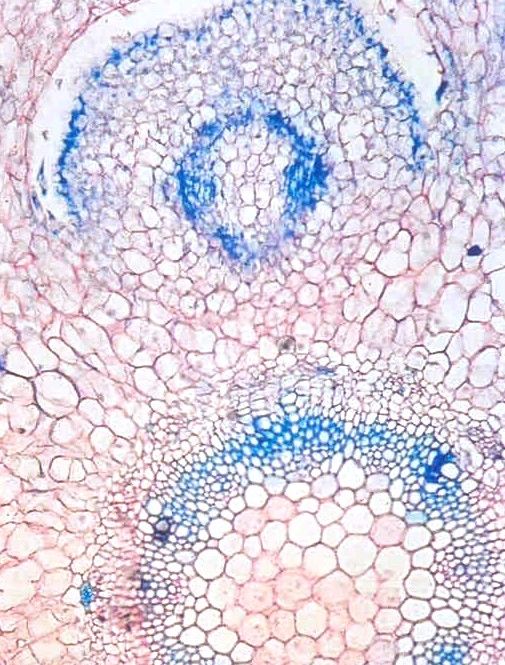
The obtained sections were colored either with fastgreen + saphranyne or with red-ruthen and
methyl-blue. After that, the sections were fixed in Canada balsam.

                                                   REGULATORI DE CREŞTERE (mg/L)
 Variants                Explant type
                                                   K    BAP    NAA    IAA   2,4-D
                   Nodal explants (basis)
    KN                                          2         -        2               -         -
                  Nodal explants (middle)
                   Nodal explants (basis)
    BI                                          -         2        -               2         -
                  Nodal explants (middle)
                   Nodal explants (basis)
    KI                                          2         -        -               2         -
                  Nodal explants (middle)
                   Nodal explants (basis)
    B2            Nodal explants (middle)       -         2        -               -         -
                      Apical explants
Table 1 – Experimantal variants used for testing the in vitro morphogenetic        reaction of
some explants frm Stevia rebaudiana Bertoni

                                     Results and discussions
          On KN medium the morphogenetic reaction of nodal explants was represented by
callus proliferation. This has different consistencies and colors: fragments of compact green
callus alternate with portions of light-yellow friable callus; is lately, a brownish friable
callus could be observed.
         The cross sections through the explant basis show the origin of this callus in the
cortical parenchyma of the stem. The general contour of the cross section is profoundly
affected by the callus proliferation. The epidermis, when is still present, have transversal
division walls.
          The areas with friable callus are formed by large cells, with very thin walls, with
large aeriferous spaces between them. In the external part these cells are elongated, and
they loose the contact one with another. In the areas with compact callus numerousness
meristematic nodules could be observed. They are formed from small, isodiametric cells,
with large nuclei. In the majority of the nodules short traheids, with reticulate thickness and
moderately lignified was differentiating.
         The central cylinder structure of the explant basis is almost intact. The xylem,
predominately by secondary origin has a ring shape. It is composed of vessels and xylemic
fibers with thick and moderate lignified walls. In some areas a proliferation of the phloem
zone could be observed. The pith is made up of isodiametric parenchymatic cells.
          The formation of new shoots is based on the presence of the axilar buds; their
growth is stimulated by the presence of the kinetine in the culture medium. The presence of
the callus in the basal part of the explant blocks the rhizogenesis process.
          The stem structure is quite normal [8]; in the cortical and in the pith parenchyma
division walls are visible. The vascular tissues have primary structure and are grouped in
collateral vascular bundles (photo 1). The epidermic cells are small, with thin walls and
                                                                                             30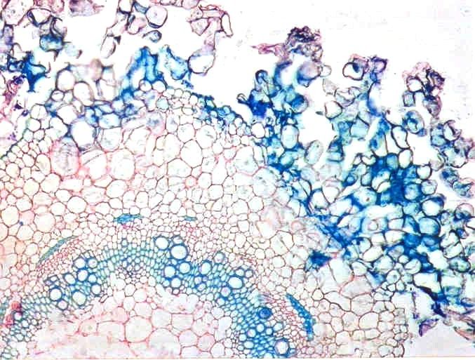
without a cuticle visible at the optic microscope. The axilar buds have a normal structure
(photo 2); the foliar primordia have a opposite disposition like at in vivo cultivated plants.
          On BI medium a small amount of callus is visible at the basis of the explant. This
is green, compact and homogenous.
          The presence of BAP in the culture medium stimulates the axilar buds growth
(photo 3); in the same time, adventitious buds are formed from the basal callus. Like on KN
medium, the presence of the callus on the explant basis block the roots formations. Sivaram
at al., (2003) [7] demonstrate that different explants (shoot apex, nodal explants, leaf
fragments) could produce shoots on a medium wit citoknins and auxins and the root
formation is induced in next subculture on a medium without auxins.
          The stem and leaf structure present small modifications. Isolately “twin apexes”
could appear because an anomalous ramification (photo 6). In a first stage, they have foliar
primordia only on external part, but subsequently their development is normal. The
anomalies of apex function could be the reason for the presence of a lot a shots on the
explant basis.
          The foliar primordia have normal structure, with a compact meristematic tissue; on
the epidermis tector and glandular hairs, in different stages of development could be
observed. The mature leaves are a quite normal structure, with no significant vitrifications
features (photo. 5). The mesophyll is differentiated into a unilayered palisade parenchyma
and a multilayered spongy one. The vascular bundle from the midvein is not prominent at
the abaxial side; it is formed by few xylem vessels, some phloemic elements and a
sclerenchyma girdle composed by cells with thick and lignified walls. The stomata are
numerousness, placed over the epidermis level. Most of them are unfunctional and have the
ostiole wide oped. However, the cuticle is thinner than that of the normal leaves.
          The structure of the new formed shoots is normal; in the central cylinder a
procambial ring could be observed (photo. 4). The vascular tissue will develop from it.
          On B2 medium a small callus could be observed at the basis of the initial explant.
Cross sections from this callus shows meristematic centers which will develop in shoots
apexes and then in stems. In the middle part of the explant, from the external part of the
cortex and from epidermic a friable callus appears (photo 7). It consists in large cells, have
no organization or meristemaric centers. The vascular bundles have secondary structure
(photo 8); the xylem is well developed, formed by vessels with thick and lignified walls,
xylem fibers and parenchyma cells. The phloem has a normal structure and is formed by
sieve tubes and companion cells.
          The mature leaves have a homogenous mesophyll, consist only in spongy
parenchyma. On KI medium the morphogenetic reaction was similar with that from BI
mediu. The process of shoots formation from axilar buds is intense. The contour of the stem
cross-section is circular; the epidermis persists on the explant exterior part, although it is
interrupted here and there, as a result of intense proliferation of the cortical parenchyma’s
cells (photo. 9). In some epidermal cells, division walls with different orientation may be
observed. Internal proliferation is more reduced than in the previously described situation.
The procambium suffers some additional divisions, yet the central cylinder maintains its
individuality (photo 10).
                                                                                              31
The cross sections from the new formed shoots show the proliferation of the
cortical parenchyma in a callus without any organization. The vascular tissues are well
developed, they have rings shapes: the internal one, of the xylem has vessels with thick and
lignified walls. At the phloem periphery several sclerenchyma fibers could be observed.
          At the basis of the explant, adventitious roots could be observed. They provided
from the stem perycicle and have a simple, diarch structure. The cortical parenchyma is
thick, with large aeriferous spaces between cells.
          The leaves structure is not constant. Some of them have a well developed
mesophyll, with one layer of elongated palisadic cells, without aeriferous spaces, and 4-5
layers of spongy parenchyma. The vascular bundles are small; the stomata are
numerousness, especially in the lower epidermis. Another has thick and undifferentiated
mesophyll consist only from spongy parenchyma.

                                               Conclusions

         In all experimental variants, at the basis of the explant a mass of callus appear.
That is more or less developed in relation with the hormonal balance. The largest callus was
formed on KN medium but only with histogenic potential. On B2 medium the callus has
organogenic capacity and was capable to regenerating shoots. On KN, BI and B2 medium
the direct caulogenesis was stimulate. Adventitious roots appear only on KI medium.

                                                References
1.   BONDAREV N. I., 2001. Peculiarities of propagation and development of Stevia rebaudiana Bertoni plants
     in vitro. Proceedings of 9th International conference of horticulture, Lednice, Czech Republic, 2: 431-434
2.   CACHITA - COSMA D., TIMOFTE V., RĂŞCĂNESCU M., 1993. Micropropagarea la Stevia rebaudiana
     Bert., Al V –lea Simp. Naţ. de culturi de ţesuturi şi celule vegetale, Bucureşti: 228 - 232
3.   CĂLIN A., BREZEANU A., 1997. Potenţialul morfogenetic in vitro la Stevia rebaudiana (Bert) Bertoni.
     Actualităţi şi perspective în biotehnologiile vegetale (Eds. Cachiţa C. D., Ardelean A., Crăciun C.) Arad:
     147 - 151
4.   MURASHIGE, T., SKOOG F., 1962. A revised medium for rapid growth and bioassays with tobacco tissue
     cultures. Physiol. Plant., 15, 473 - 497.
5.   NEPOVIM A., DRAHOSOVA H., VALICEK P., VANEK T., 1998. The Effect of Cultivation Conditions on
     the Content of Stevioside in Stevia rebaudiana Bertoni Plants Cultivated in the Czech Republic.
     Pharmaceutical and Pharmacological Letters, 8 (1): 19-21
6.   NEPOVIM, A., VANEK T., 1998. In vitro propagation of Stevia rebaudiana plants using multiple shoot
     culture. Planta Medica, 64 (8): 775-776
7.   SIVARAM L., MUKUNDAN U., 2003. In vitro culture studies on Stevia rebaudiana, In Vitro Cellular and
     Development Biology – Plant, 39 (5): 520-523
8.   TOMA I., ZBUGHIN G., IVĂNESCU L., 2003 – Histo-anatomical aspects concerning Stevia rebaudiana
     Bertoni, An. Şt. ale Univ. „Al. I. Cuza”, Iaşi, Biol. Veg., 34, (2):11 – 16
9.   YANG, Y.W., CHANG W. C., 1979. In vitro plant regeneration from leaf explants of Stevia rebaudiana
     Bertoni. Z. Pflanzenphysiol., 93: 337-343

                                                                                                           32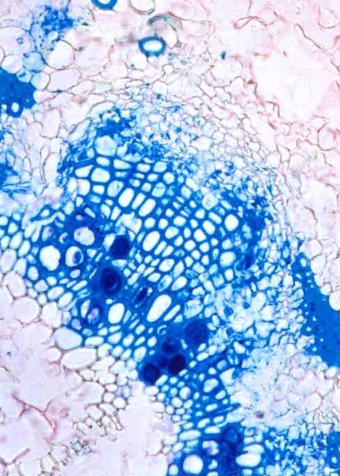
Photo 1     Photo 2

                  f.

        Photo 3    Photo 4

a. b

                  pcb

                             33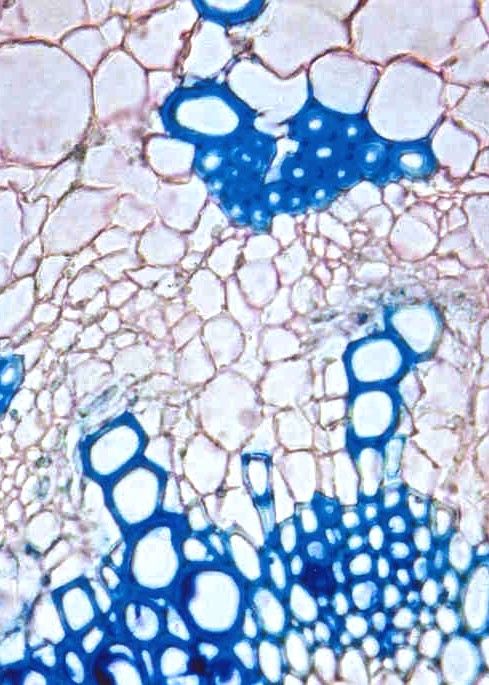
Photo 5

          p. p

                 s. p

                        st
Photo 6

                             34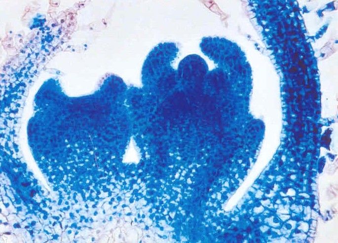
Photo 7
cl

     Photo 8

               35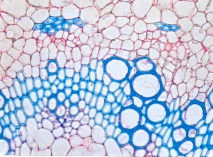
Photo 9              Photo 10

                     Photo 12
Photo 11

                                      36
Photo 13

Photo 14

            37
10. Yang Y.W., Hsing Y.I., Chang W.C., 1981. Cloning of Stevia rebaudiana through axillary shoot
    proliferation in vitro. Bot. Bull. Acad. Sin. 22: 57-62
11. Zbughin G., Cachiţa - Cosma D., Fati R., 1999. Stevia rebaudiana (Bertoni) – plantă de interes farmaceutic.
    Anal. Univ. Oradea, 4: 259 - 266

Explications of photos
Photo 1- Crossection through the shoot regenerated on KN medium
Photo 2 – Foliar primordia from a axilar bud (on KN medium)
Photo 3 - Crossection through the shoot and an axilar bud from BI medium
Photo 4 - Crossection through the shoot from BI medium
Photo 5 - Crossection through the leaf lamina (BI medium)
Photo 6 - Isolately “twin apexes” at the basis of the stem (longitudinal section)
Photo 7 – Crossection through the shoot from B2 medium
Photo 8 – Crossection through the shoot from B2 medium (detail from the vascular tissues)
Photo 9 - Crossection through the basal part of the shoot from KI medium
Photo 10 - Crossection through the basal part of the shoot from KI medium (detail from the
vascular tissues)
Photo 11 - Crossection through the leaf lamina (KI medium)
Photo 12 - Crossection through an adventitious root (KI medium)
Photo 13 -Crossection through the basal part of the initial explant from KI medium
Photo 14 – Crossection through the shoot from B2 medium (detail from epidermis –
transversals division walls could be observed)

Abreviations: a. b – axillary bud, c. c – central cylinder, cl – callus, f. p – foliar primordia,
p. p – palisade parenchyma, pcb - procambium, s.p – spongy parenchyma, x. vs – xylem
vessels (barr = 50 μm)

                                                                                                           38
You can also read