Identification and Classification of Rare Variants in NPC1 and NPC2 in Quebec
←
→
Page content transcription
If your browser does not render page correctly, please read the page content below

www.nature.com/scientificreports
OPEN Identification and Classification
of Rare Variants in NPC1 and NPC2
in Quebec
Lahoud Touma1,4,5, Marjorie Labrecque1,4,5, Martine Tetreault1,4* & Antoine Duquette1,2,3,4*
Niemann–Pick disease type C (NPC) is a treatable autosomal recessive neurodegenerative condition
which leads to a variety of progressive manifestations. Despite most cases being diagnosed at a young
age, disease prevalence may be underestimated, especially in adults, and interpretation of NPC1
and NPC2 variants can be difficult. This study aims to identify potential pathogenic variants in a large
cohort of healthy individuals and classify their risk of pathogenicity to assist with future interpretation
of variants. The CARTaGENE (CaG) cohort was used to identify possible variants of NPC1 and NPC2.
Nine-hundred and eleven RNA samples and 198 exome sequencing were screened for genetic variants
through a bio-informatic pipeline performing alignment and variant calling. The identified variants
were analyzed using annotations for allelic frequency, pathogenicity and conservation scores. The
ACMG guidelines were used to classify the variants. These were then compared to existing databases
and previous studies of NPC prevalence, including the Tübingen NPC database. Thirty-two distinct
variants were identified after running the samples in the RNA-sequencing pipeline, two of which were
classified as pathogenic and 21 of which were not published previously. Furthermore, 46 variants
were both identified in our population and with the Tübingen database, the majority of which were of
uncertain significance. Ten additional variants were found in our exome-sequencing sample. This study
of a sample from a population living in Quebec demonstrates a variety of rare variants, some of which
were already described in the literature as well as some novel variants. Classifying these variants is
arduous given the scarcity of available literature, even so in a population of healthy individuals. Yet
using this data, we were able to identify two pathogenic variants within our population and several
new variants not previously identified.
Niemann–Pick disease type C (NPC) is a rare, autosomal recessive neurodegenerative condition which leads
to a variety of progressive neurological and non-neurological manifestations. NPC is estimated to affect 1 in
100,000–120,000 live births1. It is caused by mutations of the NPC1 gene in 95% of cases while mutations of
NPC2 account for the other 5%. The spectrum of clinical presentation is wide with different onsets which have
been classified as: perinatal (shortly before and after birth, 3–12% of cases), early infantile (3 months to < 2 years,
3–37% of cases), late infantile (2 to < 6 years, 21–39% of cases), juvenile (6 to < 15 years, 21–54% of cases), and
adult (15 years and greater, 5–27% of cases)2,3. In the majority of cases, patients first develop liver, spleen or
lung involvement with a high variability of disease severity. These are then followed by progressive neurologi-
cal symptoms classically presenting with cerebellar ataxia, vertical supranuclear gaze palsy, gelastic cataplexy,
seizures and eventually dementia4. While studies of NPC2 are easily interpretable, NPC1 is highly polymorphic
where variants of unknown significance (VUS) are common, with one third of published variants being classi-
fied as VUS on ClinVar3,5.
NPC is one of the few degenerative ataxias for which a treatment is currently available. Miglustat (Zavesca)
has been approved in several countries for treating progressive neurological complications of N PC6. The drug has
been shown to slow progression of neurological manifestations in children without severe neurological symp-
toms when initiating therapy, as shown by nonsignificant improvement in horizontal saccadic eye movement
1
Départment de Neurosciences, Faculté de Médecine, Université de Montréal, CRCHUM – 900, rue Saint‑Denis,
Pavillon R, Montréal, QC H2X 0A9, Canada. 2Service de Neurologie, Département de Médecine, Unité de
Troubles du Mouvement André‑Barbeau, Centre Hospitalier de L’Université de Montréal (CHUM), Montreal,
Canada. 3Service de Médecine Génique, Département de Médecine, Centre Hospitalier de L’Université de Montréal
(CHUM), Montreal, Canada. 4Centre de Recherche du Centre Hospitalier de L’Université de Montréal (CRCHUM),
Montreal, Canada. 5These authors contributed equally: L. Touma and M. Labrecque. *email: martine.tetreault@
umontreal.ca; antoine.duquette@umontreal.ca
Scientific Reports | (2021) 11:10344 | https://doi.org/10.1038/s41598-021-89630-5 1
Vol.:(0123456789)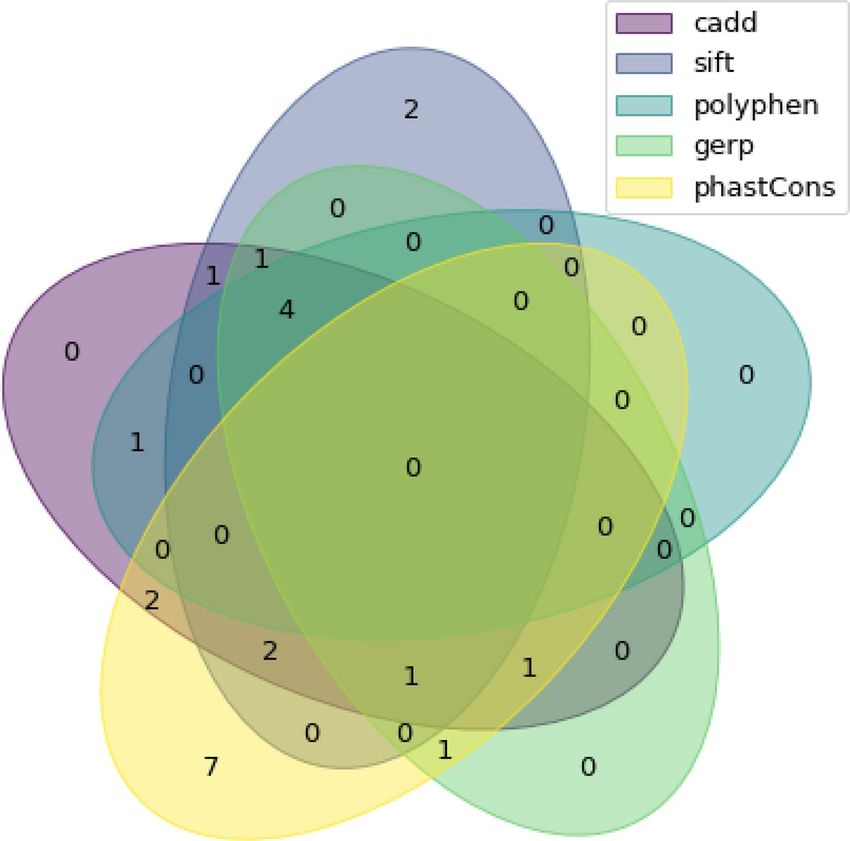
www.nature.com/scientificreports/
velocity in a preliminary open-label randomized controlled t rial7. Miglustat was also associated with improved
swallowing function and decreased aspiration risk in observational s tudies8,9.
Despite most cases being diagnosed at a young age, the adult-onset form can be more insidious and often
manifests with neuropsychiatric disturbances. The diagnosis is based on clinical evaluation and history with
biomarker screening. Several blood-based biomarkers can be used to assist with diagnosis including oxysterols,
lysosphingolipids and bile acid metabolites. Consensus guidelines still recommend completing the workup of
suspected cases with two additional diagnostic methods: filipin staining of unesterified fibroblasts or molecular
testing6,10. The former requires a skin biopsy with a specialized laboratory, but can be inconclusive in 15% of
cases without molecular t esting11. Molecular testing is more practical but can also be inconclusive in up to 15%
of cases mainly due to VUS and the absence of allele segregation s tudies2. The assessment of these variants will
require additional data input from various laboratories to allow for more specific classification.
CARTaGENE (CaG) is a cohort of healthy individuals living in Quebec12. The cohort contains a total of 43,000
individuals, including 55% of women, ranging between 40 and 69 years of age. The recruitment for this cohort
started in 2010 and participants have been followed up to this date. Genetic data is available for some of these
individuals in the form of RNA as well as exome sequencing. This data can thus be used to screen for potential
variants in a healthy pool of the population.
The study aim was to identify potential pathogenic variants in NPC1 and NPC2 in healthy individuals from the
CaG cohort and classify their risk of pathogenicity using the American College of Medical Genetics and Genomics
(ACMG) guidelines revised version of 201513 to help assist with the future interpretation of variants by providing
useful additional information derived from large databases.
Materials and methods
Initial data. This study was based on a random sample from the CaG cohort, which was representative of the
regional distribution of the Quebec population. Data acquisition was made from 911 individuals for the RNA-
sequencing (RNA-seq) and 198 individuals for exome-sequencing (exome-seq). 93 of these individuals were in
both RNA-seq and exome-seq. A bio-informatic pipeline was therefore used to analyse a total of 1016 individu-
als. Baseline characteristics as well as screening medical questionnaires were obtained for each participant from
the CaG database. Informed consent was previously obtained by CaG researchers for all study participants. The
Sample and Data Access Committee (SDAC) of CaG approved the use of the genetic and baseline characteristics
for our study. All genetic and bioinformatic analysis were carried out in accordance with relevant guidelines
and regulations. Our protocol was approved by our institution’s research ethics board (CR-CHUM REB, Project
18.116).
Bio‑informatic pipeline. The FASTQ files were aligned to the reference genome (Hg19) using BWA for
exome sequencing and STAR for RNA-sequencing14,15. In both cases, variant calling was performed with GATK
and annotated using ANNOVAR and custom scripts16,17. The bioinformatic pipeline in place allows the detection
of single nucleotide variants (SNV) either non-synonymous, splice junction or synonymous, multi-nucleotide
variants (MNV) and indels.
Since NPC1 (NM_000271) and NPC2 (NM_006432) genes are located on chromosome 18 or 14 respectively,
we only extracted variants on those chromosomes for each sample. We also added information from the NP-C
database (NPC-db2) made by the University of Tübingen, using a custom Python script. The database was last
searched in July 2019. When a variant was found in the NPC-db2, its pathogenicity classification based on their
criteria was added to the resulting file.
Analysis. As we were identifying rare variants, common variants (defined as > 1%) found in dbSNP, 1000
Genomes, Exome Variant Server, GnomAD and internal databases were filtered out. Non-synonymous, puta-
tive splicing variants and coding indels were prioritized. More specifically, we set a threshold for a CADD score
higher than 15, a Polyphen2 score higher than 0.75 with a score of one being very likely pathogenic, a SIFT score,
that was reversed to match the Polyphen2 score, with the same criteria as Polyphen2 to filter the variants18–22. For
the conservation scores, we identified the variants with a score higher than 500 for Phast cons and higher than 5
for GERP. Phast cons ranges between 0 and 1000 and the GERP score between − 12.3 and 6.17.
Classification. The ACMG 2015 revised guidelines were used to classify the different variants. The classifica-
tion uses five distinct categories: benign, likely benign, uncertain significance, likely pathogenic and pathogenic.
Each variant was analyzed using the ACMG criteria except the ones requiring segregation and laboratory data
which were not available for our dataset. We extracted the NPC-db2 classification for each variant. Thereafter,
the variants were searched on ClinVar for previous classification by other groups, using the ACMG criteria.
Classifications based on other sets of criteria were not included in our tables. Finally, we searched the largest
published study on NPC variants by Wassif et al. for identical variants already identified in their results. Previ-
ously unreported variants will be submitted to the ClinVar public database.
Results
Baseline characteristics. The total sample size for both RNA-seq and exome-seq was 1016 patients and
2032 chromosomes. Clinical data was available for 1004 patients (Table 1). Females represented 51% of our
population. The highest represented ethnicity was white from European descent (91.5%). The majority of these
individuals were employed (64.3%). Age ranged from 44 to 69, with 42% of patients in the 40–49 range. This
sample has a similar distribution as the general CaG cohort (Table 1). Additionally, the sample is also representa-
tive of the Quebec population based on the most recent epidemiological d ata23.
Scientific Reports | (2021) 11:10344 | https://doi.org/10.1038/s41598-021-89630-5 2
Vol:.(1234567890)www.nature.com/scientificreports/
Sample (%) CaG (%) Quebec (%)
Female 50.6 55 50.0
Age
40–49 42.3 42 31.3
50–59 29.0 37 32.6
60–69 28.7 25 36.1
Ethnicity
White (European Descent) 91.5 87.0b
Arab 2.2 2.7b
Black (African or Caribean descent) 1.7 4.0b
Employed 64.3 67 62.4
Highest level of education
University 37.1 45 31.2a
College 49.8 32 45.2a
Table 1. Baseline characteristics. a Data for age range of 35–64 in the Quebec population (Banque de données
des statistiques officielles sur le Québec 2015). b Data for overall province of Quebec (Statistics Canada 2016).
RNA‑seq. Our study identified 32 unique rare variants from the 911 RNA samples that were run in the bio-
informatic pipeline (Table 2, Fig. 1). Each variant was only present in one chromosome, for an allele frequency
of 0.05% in our population. None of the study participants were heterozygous for two rare variants. Twenty of
these variants were non-synonymous SNVs while the others were frameshift deletions. Among these variants,
two were classified as pathogenic. Indeed, the p.Ile1061Thr is a known protein change that leads to a change
from isoleucine to threonine. This variant has been described as causative of NPC in 15–20% of disease alleles
in the United States and Europe. Additionally, biological studies have shown that this missense change affects
proper protein localization and causes proteasomal degradation in cell culture. Another pathological variant,
p.Pro543Leu, has been identified in 1 homozygous and 4 compound heterozygous individuals with sympto-
matic disease24. It has previously been reported that this mutation leads to early-infantile form of N PC25. Both
participants were heterozygous for these mutations and were asymptomatic according to the baseline medical
screening obtained from CaG. The remaining twelve variants were indels and all were classified as VUS. Given
the limited coverage, these could represent artifacts and their exact significance is difficult to interpret.
Comparison with other databases. The NPC-db2 database was searched for identified variants which
were also present in our population. Twelve out of the 32 variants identified were also present in their sample.
Our classification based on the ACMG criteria was overall very similar to their classification for shared variants.
Notably, the p.Pro543Leu protein change was marked as potentially pathogenic in NPC-db2, while we were
able to classify it as pathogenic based on previous publications and computational/predictive data. Addition-
ally, we identified 13 variants that were filtered out by our bioinformatic pipeline, but that were present both in
our population and in the NPC-db2 database (Table 3). These were all previously classified as benign. The main
reason for their exclusion in the pipeline was a high allelic frequency (> 1%).
The variants in the study by Wassif et al. were also compared with variants identified in our study (Table 2)26.
Despite not specifically using the ACMG criteria, we were able to compare their five-level scale of classification
to our data. Twelve out of the 32 variants were also classified in their study. One notable difference in classifica-
tion in the variant p.Ile1061Thr was probably due to a mistake in their table as they present it as benign, while
describing it as on of the most common pathogenic variant in their t ext26. Moreover, five variants that were in
both our databases were excluded by our pipeline. Once again, these were classified as benign and the main
reason was a high allelic frequency (> 1%).
Exome‑seq. Exome sequencing was performed for 198 individuals, composed of 93 individuals for whom
we also had the RNA-seq and 105 new individuals for whom we only had exome-seq. Overall, 19 unique variants
were identified in the samples, 4 of which were also present in the RNA-seq (Fig. 2). In participants for whom
both RNA-seq and exome-seq data were available, the exact same sequence variants were found using both
methods. After filtering by the bioinformatic pipeline, 10 variants were identified. Four of those were already
found in our RNA-seq, including one classified as pathogenic (Table 4). The six other variants were found in
individuals for which we only had exome data. These included two variants in splicing regions, one of which
ublications24,27. The participant was heterozygous for this variant and was
was causative of disease in previous p
asymptomatic according to baseline medical screening obtained from CaG. The RNA-seq for these individuals
were not available to confirm the presence of abnormal splicing.
Discussion
Our study evaluated rare variants in NPC1 and NPC2 genes in a sample from the Quebec population in Canada.
This population is unique given the important founder effect from French colonisation in the early seventeenth
century28. We had 911 RNA samples and 198 exome samples, with an overlap of 93 individuals. This study
Scientific Reports | (2021) 11:10344 | https://doi.org/10.1038/s41598-021-89630-5 3
Vol.:(0123456789)www.nature.com/scientificreports/
Classification in
Variants Variations Ref Alt Protein Change Gene ACMG Classification NPC-db2 ClinVar (ACMG) Wassif et al
chr18:21121118 Nonsynonymous SNV C A p.V810F NPC1 3 2 3 –
chr18:21140411 Nonsynonymous SNV T C p.N222S NPC1 2 1 3 Benign
chr18:21136233 Nonsynonymous SNV G A p.P434S NPC1 2 1 – Benign
chr18:21140367 Nonsynonymous SNV G A p.P237S NPC1 2 1 1 Benign
chr18:21113406 Nonsynonymous SNV T C p.I1223V NPC1 2 2 – Benign
chr18:21119839 Nonsynonymous SNV C T p.G911S NPC1 2 1 – Benign
chr18:21131617 Nonsynonymous SNV G A p.P543L NPC1 5 4 5 Probably damaging
chr18:21121386 Nonsynonymous SNV C T p.V753M NPC1 3 2 3 Benign
chr18:21114442 Nonsynonymous SNV C T p.A1187T NPC1 3 – 3 –
chr18:21140243 Nonsynonymous SNV G A p.A278V NPC1 3 – – –
chr18:21136410 Nonsynonymous SNV T C p.T375A NPC1 3 – – –
chr18:21134806 Nonsynonymous SNV T C p.N490S NPC1 3 – – –
chr18:21134743 Nonsynonymous SNV G A p.T511M NPC1 2 2 – Probably damaging
chr18:21118536 Nonsynonymous SNV G A p.S1004L NPC1 3 2 – Probably damaging
chr14:74959920 Nonsynonymous SNV C T p.E20K NPC2 3 – – –
chr18:21136422 Nonsynonymous SNV C A p.V371F NPC1 3 – – –
chr18:21140315 Nonsynonymous SNV G T p.P254Q NPC1 3 – – –
chr18:21136439 Nonsynonymous SNV G A p.S365L NPC1 3 – – Possibly damaging
chr18:21121045 Nonsynonymous SNV A G p.M834T NPC1 3 2 – Benign
chr18:21116700 Nonsynonymous SNV A G p.I1061T NPC1 5 5 5 Benign
chr18:21153473 Frameshift deletion AT A p.N41fs NPC1 3 – – –
chr18:21125100 Frameshift deletion CA C p.F590fs NPC1 3 – – –
chr18:21141470 Frameshift deletion TC T p.D162fs NPC1 3 – – –
chr18:21121129 Frameshift deletion TC T p.D806fs NPC1 3 – – –
chr18:21140314 Frameshift deletion TG T p.P254fs NPC1 3 – – –
chr18:21119811 Frameshift deletion AC A p.V920fs NPC1 3 – – –
chr18:21120443 Frameshift deletion AT A p.I858fs NPC1 3 – – –
chr18:21153486 Frameshift deletion TC T p.D37fs NPC1 3 – – –
chr18:21124431 Frameshift deletion AG A p.A669fs NPC1 3 – – –
chr18:21116757 Frameshift deletion TG T p.H1042fs NPC1 3 – – –
chr18:21114436 Frameshift deletion CTT C p.E1188fs NPC1 3 – – –
chr18:21140211 Frameshift insertion C CA p.A289fs NPC1 3 – – –
Table 2. Rare variants in CARTaGENE sample, RNA-seq. a Classification 1: Benign 2: Likely benign 3:
Uncertain significance 4: Likely pathogenic 5: Pathogenic.
presents new variants that have not been previously described in the literature. In addition, known variants were
reclassified based on the most recent literature. In fact, we classified each individual variant based on the ACMG
2015 guidelines and compared them to the NPC-db2 database and previously published studies on such variants.
The majority of identified variants were of uncertain significance, likely benign or benign. However, we have also
identified some likely pathogenic and pathogenic variants in heterozygous individuals.
To select rare variants in our population, we used a pre-specified bioinformatic pipeline. The variants were
filtered based on their allelic frequency, with a coverage of at least 15% and where the alternative base was sup-
ported at least twice. This ensured that the classification would be applied to the most pertinent variants in our
sample. We then focused our analysis on indels and non-synonymous variants, which were more likely to lead
to pathogenic mutations. Additional variants were manually identified by comparing all variants present in our
sample to those in the NPC-db2 database. These were filtered out from our pipeline according to the aforemen-
tioned criteria but were still classified by our laboratory because they were coincidentally present in another
study population. The main reason for exclusion was allelic frequency > 1%.
With the increased use of genetic testing and the identification of more variants, it has become essential to
apply rigorous classification in clinical genetic t esting29. The set of criteria must be evidence-based, standard-
ized and o bjective30. The ACMG 2015 guidelines, used in our study, have been largely used and therefore allow
for easier comparison with previous publications. Individual laboratories also share their own classification in
large databases (including ClinVar), but it is difficult to compare with their conclusions as the set of criteria is
different. Thus, we have only compared our classification with published literature using the same set of criteria.
The CARTaGENE database encompasses a large sample of genomic data, but also baseline information
based on detailed questionnaires. Answers included demographics, socioeconomic status, education and medi-
cal surveys. Given the possible adult-onset of NPC, we searched for potential symptoms in the questionnaires of
Scientific Reports | (2021) 11:10344 | https://doi.org/10.1038/s41598-021-89630-5 4
Vol:.(1234567890)www.nature.com/scientificreports/
Figure 1. Venn diagram of included variants from RNA-seq for each bioinformatic filter.
Protein ACMG Classification ClinVar
Variants Variations Ref Alt Change Gene Classification in NPC-db2 (ACMG) Wassif et al
nonsynony-
chr18:21124945 C G p.M642I NPC1 1 1 1 –
mous SNV
nonsynony-
chr18:21112206 C T p.R1266Q NPC1 1 1 1 Benign
mous SNV
nonsynony-
chr18:21120444 T C p.I858V NPC1 1 1 1 Benign
mous SNV
nonsynony-
chr18:21140432 T C p.H215R NPC1 1 1 1 Benign
mous SNV
nonsynony-
chr18:21140367 G A p.P237S NPC1 1 1 1 Benign
mous SNV
nonsynony-
chr18:21136233 G A p.P434S NPC1 1 1 – Benign
mous SNV
nonsynony-
chr18:21140411 T C p.N222S NPC1 1 1 2, 3 Benign
mous SNV
synonymous
chr18:21148863 A G p.Y129Y NPC1 1 1 – Benign
SNV
synonymous
chr18:21115579 G A p.L1111L NPC1 2 1 – –
SNV
synonymous
chr18:21124335 G A p.N701N NPC1 1 1 – –
SNV
synonymous
chr18:21134772 G A p.D501D NPC1 2 1 – –
SNV
synonymous
chr18:21114440 C A p.A1187A NPC1 2 1 – –
SNV
synonymous
chr18:21124365 C T p.P691P NPC1 2 1 – –
SNV
Table 3. Variants in the CARTaGENE RNA-seq sample excluded from the pipeline but identified in NPC-db2.
a
Classification 1: Benign 2: Likely benign 3: Uncertain significance 4: Likely pathogenic 5: Pathogenic.
Scientific Reports | (2021) 11:10344 | https://doi.org/10.1038/s41598-021-89630-5 5
Vol.:(0123456789)www.nature.com/scientificreports/
Figure 2. Venn diagram of included variants from exome-seq for each bioinformatic filter.
Classification in
Variants Variations Ref Alt Protein Change Gene ACMG Classification NPC-db2 ClinVar (ACMG) Wassif et al
chr18:21118536 nonsynonymous SNV G A p.S1004L NPC1 3 2 4 Probably damaging
chr18:21134806 nonsynonymous SNV T C p.N490S NPC1 3 – – –
chr18:21121118 nonsynonymous SNV C A p.V810F NPC1 3 2 3 –
chr18:21118618 nonsynonymous SNV C T p.V977I NPC1 3 – – –
chr18:21116700 nonsynonymous SNV A G p.I1061T NPC1 5 5 5 Pathogenic
chr18:21115615 nonsynonymous SNV T C p.I1099V NPC1 3 – – –
chr14:74947404 splicing C T NPC2 3 – 3 Strong negative
chr14:74953027 splicing-extended C T NPC2 3 – 3, 5 –
chr14:74959920 nonsynonymous SNV C T p.E20K NPC2 3 – – –
chr14:74951269 nonsynonymous SNV T C p.K71R NPC2 3 – – Probably damaging
Table 4. Rare variants in CARTaGENE sample, exome-seq. a Classification 1: Benign 2: Likely benign 3:
Uncertain significance 4: Likely pathogenic 5: Pathogenic.
patients with pathogenic or likely pathogenic variants. None of the identified individuals presented symptoms
suggestive of the disease, as we had expected given the heterozygosity of the alleles. These pathogenic variants
will allow us to estimate the carrier frequency in the Quebec population.
Our study has several potential limitations. First, our dataset is based on a relatively small sample size of
1016 individuals. However, given the important founder effect in our Quebec population pool, genetic variation
is relatively lower when compared to other p opulations31. Second, we did not perform any functional biology
experiments which limits our ability to classify some of these mutations based on functional criteria. Third, no
segregation data was available in the database, which can often provide strong evidence for a benign variant in
a new mutation.
In brief, this study analyzed variants in the NPC1 and NPC2 genes from a representative sample of the Quebec
population. The results described novel variants that were not previously described in the literature. In addition,
known variants were reclassified using the ACMG guidelines. Despite identifying pathogenic or likely pathogenic
variants, the individuals were heterozygous and asymptomatic based on baseline questionnaires. Classifying
these variants is arduous given the scarcity of available literature, even so in a population of healthy individu-
als, leading to a large proportion of variants of uncertain significance. Using this data, we were able to identify
three pathogenic variants within our population and several new rare variants in NPC1 and NPC2 which had
Scientific Reports | (2021) 11:10344 | https://doi.org/10.1038/s41598-021-89630-5 6
Vol:.(1234567890)www.nature.com/scientificreports/
not previously been identified. This additional information should help clinicians interpret the pathogenicity of
variants identified in these two genes moving forward.
Data availability
Data was provided by the CARTaGENE database from a sample from the Quebec population. The data gener-
ated or analyzed during this study are included in this published article and its supplementary information files.
Code availability
We used publicly and freely accessible codes, referenced throughout our method section. Our custom Python
scripts are available if necessary.
Received: 4 March 2021; Accepted: 29 April 2021
References
1. Garver, W. S. et al. The national Niemann–Pick C1 disease database: report of clinical features and health problems. Am. J. Med.
Genet. A 143a, 1204–1211. https://doi.org/10.1002/ajmg.a.31735 (2007).
2. Vanier, M. T. Niemann–Pick disease type C. Orphanet J. Rare Dis. 5, 16. https://doi.org/10.1186/1750-1172-5-16 (2010).
3. Nadjar, Y. & Vanier, M. T. In Neurometabolic Hereditary Diseases of Adults (ed. Burlina, A. P.) 121–146 (Springer, 2018).
4. Yanjanin, N. M. et al. Linear clinical progression, independent of age of onset, in Niemann–Pick disease, type C. Am. J. Med. Genet.
B Neuropsychiatr Genet. 153b, 132–140. https://doi.org/10.1002/ajmg.b.30969 (2010).
5. Information., N. C. f. B. (ClinVar, 2020).
6. Patterson, M. C. et al. Recommendations for the diagnosis and management of Niemann–Pick disease type C: an update. Mol.
Genet. Metab. 106, 330–344. https://doi.org/10.1016/j.ymgme.2012.03.012 (2012).
7. Patterson, M. C., Vecchio, D., Prady, H., Abel, L. & Wraith, J. E. Miglustat for treatment of Niemann–Pick C disease: a randomised
controlled study. Lancet Neurol. 6, 765–772. https://doi.org/10.1016/s1474-4422(07)70194-1 (2007).
8. Fecarotta, S. et al. The videofluoroscopic swallowing study shows a sustained improvement of dysphagia in children with Nie-
mann–Pick disease type C after therapy with miglustat. Am. J. Med. Genet. A 155a, 540–547. https://d oi.o
rg/1 0.1 002/a jmg.a.3 3847
(2011).
9. Solomon, B. I. et al. Association of miglustat with swallowing outcomes in Niemann–Pick disease, type C1. JAMA Neurol. 77,
1564–1568. https://doi.org/10.1001/jamaneurol.2020.3241 (2020).
10. Porter, F. D. et al. Cholesterol oxidation products are sensitive and specific blood-based biomarkers for Niemann–Pick C1 disease.
Sci. Transl. Med. 2, 56ra81. https://doi.org/10.1126/scitranslmed.3001417 (2010).
11. Vanier, M. T. & Latour, P. Laboratory diagnosis of Niemann–Pick disease type C: the filipin staining test. Methods Cell Biol. 126,
357–375. https://doi.org/10.1016/bs.mcb.2014.10.028 (2015).
12. Godard, B., Marshall, J. & Laberge, C. Community engagement in genetic research: results of the first public consultation for the
Quebec CARTaGENE project. Community Genet 10, 147–158. https://doi.org/10.1159/000101756 (2007).
13. Richards, S. et al. Standards and guidelines for the interpretation of sequence variants: a joint consensus recommendation of the
American college of medical genetics and genomics and the association for molecular pathology. Genet. Med. 17, 405–424. https://
doi.org/10.1038/gim.2015.30 (2015).
14. Li, H. & Durbin, R. Fast and accurate short read alignment with Burrows–Wheeler transform. Bioinformatics 25, 1754–1760.
https://doi.org/10.1093/bioinformatics/btp324 (2009).
15. Dobin, A. et al. STAR: ultrafast universal RNA-seq aligner. Bioinformatics 29, 15–21. https://d oi.o
rg/1 0.1 093/b ioinf ormat ics/b
ts635
(2012).
16. McKenna, A. et al. The genome analysis toolkit: a MapReduce framework for analyzing next-generation DNA sequencing data.
Genome Res. 20, 1297–1303. https://doi.org/10.1101/gr.107524.110 (2010).
17. Wang, K., Li, M. & Hakonarson, H. ANNOVAR: functional annotation of genetic variants from high-throughput sequencing data.
Nucleic Acids Res. 38, e164–e164. https://doi.org/10.1093/nar/gkq603 (2010).
18. Rentzsch, P., Witten, D., Cooper, G. M., Shendure, J. & Kircher, M. CADD: predicting the deleteriousness of variants throughout
the human genome. Nucleic Acids Res. 47, D886–D894. https://doi.org/10.1093/nar/gky1016 (2018).
19. Adzhubei, I., Jordan, D. M. & Sunyaev, S. R. Predicting functional effect of human missense mutations using polyPhen-2. Curr.
Protoc. Hum. Genet. 76, 7.20.21-27.20.41. https://doi.org/10.1002/0471142905.hg0720s76 (2013).
20. Ng, P. C. & Henikoff, S. SIFT: predicting amino acid changes that affect protein function. Nucleic Acids Res. 31, 3812–3814. https://
doi.org/10.1093/nar/gkg509 (2003).
21. Siepel, A. et al. Evolutionarily conserved elements in vertebrate, insect, worm, and yeast genomes. Genome Res. 15, 1034–1050.
https://doi.org/10.1101/gr.3715005 (2005).
22. Davydov, E. V. et al. Identifying a high fraction of the human genome to be under selective constraint using GERP++. PLOS
Comput. Biol. 6, e1001025. https://doi.org/10.1371/journal.pcbi.1001025 (2010).
23. Québec, I. d. l. s. d. (2019).
24. Millat, G. et al. Niemann–Pick disease type C: spectrum of HE1 mutations and genotype/phenotype correlations in the NPC2
group. Am. J. Hum. Genet. 69, 1013–1021. https://doi.org/10.1086/324068 (2001).
25. Verot, L. et al. Niemann–Pick C disease: functional characterization of three NPC2 mutations and clinical and molecular update
on patients with NPC2. Clin. Genet. 71, 320–330. https://doi.org/10.1111/j.1399-0004.2007.00782.x (2007).
26. Wassif, C. A. et al. High incidence of unrecognized visceral/neurological late-onset Niemann–Pick disease, type C1, predicted by
analysis of massively parallel sequencing data sets. Genet. Med. 18, 41–48. https://doi.org/10.1038/gim.2015.25 (2016).
27. Yang, Y. et al. Molecular findings among patients referred for clinical whole-exome sequencing. JAMA 312, 1870–1879. https://
doi.org/10.1001/jama.2014.14601 (2014).
28. Moreau, C., Vézina, H. & Labuda, D. Founder effects and genetic variability in Quebec. Med. Sci. (Paris) 23, 1008–1013. https://
doi.org/10.1051/medsci/200723111008 (2007).
29. Kumar, D. From evidence-based medicine to genomic medicine. Genom. Med. 1, 95–104. https://doi.org/10.1007/s11568-007-
9013-6 (2007).
30. Rehm, H. L. et al. ClinGen–the clinical genome resource. N. Engl. J. Med. 372, 2235–2242. https://doi.org/10.1056/NEJMsr1406
261 (2015).
31. Tremblay, M., Vézina, H., Desjardins, B. & Houde, L. In Kinship and Demographic Behavior in the Past (eds Bengtsson, T. & Mineau,
G. P.) 259–277 (Springer, 2008).
Scientific Reports | (2021) 11:10344 | https://doi.org/10.1038/s41598-021-89630-5 7
Vol.:(0123456789)www.nature.com/scientificreports/
Acknowledgements
The authors would like to highlight the collaboration with the CARTaGENE database for the genetic and survey
data used in this study. The authors would also like to express their appreciation to Dr Eric Bareke and Dr Alina
Levtova for their great support in designing the methodology of this study
Author contributions
L.T. and M.L. wrote the main manuscript text and designed the tables. M.L., M.T. and A.D. designed the protocol.
M.L. and M.T. ran the data through the bio-informatic pipeline and performed the analysis. L.T. completed the
classification. All authors reviewed the manuscript.
Funding
We received an unrestricted educational grant from Actelion Pharmaceutiques Canada, RMGA. M.T. received
a Junior 1 salary award from Fond de recherche du Québec—Santé. M.L. received an Excellence bursary from the
Bioinformatic program, Université de Montréal.
Competing interests
The authors declare no competing interests.
Additional information
Correspondence and requests for materials should be addressed to M.T. or A.D.
Reprints and permissions information is available at www.nature.com/reprints.
Publisher’s note Springer Nature remains neutral with regard to jurisdictional claims in published maps and
institutional affiliations.
Open Access This article is licensed under a Creative Commons Attribution 4.0 International
License, which permits use, sharing, adaptation, distribution and reproduction in any medium or
format, as long as you give appropriate credit to the original author(s) and the source, provide a link to the
Creative Commons licence, and indicate if changes were made. The images or other third party material in this
article are included in the article’s Creative Commons licence, unless indicated otherwise in a credit line to the
material. If material is not included in the article’s Creative Commons licence and your intended use is not
permitted by statutory regulation or exceeds the permitted use, you will need to obtain permission directly from
the copyright holder. To view a copy of this licence, visit http://creativecommons.org/licenses/by/4.0/.
© The Author(s) 2021
Scientific Reports | (2021) 11:10344 | https://doi.org/10.1038/s41598-021-89630-5 8
Vol:.(1234567890)You can also read