Introduction to the principles of small animal surgery - Daniel ...
←
→
Page content transcription
If your browser does not render page correctly, please read the page content below
Universität Zürich
Introduction to the principles of small animal surgery   page 1
    Kleintierchirurgie

    Introduction to the principles of
    small animal surgery

   1st Continuing Education Course for Japan Small
   Animal Surgeons at the Small Animal Surgery Clinic,
   University of Zürich, Switzerland
   July 13 – 18, 2001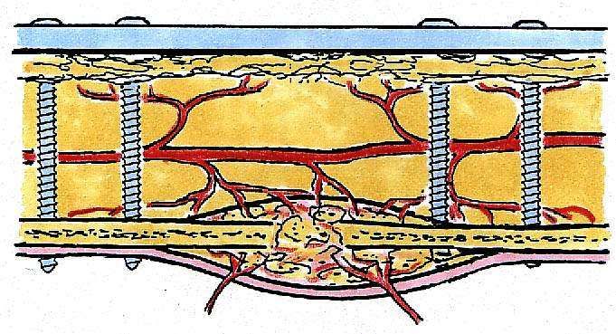
Introduction to the principles of small animal surgery                                         page 2

Contents:

Course overview                                                                                3
Halsted's principles of surgery                                                                5
Suture techniques                                                                              8
Wound debridement and wound dressings                                                         13
Orthopaedic bandages and slings                                                               16
Basic skin course                                                                             20
Introduction to the principles of simple osteosynthesis                                       23
Basic course in fracture treatment                                                            26
Balanced anaesthesia                                                                          32
Oesophagostomy and gastrostomy feeding tubes                                                  35
Celiotomy, exploration, biopsy techniques                                                     37
Splenectomy                                                                                   42
Enterectomy, anastomosis, bowel plication                                                     44
Gastropexy                                                                                    47
Diaphragmatic hernia                                                                          50
Reconstruction techniques                                                                     52
External fixator on the humerus                                                               54
Tension band wiring and parallel pinning on proximal humerus fracture                         56
Metacarpal and metatarsal fractures                                                           58
Fixation of a radius / ulna fracture with a tubular fixateur externe                          60
Femoral head and neck fractures                                                               62
Distal femoral fractures                                                                      65
Trilam nail                                                                                   67

Impressum

The authors of this handout are the assistants of the small animal surgery clinic of the University of
Zurich. The work was revised by Prof. P. M. Montavon and edited by Dr. D. Koch. Most of the pictures
were drawn by M. Haab.
First edition: June 2001, copyright by Small animal surgery clinic.
Introduction to the principles of small animal surgery                                           page 3

Course overview

 13.07.01 Introduction to the principles of small animal surgery
          Zurich University
          Chair: Montavon / Koch

              0900      Tour in Zurich                                            De Robillard
              1200      Lunch
              1300      Welcome, goal of the course                               Montavon
              1315      Halsted principles                                        Voss
              1345      Suture techniques                                         Keller
              1515      Break
              1530      Basic skin course                                         Grundmann
              1730      End of the day                                            Montavon
              1830      Apero at the hotel                                        Koch

 14.07.01 0830          Wound toilet and wound dressings                          Dennler
          0930          Orthopaedic bandages and slings                           Koch
          1100          Introduction to the principles of fracture repair         Koch
          1200          Lunch, end of morning session
                        Afternoon: shopping in Zurich

 15.07.01 Social programm
          Bernese mountains
          Chair: NN

              0800      Departure at the hotel
              All       Visit in the Bernese mountains, Jungfraujoch              Faculty
              day

 16.07.01 Basic course in fracture treatment
          Bettlach
          Chair: Schwab (AO)

              0730      Departure at the hotel
              0900      Basic course in fracture treatment:                         Schwab
              –         Several excercises on plastic bones with original material, Montavon
              1700      short visit at the factory                                  Koch
              1830      Arrival at the hotel
Introduction to the principles of small animal surgery                                            page 4

 17.07.01 Introduction to anesthesia, abdominal surgery on cadavers
          Zurich University
          Chair: Montavon / Koch

              0830      Introduction to anesthesia                               Boller
              0930      Feeding tubes                                            Keller
              1030      Break
              1050      Celiotomy, exploration, biopsy techniques                Dennler
              1200      Lunch
              1300      Splenectomy                                              Damur
              1345      Enterectomy, anastomosis                                 Voss
              1445      Gastropexy                                               Haas
              1545      Hernia diaphragmatica                                    Dennler
              1630      Reconstruction techniques (omentum, serosal patching,    Koch
                        mesh, allografts)
              1715      End of the day

 18.07.01 Simple fracture treatment on cadavers
          Zurich University
          Chair: Montavon / Koch

              0830      External skeletal fixator on the humerus (tie-in)        Bass
              0930      Tension band wiring and parallel pinning on proximal hu- Bass
                        merus fracture
              1030      Break
              1045      Metacarpal fracture treatment with pinning               Keller
              1130      Lunch
              1230      External fixator (tubular) radius / ulna fracture        Voss / Dennler
              1330      Pinning technique for femoral neck fracture              Koch
              1430      Cross-pinning for distal femoral Salter Harris fracture  Damur
              1530      Break
              1600      Tibia fracture: it is your decision                      Koch
              1700      Assessment of postoperative radiographs                  Montavon
              1730      End of the day
              1900      Farewell dinner, certificates                            Faculty
Introduction to the principles of small animal surgery page 5
Halsted’s Principles of Surgery
Katja Voss, Dr. med. vet.; Curzio Bernasconi, Dr. med. vet. FVH
Introduction
Around the middle of the 19th century two important discov-
eries were made to bring surgery to the present state: the
discovery of general anesthesia and the recognition of bacte-
ria as a source of infection. These changes enabled surgeons
to pay more attention on asepsis and surgical techniques.
instead of having to operate as quick as possible on a con-
scious patient.
William S. Halsted (1852-1922) was a famous surgeon living
in that period. His contributions to surgery were in the fields
of asepsis, local and regional anesthesia and vascular, thy-
roid, intestinal and cancer surgery. Halsted propagated the
importance of asepsis and the need for gentle tissue handling
during operations. His surgical principles are still true at pre-
sent. Their common goal is the prevention of infections and
the promotion of rapid tissue healing:
1. Gentle tissue handling
Every operation causes damage to the tissues, inflammatory reactions and pain. A certain degree
of inflammation is part of the healing process. But rough tissue handling leads to tissue necrosis,
the accumulation of fluid and debris in the wound bed and to a decrease in vascularization. The
larger the amount of debris the longer it will take phagoycytic cells to remove it. Migration of fi-
broblasts and capillaries takes place after the “cleaning-up” of a wound and will therefore also be
delayed. In addition to the prolonged healing time infections are more likely to occur.
The use of appropriate instruments and correct surgical techniques help to treat tissues as gentle
as possible and to minimize tissue damage, pain and infections. Some examples are:
- Sharp dissection whenever possible (scalpel blade instead of scissors)
- The least possible manipulation of tissues with appropriate instruments (Adson Brown forceps
for skin, De Bakey forceps for abdominal surgery, oscillating drill for pelvic and spinal fracture
repair)
- Gentle retraction of important structures like nerves and vessels
- Avoidance of drying out of tissues by frequent flushing
- Heat reduction during drilling or sawing bone by flushing
2. Accurate hemostasis
Intraoperative advantages of a good hemostasis are obviously a better view in the operation field
and a lesser amount of blood loss. Postoperative advantages are the avoidance of hematomas,Introduction to the principles of small animal surgery page 6
which are a potential source for infections. Blood clots are a perfect media for bacterial growth
because the host defense mechanisms and antibiotic agents cannot reach the center of a fluid
filled space.
Hemostasis can be achieved by two techniques, electrocoagulation and ligation. Veins smaller
than 2 and arteries smaller than 1 mm in diameter can be electrocoagulated. The electric current
causes denaturation of proteins in the intima and seals the vessel. Care is to be taken to grasp
only the vessel and not the surrounding tissue with the forceps. Larger vessels should be ligated,
because this is more secure. Appropriate suture materials are catgut and silk because of their
good knot security.
3. Preservation of adequate blood supply
An intact blood supply is the cornerstone for healing of soft tissues and bones. In regions with
impaired vascular supply, host defense mechanisms cannot reach the wound and the growing of
bacteria cannot be hindered. Also the migration of fibroblasts and new capillaries cannot take
place without adequate blood supply. A good knowledge of the regional anatomy helps to identify
and retract important vessels. Many techniques have been developed in the different surgical
fields to enhance regional blood supply. Some examples are:
- “Biologic Fracture Repair”: no attempt is made to reduce small bone fragments in comminuted
fractures anatomically, only the main fragments are stabilized (fixateur externe, plate and
rod). Like this the small fracture fragments remain attached to the surrounding soft tissue and
the extraosseus blood supply. This leads to a rapid callus formation and therefore to early
stability.
- Using implants that preserve blood supply in fracture repair (fixateur externe, special plates
(Point Contact Plate, Low Contact Plate), interlocking nail…
- Avoidance of implants that destroy the regional blood supply (cerclage wiring)
- Angling the cut in enterectomies towards the antimesenteric side of the intestine
- Axial pattern skin flaps: transposition of a skin flap with its main vessel
- Tying sutures too tight strangulates the incorporated tissue, impairs blood flow and can lead
to dehiscence.
4. Strict Asepsis
Factors leading to wound infection are contamination of the wound with a certain number of bac-
teria and impaired host defense mechanisms. Contamination can’t be prevented, but minimized by
aseptic pre- and perioperative techniques, host defense mechanisms can be influenced by proper
surgical techniques. More than 106 bacteria per gram tissue in a closed wound generally lead to
postoperative infections. This number is even lower in wounds with necrotic tissue, blood clots,
dead space and impaired blood supply.
Prevention of surgical infections can be achieved by the following aseptic techniques:
- Aseptic preparation of the patient, the surgical team, the instruments and the operating room
- Proper surgical technique (hemostasis, gentle tissue handling, changing gloves and instru-
ments after opening and closure of contaminated organs)
- Copious wound lavage with sterile solutions
- Reduction of operation time
- Sterile dressings on the surgical wound postoperatively and clean waking-up boxesIntroduction to the principles of small animal surgery page 7
5. No tension on tissues
Too much tension on tissues leads to occlusion of underlying blood vessels by pressure elevation
and therefore to ischemia. This problem is often encountered in closure of skin defects under ten-
sion, which leads to dehiscence of the suture. The problem can be avoided by placing tension re-
lieving sutures or by using skin flaps in large defects.
6. Careful approximation of tissues
The careful approximation of tissue layers during wound closure promotes wound healing and al-
lows restoration of normal functionality. The tissue planes also serve as a natural barrier against
infections and should be respected.
- Avoidance of seromas by closing subcutis and cutis
- Allowing normal function of muscles or muscle groups through adapting corresponding fascial
layers
- Quicker initial healing of intestinal wounds with adaptive suture techniques compared to in-
verting or everting techniques
7. Obliteration of dead space
Dead spaces fill with blood or exsudative fluid and are prone to the development of infections.
Smaller dead spaces can be avoided by a 3-point suture pattern (for example during closure of
the subcutis in after midline coeliotomies, or mastectomies) or by placing a pressure bandage
postoperatively (for example after fracture repair). Large dead spaces or already infected or fluid
filled areas must be treated by drainage (bite wounds).Introduction to the principles of small animal surgery page 8
Suture techniques
Marcel Keller, Dr. med. vet.
Selection of the Suture Material
Sutures are either absorbable or nonabsorbable. Absorbable sutures undergo degradation and rapid
loss of tensile strenght within 60 days. Sutures are also natural (e.g., surgical gut/catgut) or synthetic
(e.g., polydioxanone/PDS). One advantage of the synthetic suture material is the minimal foreign
body reaction. A further classification exists of braided and monofilament sutures. Braided sutures
have better handling properties and knot security, but they have the disadvantage of capillarity and
dragging bacterias into deeper tissues.
As a general rule sutures should be at least as strong as the normal tissue through which they are
placed. The relative rates at which suture loses strenght and the wound gains strenght should be
compatible. A further important factor is if the suture biologically alters the healing process seen as an
example in infected wounds. Monofilament sutures are more infection resistant, whereas catgut loses
rapid its strenght when placed in infected wounds.
The smallest suture size which is appropriate should be used. Use of too large a suture results in ex-
cessive foreign material in the wound and needlessly alters the architecture of the sutured tissue.
Guidelines for Selection of Sutures
Skin: Monofilament sutures like polymerized caprolactam (Supramid!) or Polypro-
pylene (Prolene!) size 4-0 to 3-0.
Subcutis: Synthetic absorbable sutures like Poyglactin 910 (Vicryl!) or Polydioxanone
(PDS!) size 4-0 to 3-0.
Fascia: Synthetic nonabsorble sutures like Polypropylene (Prolene!) or absorbable
sutures like Polydioxanone (PDS!) size 3-0 to 0.
Hollow viscus: Monofilament absorbable sutures like Polydioxanone (PDS!) or surgical gut
(Catgut!), or nonabsorbable like Polypropylene (Prolene!) size 5-0 to 2-0.
Tendon: Nonabsorbable sutures like Polypropylene (Prolene!) size 3-0
to 0, or stainless steel.
Handling of Instruments
Good handling of instruments is important to minimize iatrogenic injury to tissues. With practice, time
can be saved when respecting the following rules.
1. Positioning the needle: Generally grasped perpendicular to the long axis of the needle holder
and near the tip of the needle for greatest driving force (dense tissue), near the midpoint of
the needle for general purpose suturing and near the eye of the needle when suturing delicate
tissues.Introduction to the principles of small animal surgery page 9
2. Properly grasping the needle holder: Use of the modified thenar eminence grip for suturing
speed and the thumb-third finger grip for suturing precision.
3. Positioning of the free end of the suture: Generally placed on the far side of the field or car-
ried by an assistant.
4. Placing the needle point: Forehand sewing is easiest (toward the surgeon or from right to
left). The distance between the needle puncture site and the wound edge should approximate
the thickness of the tissue layer being sutured.
5. Driving the needle: A single rotating motion of the hand with an arc similar to that of the nee-
dle is most efficient.
6. Releasing the needle: Use of tissue forceps to stabilize the tissue layer being sutured during
release of the needle holder decreases the chance of needle dislodgement.
7. Regrasping the needle: Perpendicular regrasping is most efficient.
8. Extracting the needle: Extraction with the hand supinated often allows the next suture of a
continuous pattern to be placed without having to reposition the needle. Extraction with the
hand pronated facilitates more precise extraction.
9. Pulling the desired length of suture trough the wound: One uninterrupted motion with the
needle holder away from the wound is preferable to hand-over-hand pulling of the suture by
an assistant.
10. Tying the knot or repositioning the needle for the next suture.
Smooth execution of these ten steps is greatly fascilitated by efficient use of tissue forceps with the
nondominant hand. Tissue forceps initially grasp the tissue layer above the one being sutured and
retract it upward and outward to provide exposure for needle placement. As the needle is driven, the
tissue forceps are moved to the layer being sutured, which is elevated to expose the needle exit point.
The forceps are then moved to the near side or left side of the wound to expose the needle entrance
site. As the needle penetrates the second site, the tissue forceps are moved to retract the layer
above. Visibility of the needle is maximized throughout, thus aiding accurate suture placement. Al-
though time can be saved by penetrating both sides of the wound with one motion of the needle,
more accurate approximation of wound margins is often obtained by taking seperate bites on each
site.
General Principles of Knot Tying
Simple Square Half-hitch Granny
Surgeon‘sIntroduction to the principles of small animal surgery page 10
The knot is the weakest point of each suture and therefore important to prevent losening of the su-
ture.
The simple knot is the basic component of three different types of knots. Depending on how they are
thrown, two consecutive simple knots can result in a square knot, a granny knot, or a half-hitch.
Square knots are produced by reversing direction on each successive simple knot and maintaining
even tension on both strands parallel to the plane of the knot as each throw is tightened. Failure to
reverse direction on successive throws results in a granny knot. Failure to maintain even tension on
both strands or applying upward tension on the strands (pulling the strands away from the plane of
the knot) often results in a half-hitch. Granny knots and half-hitches are not generally recommended,
because both are subject to slippage. They are sometimes intentionally applied as slipknots and tight-
ened to overcome tension. When this is done, the knot should be covered with several square knots
to prevent loosening.
A surgeon’s knot is similar to a square knot except that one strand is passed through the loop twice
on the first throw. This produces increased friction and can be used when tissue tension precludes
adequate tightening of the first throw of a square knot. A surgeon’s knot is not recommended when
using surgical gut, because increased friction tends to fray the material at the knot, weakening it.
1. Knot security is inversely proportional to suture diameter; thus the smallest suture material provid-
ing adequate strenght is used. Sutures no larger than 3-0 for isolated vessels and no larger than 0 for
tissue pedicles are used. Polydioxanon or silk are recommended for ligatures.
2. Inadequate tightening of each throw results in a bulkier and less secure knot. Adequate tension
must be applied evenly to both strands in a controlled manner to produce secure square knots and to
avoid damage to suture material.
3. To minimize foreign body reaction, the completed knot is small with the ends cut short, about 3
mm long for synthetic sutures and 6 mm long for surgical gut. Gut must be cut longer because its
tendency to swell.
4. Needle holders or hemostats are never placed on any portion of the suture that is to remain in a
patient, especially the knot.
5. Only the minimum throws needed to produce a secure knot are used. Single interrupted sutures
need about 4 throws . Simple continous sutures for an example closure of the linea alba with PDS! 0,
need 7 throws.
Selected Suture Patterns
Suture patterns are categorized into continuous or interrupted, and into their tendency to appose
tissue in appositional, inverting and tension sutures.
Single interrupted suture Continuous suture
Advantage precisely adjust tension at each point speed
air and watertight seal
Disadvantage time consuming suture breakage may lead to disruption
poor suture economyIntroduction to the principles of small animal surgery page 11
Appositional Sutures
single interrupted
Skin, subcutis
single continuous
linea alba, anastomosis of the intestines
interrupted cruciate
fasciaIntroduction to the principles of small animal surgery page 12
Inverting Sutures
continuous vertical matress (Lembert)
outer layer of stomach
Tension Sutures
continous horizontal matress
inner layer of stomachIntroduction to the principles of small animal surgery page 13
Wound debridement and wound dressings
Renate Dennler, Dr. med. vet.
Wounds may be closed immediately after injury (primary wound closure), within 1 to 3 days after
injury when they are free of infection but before granulation tissue has appeared (delayed primary
wound closure), after the formation of granulation tissue (secondary closure) or they may be allowed
to contract and epithelialize (second intention healing). If there is any doubt as to whether a wound
should be closed it is best to leave it open. The following list shows factors that affect the decision to
close wounds:
1. Time since injury: Wounds older than 6 to 8 hours are initially treated with bandages.
2. Degree of contamination: Obviously contaminated wounds should be thoroughly cleansed and
initially treated with bandages.
3. Amount of tissue damage: Wounds with substantial tissue damage are more likely to become
infected, therefore they should initially be left open and treated with bandages.
4. Completeness of debridement: Wounds should remain open if the initial debridement was con-
servative and further debridement is needed.
5. Blood supply: A wound with critical blood supply should be observed until the extent of nonvi-
able tissue is determined.
6. The animal's health: Animals unable to tolerate anesthesia should be managed with bandages
until their health has improved.
7. Closure without tension or dead space: Wounds should be managed with bandages if excessive
tension or dead space is present in order to prevent dehiscence, fluid accumulation, infection,
and delayed wound healing.
8. Location of the wound: Large wounds in some areas (e. g. limbs) are not amenable to closure.
Degloving injuries are the best example for wounds that should be treated with conservative man-
agement as wound debridement and bandages. Degloving injuries result from rotational forces for
example in car accidents. The sudden shearing strain leads to excessive skin loss (anatomical flaying)
in combination with damage to soft tissue and bone. The skin surface can also stay intact (physiologi-
cal flaying) while there is complete disruption of the less resistant subcutaneous vascular network.
This leads to ischemic necrosis of the skin several days after wounding. In most instances degloving
injuries affect the distal limbs.
Emergency Wound Care
In general, there should be minimal interference with a wound before definitive treatment is per-
formed. Harsh antiseptic agents, ointments, solutions, and powders may inhibit normal wound healing
and cause chemical injury to tissue. They should therefore be avoided.
1. Wear gloves! Bacteria from ungloved hands can easily cause infection.
2. Apply a temporary bandage to prevent further contamination and self-mutilation and to control
local hemorrhage.
3. Systemic broad spectrum antibiotics.
4. Take radiographs to evaluate bone damage or joint instability.
Wound Cleansing
1. Protect the wound with sterile, water-soluble, lubricating jelly12.
1
IntraSite , Smith & Nephew, England
"Introduction to the principles of small animal surgery page 14
2. Clipp the hair around the wound (electric clipper).
3. Rinse the wound with physiological solutions and gently remove jelly and adherent dirt.
Wound Debridement
1. Use a mosquito to remove foreign bodies, fibrin or necrotic tissue.
2. Use the scalpel for incisions at the wound edges if necessary
3. Irrigate the wound with moderate pressure (use wound lavage solution, syringe and needle. In
open joints use Ringer solution or saline only!)
Material:
Sponges, Mosquito, forceps, syringe (20 ml), 20 gauge needle, wound lavage solution (Ringer solu-
tion, saline)
Wet and Dry Bandaging
1. Dress the wound with sterile sponges and soak it with wound lavage solution.
2. Wrap one layer of hydrophilic cotton wool loosely from distal to proximal (distal extremity is
closed by the bandage).
3. Pour wound lavage solution over the cotton wool.
4. Wrap 1-2 layers of hydrophobic gauze.
5. Finish with a water repellent elastic tape.
Technique of bandaging compare chapter “Slings and Supportive Bandaging”.
Material:
Sterile sponges, hydrophilic cotton wool, hydrophobic gauze3, elastic tape4, wound lavage solution5.
Aftercare:
Change wet and dry bandage once or twice a day, depending on secretion. When the wound bed is
covered by healthy granulation tissue, wet and dry bandage is replaced by a nonadherent dressing67
until closure either by second intention or flap or grafts. Change dry, nonadherent bandages every 2-4
days.
Wound Drainage
Drainage is performed when contaminated or infected wounds are closed (e.g. bite wounds). Another
indication for drainage is the accumulation of fluid in a large dead space (e.g. tumor resection). We
prefer latex tubes, as they are well tolerated. The tube is fixed to the skin as proximal as possible and
its exit is as distal as possible through a separate opening. The drainage should cross the whole dead
space, but should not interfere with the skin sutures wound as it does disturb healing. To prevent
contamination and self-mutilation, a protecting bandage is necessary consisting of the same layers as
a wet and dry bandage but without soaking.
2
K-Y lubricating jelly, Johnson & Johnson, USA
"
3
Soffban , Smith & Nephew, England
"
4
Easifix , Smith & Nephew, England
"
5
e.g. Lavasept , Fresenius Pharma AG, Switzerland,
"
6
Adaptic , Johnson & Johnson, USA
"
7
Telfa , The Kendall Company, USA
"Introduction to the principles of small animal surgery page 15
After 3-5 days secretion normally reduces so that the drainage tube can be taken out. As every
drainge configuration acts as a foreign body, secretion will never stop totally with the drainage left in
place.
Fig.1: Latex tube placed in a wound as drainage. Note that the drainage does not lie under the skin
suture.Introduction to the principles of small animal surgery page 16
Orthopaedic bandages and slings
Daniel Koch, Dr. med. vet. ECVS
Slings and supportive bandages provide protection, immobilisation and even stabilisation to the in-
jured limb. In some instances they also counteract to swelling after surgery. Supportive bandaging,
with or without splints added, are normally used for injuries or postoperative treatment as proximal as
the stifle joint or the elbow joint. Only a spica splint may be used for more proximal forelimb prob-
lems. Slings however are excellent immobilisation techniques for the shoulder or hip joint. Care must
be taken not to impair blood flow in the distal extremity. Animals with slings are therefore favourably
stationary patients.
Modified Robert Jones bandage:
Indications: Postoperative bandaging of any fracture treatment or arthrotomy or soft tissue
surgery as proximal as elbow or stifle joint;
emergency treatment for fractures in con-
junction with additional stabilizers (casts,
metal, wood etc);
Technique: 1. Place tape stirrups on the distal part of
the extremity and support the free end
with a short piece of wood
2. Protect the interdigital spaces and the
carpal pad with cotton
3. Wrap cotton band from distal to proxi-
mal in endorotation around the extrem-
ity (exception exorotation: postoperative medial patellar luxation and extracap-
sular stabilization of the cruciate ligament rupture). The distal end of the ex-
tremity must be left open for bandage control. The number of cotton layers ap-
plied depends on the stability required.
4. Wrap 1 or 2 layers of gauze on top of the
cotton
5. Reflect the tape on the bandage
6. Protect the padding with waterresistant
elastic bandaging
7. Control swelling, sensibility and tempera-
ture of the third and fourth toe daily and
remove the padding if viability of the limb
is of concernIntroduction to the principles of small animal surgery page 17
Spica splint
Indications: Immobilisation of humerus, shoulder joint
and elbow joint; postoperative to elbow
luxation
Technique: 1. Apply tape stirrups, cotton and gauze
to the forelimb (in the same manner
as with a modified Robert Jones ban-
dage) as proximal as the withers or
around the thorax
2. Support the padding with a synthetic
cast while the limb is extended
3. Protect the splint with waterresistent gauze
Cast
Indication: Fractures of radius / ulna or tibia / fibula, especially in young animals; the frac-
tures ideally have rotational stability and the fragments are minimally displaced
Technique: 1. The animal is put under anesthesia and placed in lateral recumbency
2. Place wo tape stirrups on the palmar and dorsal distal extremity
3. Protect interdigital space, if possible, and carpal pad with cotton
4. A stockinette is applied, long enough to reflect the ends
5. Wrap maximal two layers of cotton
around the prominences (os carpi ac-
cessorium, olecranon, calcaneus, fibula
head, patella). The joints adjacent to
the fractures must be enclosed; during
the procedure, the limb is held in
slight varus position by means of the
tape stirrups
6. Wrap gauze or a special nonadhesive
material around the padding o fix it
7. Synthetic cast is prepared (follow the
instructions of the manufacturer) and
wrapped around the limb
8. Mold the cast with flat hands (no
wrinkles) until it is hardened
9. Reflect the tape stirrups and stick it
onto the cast
10. Control reduction with radiographsIntroduction to the principles of small animal surgery page 18
11. Protect the cast with waterresistant wrapping
12. Change cast (with the help of an oscillating saw) every 7 to 14 days and
check reduction radiographically
13. Special note on casts on the hindlimb: Be aware that the Achilles tendon in
young animals may shorten very fast; therefore casts on the hindlimbs should
always be applied in maximal tarsal flexion.
14. Special note on casts on the forelimb: slight carpal and elbow flexion helps
maintaining the cast on the limb
Velpeau sling
Indications: A Velpeau sling maintains the carpus, elbow, and shoulder joints in flexed position
and prevents weightbearing on the forelimb. Therefore indications are: Scapular
and humerus fractures, arthopathies in the shoulder and elbow joint. The Velpeau
sling is best applied with the animal awake.
Technique: 1. A selfsticking broad tape is used
2. Enclose the antebrachium in the sling
3. Wrap the tape around the thorax, achieving flexion in the elbow joint
4. Use a second selfsticking tape;
wrap it alongside the antebra-
chium, enclosing elbow and
carpal joint; avoid excessive
carpal joint flexion; that way,
cranial displacement of the
forelimb is prevented
4. Complete the sling with your
first tape by including the sec-
ond tape in the thoracal encir-
cling
5. Check the limb daily for sings of obstruction
Carpal sling
Indications: Same indications as Velpeau sling, but the elbow
joint maintains full range of motion. Cats tolerate
carpal slings better than Velpeau slings
Technique: 1. Flex the carpus and wrap cotton bandage
around it
2. Maintain flexion with an elastic tape
3. Do not flex the carpus above 90 degreesIntroduction to the principles of small animal surgery page 19
Ehmer sling
Indications: Craniodorsal hip luxations (after reposition); post-
operative to internal fixation of femoral head frac-
tures. The non-weightbearing Ehmer sling pro-
vides flexion of the hip and stifle joint, internal ro-
tation and abduction of the hindlimb
Technique: 1. A selfsticking broad tape is used
2. Enclose the metatarsals in the tape
3. The tape is directed medial to the crus, then
lateral to the quadriceps muscle and is
wrapped around the caudal abdomen. By
means of that, the hindlimb is held in the
forementioned position.
4. Check the limb daily for correct placement of the sling and signs of obstruc-
tion
Hobble sling
Indications: Ventral hip luxation (after reposition), fractures of
the plevic floor
Technique: 1. A selfsticking tape is used
2. Enclose both distal tibiae in either a tape.
3. Stick the tapes together, so that the cat may
walk, but straddling is prevented
4. Secure the sling with two pieces of tape
placed on both medial aspects of the tibiae.Introduction to the principles of small animal surgery page 20 Basic skin course: Tension relieving techniques and skin flaps Stefan Grundmann, Dr. med. vet. Introduction Most skin wounds which require reconstructive surgery are due to vehicular or thermal trauma. Surgi- cally created wounds may also need reconstructive surgery. When dealing with malignant skin tumors the goal should be to get wide margins free of tumor without regard for problems with primary skin closure. Problems associated with the above mentioned cases are excessive tension during wound closure, which can result in circulatory compromise, dehiscence and skin necrosis. Tension relieving techniques Tension relieving procedures are performed to reduce tension in wounds which do not require any other reconstructive procedures. Z–plasty is a very effective technique to release tension adjacent to an incision or to increase the length of skin along linear scars across curved flexor surfaces. Local skin flaps Skin flaps offer a reconstructive method without the undesirable effect of second intension healing. The vascular supply of subdermal plexus flaps depends on the deep or subdermal plexus of the skin. Because of the indirect blood supply they are limited in size and not appropriate for large skin wounds and defects involving the lower extremities. According to the method of transfer they can be classified as advancement, rotation and transposition flaps. Instruments: scalpel Adson tissue forceps Metzenbaum scissors needle holder, suture material Majo scissors Technique Tension relieving technique: Z-plasty (Fig.1) A „Z"-shaped incision is drawn onto the skin with equal length and angles of 60 degrees to the central limb. The lengthening will take place in direction of
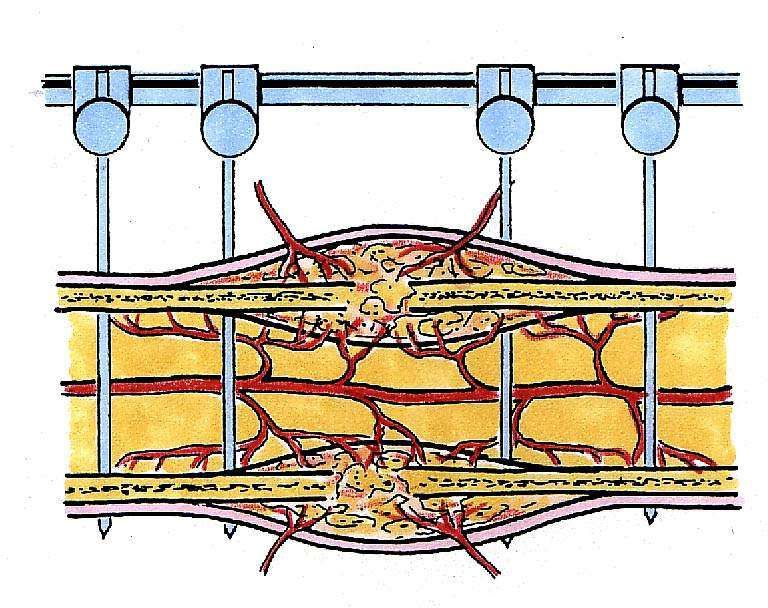
Introduction to the principles of small animal surgery                                            page 21

the central limb.
Incision of the skin and undermining the two triangular shaped flaps.
Transposition into the opposing donor beds and suturing with interrupted suture pattern.

Local skin flaps:
A. Advancement flap (Fig.2)

The skin flap is moved forward into the defect without lateral movement. This technique is mostly
used in square or rectangular wounds.

A rectangular skin defect is created.
The orientation of the flap along the tension lines is determined.
Slightly diverging skin incisions are made and the flap is undermined.
Stepwise progression until the flap can be advanced over the defect.
Suturing of the skin with interrupted suture pattern. In the edges horizontal matrass sutures can be
used to minimize circulatory compromise of the tips .

Alternative: H-plasty
Two single pedicle advancement flaps from each side of the wound.

B. Rotation flap (Fig.3)

Semicircular pedicle flap, commonly used to close triangular defect, rotating the flap over the defect.
Creation of a triangular skin defect.
Planning the position of the flap.
A curved incision is made and stepwise undermined until the flap can be rotated over the defect.
Suturing of the skin with single interrupted suture pattern.
Introduction to the principles of small animal surgery                                          page 22

C. Transposition flap (Fig.4)

Rectangular pedicle grafts, which are applicable in various locations.

Creation of a skin defect.
Local skin tension is assessed.
The base of the flap is measured and marked on the skin. The width equals the width of the defect
and is usually taken within 90 degrees to the long axis of the defect. The length is determined by the
distance of the pivot point to the most distant part of the defect. The outline of the flap is drawn on
the skin prior to the incision.
After skin incision the flap is undermined, transposed and sutured into place. The donor bed is pri-
mary closed in a similar fashion.
Introduction to the principles of small animal surgery                                             page 23

Introduction to the principles of simple osteosynthesis
Daniel Koch, Dr. med. vet. ECVS

Biological fracture healing

Principles of modern fracture healing are guided by providing maximal blood supply at the fracture
site. Therefore, perfect alignment of the fragments should not be accompanied by additional vascular
damage, which could impair fracture healing. Minimal requirements for biologoical fracture fixation
are: correct length of the extremity, correct rotational and axial alignment and interfragmentary sta-
bility. Methods, which do not (cast, closed application of a external fixator) or minimally (OBDT, "open
but don't touch", external fixator, intramedullary pin, Trilam nail) interfere with the fracture zone, are
preferred. Cerclage wires around the whole bone are not indicated with young animals, because two
thirds of the bony blood supply are provided by periosteal vessels. In adult animals, periosteal blood
supply is one third, endostal is two thirds.

Implant design shows the same evolution with some retardation. New implants with lesser contact
area with the bones are now available (LC-DCP, PC Fix, no contact fixateur, Vet-Fix). We also notice a
revival of the external fixator or the external coaptation with cast or plaster of Paris.

The sequence of fracture healing steps follows the rules of the interfragmentary strain theory. The
more micromotion a fixation system allows, the more healing tissues with properties that can stand
this strain are built. Granulation tissue can be deformed to 100 %, cartilage and connective tissue to
10 % and bone to 2 %. When choosing a fixation with high stability (e.g. plates), a direct fracture
healing is therefore expected. In contrast, when choosing a cast or am external fixator system, first a
callus is built. It further stabilizes the fragments and allows the development of cartilage and bone
(indirect fracture healing).

Indications

There are no strict rules for the choice of an implant and the way of approaching a fracture. Factors
as personal preferences, education level, economics, patient and kind of trauma all give their contribu-
tion. The following is thought as a guideline:
Introduction to the principles of small animal surgery                                                         page 24

General risk parameters

High risk                                                                                                  Low risk

Old patient               Middleaged patient                Young patient                  Juvenile patient
Large breed                                                                                    Small breed
Several extremities                                                                          One extremity
Bad health                Preexisting disease                                                  Good health
Thin soft tissue coverage                                                        Good soft tissue coverage
Cortical bone                                                                             Cancellous bone
High trauma velocity                                                                  Low trauma velocity
Large approach                                              Mini-Approach                           Closed
                                                                                  From: Fossum, Small animal surgery, 1997

Fracture assessment:

Criterium                     Fact                                     Rule

Soft tissue trauma            Open degree 1                      As closed
                              Open degree 2                      Drainage, external fixator
                              Open degree 3                      Open, external fixator
Fracture type                 Simple, fragment apposition        Smaller implants, lesser stability re-
                                                                 quired
                              Comminuted fracture, no fragment Bigger implants, high stability devices
                              apposition                         required, intramedullary implants
Localisation                  Diaphysis                          Plates, external coaptation, intrame-
                                                                 dullary implants with rotational sta-
                                                                 bility and others.
                              Epiphysis, Salter-Harris fractures Pinning, cross pins
                              Apophysis                          Tension band
                              Artikular                          Plates, screws
Bone                          Humerus                            Special: medial plating, external fixa-
                                                                 tor type I and Ib, Trilam nail, N. ra-
                                                                 dialis injury
                              Radius                             Special: medial plating, external fixa-
                                                                 tor type I, II, III, no IM nails, slow
                                                                 healing with toy breeds
                              Femur                              Special: lateral plating, external fixator
                                                                 type I and Ib, Trilam nail, watch out
                                                                 muscle contractures in young animals
                              Tibia                              Special: plate medial, external fixator
                                                                 type I, II , III; nailing possible, thin
                                                                 soft tissue coverage
Infection                     Infection and instabilty           Maximal stability required
Introduction to the principles of small animal surgery page 25
Decision making:
Diaphysal epiphysal articular apophysal
Young animal Adult animal
closed open closed
simple comminuted simple comminuted
Splint # -
Cast # ( ) ( ) -
Cross Pins, Pinning #
External fixator # # # # #
Tension band ( ) #
Trilam nail # # -
Screws, plates # # # # #
Plate - Rod ( ) ( ) # ( )Introduction to the principles of small animal surgery page 26 Basic course in fracture treatment Pierre M. Montavon, Prof. Dr. med. vet.; Daniel Koch, Dr. med. vet. ECVS Cerclage Wire Application. A) Cerclage wire with “Eye” Secure the bone model in the vice, place the wire passer around the bone staying close to the bone’s surface. Insert the straight end of the wire into the end of the wire passer. Withdraw the wire passer and remove it from the wire. Insert the straight end of the wire through the eye. The straight end of the wire is passed through the opening in the wire tightener. The wire is then passed through the hole in the center of the peg and the peg turned to tighten the wire. Once the wire is secured around the bone, it must be bent away from the eye to lock it in place. To achieve this, the peg is turned in the opposite direction, and tension is applied to the instrument to expose approximately 1 cm of wire. The wire is then bent over and cut off with the wire cutter. B) Cerclage wire without “Eye” A 15 cm strand of wire has been cut from the coil. For an alternative method of wire passing, curve one end of the wire and pass it around the bone. The two ends of the wire are twisted together two or three times by hand. The two free ends of the wire are bent 90° and held securely while excess wire ends are cut off. The thumb is being used to help keep the wire 90° to the long axis of the bone. In surgery, an instrument would be used for this purpose. The wire is twisted with even tension ap- plied until it engages the bone. To lay the twisted end of the wire close to the bone the twisting action is continued (in the same direction) without any tension being applied to the wire. The excess wire is cut off leaving three or four twists. Fracture of the olecranon with tension band wiring. Place the proximal fragment in the vice. Starting at the caudal lateral point of the olecranon drill a Kirschner wire, also called K-wire, through the fragment until it penetrates the fracture site. Start a second K-wire at the caudal medial point of the olecranon and advance it to the fracture site with the air drive. If necessary, withdraw the wires to a point just below the fracture surface. Reduce the frac- ture and hold the proximal fragment with the pointed reduction forceps. Advance the K-wires into the shaft of the distal fragment. With a K-wire drill a transverse hole in the distal fragment approximately 1.5 cm distal to the fracture site. Cut 15 to 20 cm of cerclage wire from the coil. Pass 10 to 12 cm through the transverse hole. The wire is brought tightly round the K-Wires at the point of the olecra- non and while applying even tension to the free ends, twist them together forming a figure eight. Continue to apply tension while twisting until the wire is tight. Now release the tension on the wire and continue to twist. The wire twist will come to lie flush with the straight portion of the wire. Cut the excess wire off at the level of the fourth twist. Bend the K-wires at right angles with the bending iron. Cut the excess off with the wire cutter. Note that the final position of the wire’s twisted end is centrally located in the tension band.
Introduction to the principles of small animal surgery                                            page 27

Principles of the Lag Screw and the Position Screw

Exercise a) Lag Screw
A lag screw will be placed in the bone model perpendicular to the fracture line. Be sure to choose the
end of the bone that has a simulated fracture matching the one shown on the screen. The 3.5 mm
drill bit is placed through the appropriate end of the double drill sleeve. The glide hole is drilled
through the near cortex only. This hole has the same diameter as the outer diameter of the threads of
the 3.5 mm screw. The 2.5 mm end of the double drill sleeve is placed through the glide hole until it
engages the far cortex. The 2.5 mm drill bit is passed through this end of the double drill sleeve and
the thread hole is created in the far cortex. Remove the double drill sleeve. Attache the countersink to
the T-handle. Countersink the glide hole in a clockwise direction. Using this instrument increases the
contact interface between the bone and the screw head. The depth gauge has a rod with a hooked
end, and a measurement bar which is used to determine the length of the screw needed. The meas-
urement bar is marked in 2 mm increments. The hooked rod is passed through both holes in the
bone. It is tilted slightly and withdrawn until the hook engages the surface of the far cortex. The
rounded end of the measuring device is pushed tightly against the near cortex and the length of
screw is determined from the measuring scale. Measurements are rounded up to the nearest 2 mm
mark. The 3.5 mm tap is placed through the appropriate end of the double drill sleeve and passed
through the glide hole until it engages the thread hole. Turning the tap cuts the thread in the thread
hole. The tap should be turned until at least two or three threads have exited the far cortex. The tap
is removed, and a 3.5 mm cortex screw of appropriate length inserted. Compression is achieved
across the fracture line as the screw is tightened. Note that the long axis of the screw is perpendicular
to the long axis of the fracture line. The screw is removed and the bone model rotated 90°. A lag
screw is inserted as before with the only exception being the orientation of the lag screw relative to
the fracture line. When the lag screw is not placed perpendicular to the fracture line shear forces de-
velop along the fracture line, which can cause displacement of the fracture fragments as is shown in
this demonstration.

Excercise b) The position screw
If the configuration of the fracture is such that the fracture fragment will collapse into the medullary
canal when compression is applied, then a screw with position function should be used. A thread hole
is drilled through both the near and far cortices using the 2.5 mm drill bit through the appropriate end
of the double drill sleeve. In the bone model, it will be necessary to support the fracture fragment
while drilling. The length of the screw is determined by using the depth gauge. Note that the counter-
sink was not used. The 3.5 mm tap is used to cut the threads in both the far and near cortices.Now
the appropriate 3.5 mm cortex screw is inserted. The position of the near fragment is maintained by
the screw, and the fragment can no longer be collapsed into the medullary canal. Remove the screw.
The effect of using a screw with lag function in this type of fracture configuration will be demon-
strated by creating a glide hole in the near cortex with the 3.5 mm drill bit. The same 3.5 mm screw is
now re-inserted. Because threads no longer engage the near cortex, the fracture fragment collapses
into the medullary canal as the screw is tightened.

Interfragmentary Compression Fixation of the Lateral Portion of the Humeral Condyle
with a Lag Screw and Anti-rotation Pin
The lateral portion of the humeral condyle has been placed in the vice and a 3.5 mm glide hole is
Introduction to the principles of small animal surgery                                               page 28

drilled from the outside-in, starting at a point distal and cranial to the epicondyle. The drill should be
oriented so that it will exit in the center of the condyle. An alternative method is to drill this hole from
the inside-out. When this is done, the glide hole is drilled from the center of the condyle, exiting the
bone, just distal and cranial to the lateral epicondyle. The fracture is reduced and held in place with
the large pointed reduction forceps. The 2.5 mm end of the double drill sleeve is inserted in the glide
hole and the thread hole is drilled with the 2.5 mm drill bit. In hard bone, the countersink is used. The
correct screw length is determined with the depth gauge. he 3.5 mm tap through the double drill
sleeve is used to cut the thread in the thread hole. The appropriate 3.5 mm cortex screw is inserted
achieving compression along the fracture line. To provide additional stability to the fracture, a Kirsch-
ner wire is inserted as an anti-rotation pin, starting at a point just distal and caudal to the epicondyle.
The pin should be driven from the lateral fragment through the medial cortex. The wire is bent with
the bending iron and cut off. When the bone is soft, as it might be in a young animal, the countersink
is not used. In this case a washer is used with the screw to keep the screw head from pulling through
the bone as the screw is tightened. The critical part of the repair is restoring the articular congruency
utilizing interfragmentary compression.

An oblique fracture of the humerus will be stabilized with a 3.5 mm lag screw. This sta-
bilization will be protected with a 3.5 dynamic compression plate used as a neutralization
plate.
Place the proximal fragment in the vice and reduce the fracture using the large pointed reduction
forceps. With the 3.5 mm drill bit through the double drill sleeve, drill the glide hole perpendicular to
the fracture line in the near cortex. Insert the 2.5 mm end of the double drill sleeve through the glide
hole until it engages the far cortex. Use the 2.5mm drill bit to create the thread hole in the far cortex.
The countersink is used to increase the bone - screwhead contact. Determine the screw length with
the depth gauge. Place the 3.5 mm tap through the double drill sleeve and cut the thread in the far
cortex. Insert the 3.5 mm cortex screw. This provides compression along the fracture line. Adjust the
bone in the vice so that the medial side is upper most. Contour the bending template to the medial
side of the bone. The bending pliers are used to contour the plate to the shape of the bone. The me-
dium sized anvil is inserted into the bending pliers and secured in place. The bending bar has a con-
vex surface,a flat surface, and a concave surface. The convex surface is used on the concave side of
the plate. The position of the handles is adjusted by turning the knob at the end of the pliers. The
bending template is used to indicate where the plate needs to be bent to match the shape of the
bone. The plate is bent along its length at multiple sites between the plate holes until its shape
matches that of the bending template. The plate is secured to the medial side of the humerus with the
reduction forceps. The green neutral drill guide is centered in the plate hole at one end of the plate,
and a 2.5mm hole is drilled through both cortices. The depth gauge is used to determine the length of
the screw. The 3.5 mm tap through the double drill sleeve is used to cut the thread in both cortices.
Insert the 3.5 mm cortex screw. A second screw is inserted in the same manner at the other end of
the plate. The rest of the screws are inserted on either side of the fracture repeating the same se-
quence of drilling, measuring, tapping, and inserting the screw. The reduction forceps are removed
and each screw is retightened. The fracture has been anatomically reduced and stabilized with a lag
screw. The plate protects the fracture reduction and has a neutralization function.

Axial Compression Fixation of the Radius with the Dynamic Compression Plate
A straight uncontoured plate has been fixed to the two fragments of the radius with reduction forceps.
The first screw will be inserted in one end of the plate using the green neutral end of the DCP drill
Introduction to the principles of small animal surgery                                               page 29

guide. Using this guide will place the screw in the center of the plate hole. The 2.5 mm drill bit is used
to create the hole in the bone. The depth gauge determines the screw length. The 3.5 mm tap, placed
through the double drill sleeve, cuts the thread in the hole. The appropriate length screw is inserted.
An other screw is inserted at the other end of the plate in exactly the same manner. Axial alignment
of one fragment is secured by inserting a second screw in this fragment using the neutral drill guide.
Compression will now be achieved by using the yellow-load end of the drill guide. The arrow on the
guide should point towards the fracture line. Using this guide will cause the screw to be placed eccen-
trically in the plate hole. The 2.5 mm drill bit is used to create the hole. The hole is measured and
tapped as before and the screw is inserted. As the screw head engages the gliding slope of the plate
hole it pushes the plate to our left. Compression is created at the fracture line in the cortex under-
neath the plate. Note, that a gap remains in the far cortex. Remove the plate and contour a bending
template to the cranial surface of the radius. Bend the plate with the bending pliers. Slightly over-
bending the plate at the fracture site will pre-stress the plate. The over-bending is observed when the
plate is compared to the bending template. Reinsert the screws in the holes in the order they were
originally placed. Note, with the plate being pre-stressed, the eccentrically positioned screw provides
compression along the entire fracture line. The remaining screws are inserted as before using the
neutral drill guide. After all the screws are in place they should be retightened. The plate hole design
allows the yellow-load drill guide to be used in any plate hole, once the fragments are axially aligned.

In this exercise an oblique fracture will be fixed with a lag screw through a plate that has
a neutralization function.
The fracture has been reduced and the bending template is contoured to the lateral surface of the
bone. The extent of plate contouring is determined by comparing the bending template to the plate.
Contour the plate with the bending pliers. The plate is again compared to the bending template. The
plate is now held in place with a reduction forceps and the bone holding forceps, while the oblique
fracture is stabilized with the pointed reduction forceps. The first hole is drilled in one end of the plate
using the 2.5 mm drill bit through the green neutral drill guide. The screw length is determined with
the depth gauge. The 3.5 mm tap through the double drill sleeve cuts the thread in the bone. A
3.5 mm cortex screw is now inserted. The next screw is placed in the same manner at the other end
of the plate. A second screw in the proximal fragment is inserted to fix this fragment’s axial alignment.
A lag screw will now be placed through the plate. The glide hole is created with the 3.5 mm drill bit
through the double drill sleeve. The glide hole should be aligned perpendicular to the fracture line.
The 2.5 mm end of the double drill sleeve is inserted through the glide hole until the far cortex is en-
countered. The 2.5 mm drill bit is used to create the thread hole. The depth gauge determines the
screw length. The thread is cut in the thread hole with the tap placed through the double drill sleeve.
The lag screw is inserted. The remaining screws are inserted. Although it appears from this view that
the fracture line may go through one of the plate holes. When the plate hole is viewed from above, it
is clear that this was not the case. Had the fracture line gone through the plate hole this hole would
not have been filled with a screw. The screws are retightened after they have all been inserted. A
view of the medial side of the bone shows anatomic reduction of the fracture.

Buttress Plate Fixation of a Midshaft Femoral Fracture
We will use the fractured femur from the previous exercise. Reduce the fracture and apply the long
bending template to the lateral side of the femur. Because there is a medial deficit, a longer and
stronger plate will be used in this fracture stabilization. Contour the broad 3.5 dynamic compression
Introduction to the principles of small animal surgery                                              page 30

plate to the configuration of the bending template. Fix the plate to the lateral side of the femur with
the forceps. In the first plate hole drill a hole with the 2.5mm drill bit through the green neutral drill
guide. Determine the screw length with the depth gauge. With the 3.5 mm tap through the double
drill sleeve, cut the thread in both cortices. Insert the 3.5 mm cortex screw. Following the same steps,
a screw is now inserted in the most distal plate hole. Screw holes are alternately filled throughout the
length of the plate. Screw holes that lie over the area of the deficit will either be filled with screws
that go through only the near cortex, or not filled at all, depending on the position of the fracture line.
Clinically, the bone defect is filled with cancellous bone graft. Because of the deficit in the medial
cortex, the longer and stronger, broad plate has a buttress function and provides adequate stabiliza-
tion of this fracture.

In this exercise a comminuted fracture will be reconstructed with 2.7 mm cortex screws
functioning as lag screws, and a 3.5 dynamic compression plate with neutralization func-
tion will be applied to protect the reconstructed fracture.
Secure one end of the tibia in the vice and reduce the comminuted segment to this fragment using
the pointed reduction forceps. Starting on the cranial surface, drill a 2.7 mm glide hole perpendicular
to the fracture line with the 2.7 mm drill bit through the appropriate double drill sleeve. Insert the
2 mm end of the double drill sleeve through the glide hole until the far cortex is contacted. With the
2 mm drill bit, create the thread hole. Countersink the glide hole. Determine the appropriate screw
length with the depth gauge. With the 2.7 mm tap through the double drill sleeve, cut the thread in
the thread hole. Insert a 2.7 mm cortex screw, to provide compression at the fracture line. Reduce
the third fracture segment with the pointed reduction forceps. Insert the second lag screw, by creat-
ing the glide hole, and the thread hole, using the double drill sleeve, countersinking the glide hole, ..
measureing the screw length, tapping the thread hole and inserting a 2.7 mm cortex screw. Rotate
the reconstructed fracture so that the medial surface is upper most. Contour the bending template to
the medial surface of the tibia. Secure the contoured plate to the bone and with the 2.5 mm drill bit
through the green neutral drill guide, drill a hole at one end of the plate. Measure the screw length
with the depth gauge. And with the 3.5 mm tap through the double drill sleeve cut the thread in the
bone. Insert the appropriate 3.5 mm cortex screw. These steps are repeated at the opposite end of
the plate. In each main fragment, a second screw is placed to fix the axial alignment of the bone.
Remove the reduction forceps and insert the remaining screws following the same steps of drilling,
measuring, tapping, and inserting the screw. Retighten all the screws. One plate hole is left empty
because the fracture line crosses it, which is clearly seen when the hole is viewed from above.

An obliquely fractured ilium will be stabilized with a 2.7 dynamic compression plate.
Fix the caudal fragment of the ilium in the vice and reduce the cranial fragment. Fracture reduction is
maintained with the pointed reduction forceps. Contour the bending template to the lateral surface of
the ilium. Using the bending irons, duplicate the contour of the bending template on a 2.7 dynamic
compression plate. With the 2 mm drill bit through the double drill sleeve, drill a hole 5 mm from the
fracture line in the caudal fragment. Measure the depth of the hole and add 2 mm to compensate the
thickness of the plate. Cut the thread in the hole with the 2.7 mm tap through the double drill sleeve.
Position the plate over the hole and insert the appropriate 2.7 mm cortex screw. With the 2 mm drill
bit through the green neutral drill guide, drill a hole in the cranial fragment through the plate hole
next to the fracture line. After measuring and tapping the appropriate 2.7 mm screw is inserted. The
You can also read