IS THERE A HIGH-RISK MAMMOGRAPHIC PATTERN IN B3 LESIONS?
←
→
Page content transcription
If your browser does not render page correctly, please read the page content below
April, 2020

                                       Special issue • 2020 • vol.1 • 39-45
           IS THERE A HIGH-RISK MAMMOGRAPHIC PATTERN IN B3 LESIONS?
           Crivelli P.1, Ledda R.E.2, Piga G.3, Lampus M.L.3, Sotgiu M.A.4, Soro D.1, Conti M.3
                           1
                          AOU Sassari, Institute of Diagnostic Imaging 2, Italy
       2
    Section of Radiology, Unit of Surgical Sciences, Department of Medicine and Surgery (DiMeC),
                                   University of Parma, Parma, Italy
  3
   Department of Clinical and Experimental Medicine, Institute of Diagnostic Imaging 2, University of
                                              Sassari, Italy
      4
        Department of Biomedical Sciences, Medical School, University of Sassari, Sassari, Italy.
                                    Email: paocri2000@gmail.com

Abstract
Introduction: Breast lesions of uncertain malignant potential (B3) include different histopathological
subtypes. Vacuum assisted biopsy (VAB) excision and surveillance have been more recently proposed
as a valid alternative to the more traditional surgical approach. A significant association between
radiological findings and malignancy in excision biopsy has not been proved as yet.
Aim of this paper is to find a prevalent mammographic pattern for each histological B3 subtype in order
to identify a “high-risk mammographic pattern”.
Methods and materials: We retrospectively included all B3 patients referred from spontaneous
screening to the Breast Radiology Service at the University Hospital of Sassari, Italy, from January 2012
to June 2018. All patients underwent a mammography and a histological characterization. Six different
mammographic patterns and six histological subtypes were identified.
Results: 69 patients were enrolled. Median (IQR) age was 50 (44-57) years; the majority of lesions was
localized in the left breast (37, 53.6%). Clustered microcalcifications was the most prevalent
mammographic pattern in our series, whereas ADH was the most common histological subtype. A
prevalent mammographic pattern was found within each pathological subtype (p-value=0,02).
Discussion: B3 subtypes, whose cellular atypia is the characteristic feature, showed clustered
microcalcifications as their prevalent mammographic pattern (“high-risk mammographic pattern”). In
this context, the VAB biopsy is likely to change the management of B3 lesions, assuming a therapeutic
role too and, thus, reducing both biological and economic costs.
Larger and multicentric studies are necessary to find a generalizable mammographic pattern for the
lesser common B3 histological subtypes.

Keywords: B3 lesions, atypia, biopsy, therapeutic approach

   .

                                        http://pharmacologyonline.silae.it
                                                 ISSN: 1827-8620
PhOL                                        Crivelli, et al.                 40 (pag 39-45)

Introduction                                                        microcalcifications,    mass      with     parenchymal
                                                                    distortion and parenchymal distortion with
Breast cancer is the most common female tumour, it
                                                                    microcalcifications. Histological samples were
manifests at different ages with different histotypes
                                                                    obtained by a 14-16G spring-loaded CNB under
1-3], and several factors are involved in the
                                                                    ultrasound or by VAB, using a 9-11G device under
carcinogenesis, even environmental [4, 5]. Early
                                                                    stereotactic mammography guidance. All specimens
diagnosis, performed by conventional and
                                                                    were evaluated by two expert breast pathologists
innovative imaging techniques, is mandatory to
                                                                    and results classified into 6 subtypes as per the
prevent metastasis, improving clinical outcome [6-
                                                                    European guidelines [9] (atypical ductal hyperplasia,
8]. Breast lesions of uncertain malignant potential
                                                                    ADH, flat epithelial atypia, FEA, lobular
(B3) encompass different histological entities [9-12].
                                                                    intraepithelial neoplasia, LIN 1/2, papillary lesion, PL,
The number of B3 diagnoses, often incidental, has
                                                                    benign phyllodes tumours, PT, and radial scar, RS).
been increasing over the last decades due to the
                                                                    Following a multidisciplinary (MDT) discussion,
implementation and scaling up of mammographic
                                                                    involving      breast      surgeons,        radiologists,
screening programs [13] and to a larger availability
                                                                    radiotherapists and pathologists, all patients
of minimally invasive biopsy techniques (core-needle
                                                                    underwent surgery.
biopsy, CNB, and vacuum assisted biopsy, VAB) [14-
16]. Although surgical excision remains the                         Data were retrieved from medical files using an ad-
preferred therapeutic option, VAB excision and                      hoc electronic form, including demographic, clinical,
surveillance have been more recently proposed as                    epidemiological, radiological and histological
alternative approaches in some selected cases [11].                 variables. Quantitative covariates were summarized
A significant association between radiological                      with means and standard deviations (SD) or
findings and malignancy in excision biopsy has never                medians and interquartile ranges (IQR) in case of a
been proved, assigning the histology the exclusive                  parametric     or     non-parametric    distribution,
role to orientate the management of these                           respectively, whereas qualitative variables were
heterogeneous lesions [15, 18, 19].                                 described using absolute frequencies and
                                                                    percentages. Statistical computations were
This paper aims to find a prevalent mammographic
                                                                    performed with the statistical software Stata13.0
pattern for each histological B3 subtype.
                                                                    (StataCorp, College Station, TX, USA).
Methods
                                                                    Results
It was carried out a retrospective study including all
                                                                    Sixty-nine female patients were retrospectively
patients diagnosed with B3 lesions who referred
                                                                    enrolled. Median (IQR) age was 50 (44-57) years; the
from spontaneous screening to the Breast
                                                                    majority of lesions was localized in the left breast
Radiology Service at the University Hospital of
                                                                    (37, 53.6%). Mammographic patterns were as
Sassari, Italy, from January 2012 to June 2018. All
                                                                    following: 54 (78.3%) clustered calcifications, 8
patients       underwent      a    bilateral     digital
                                                                    (11.6%) mass, 4 (5.8%) mass with microcalcifications,
mammography in standard projections (cranio-
                                                                    1 (1.5%) parenchymal distortion, 1 (1.5%) parenchymal
caudal and oblique) using Selenia® Dimensions®
                                                                    distortion with calcifications, and 1 (1.5%) mass with
Mammography System (Hologic). Where required,
                                                                    parenchymal distortion. Histology showed 44
radiological exam was completed with an
                                                                    (63.8%) cases of ADH, 10 (14.5%) of PL, 7 (10.1%) of
ultrasound (US) breast scan using a linear probe of a
                                                                    FEA, 4 (5.8%) of RS, 3 (4.4%) of LIN 1/2, and 1 (1.5%) of
MyLab™ClassC (Esaote). Images were reviewed by
                                                                    PT (Table 1). A prevalent mammographic pattern
two expert breast radiologists who classified B3
                                                                    was found within each pathological subtype (p-
lesions      in    patterns,     considering     single
                                                                    value=0,02). All ADHs presented with calcifications,
mammographic findings as described in previous
                                                                    either in isolated clusters (86.4%) or associated with
literature, and their possible combination [11, 15]. Six
                                                                    mass or parenchymal distortion (4.6%) (Table 2)
different mammographic patterns were identified
as following: clustered microcalcifications, mass and
parenchymal          distortion,      mass         with

                                         http://pharmacologyonline.silae.it
                                                  ISSN: 1827-8620
PhOL Crivelli, et al. 41 (pag 39-45)
Discussion women. Our experience. G Chir 2011 Oct; 32 (10):411-
6.
Six different mammographic patterns (Figure 1)
were identified and their prevalence within the six 3. Amadu AM, Marras V, Crivelli P, Soro D,
histological subtypes investigated (Figure 2). Conti M, Meloni GB. Isolated breast metastasis 4
Clustered microcalcifications represented the most years after nephrectomy. Breast J. 2018 Jan; 24
prevalent mammographic pattern in our series, (1):85-87.
whereas ADH was the most common histological 4. Oggiano R, Solinas G, Forte G, Bocca B,
subtype. Farace C, Pisano A, Sotgiu MA, Clemente S,
As previously demonstrated, ADH, LIN 1/2 and FEA Malaguarnera M, Fois AG, Pirina P, Montella A,
are associated with an increased risk for malignancy Madeddu R. Trace elements in ALS patients and
presenting cellular atypia as their characteristic their relationships with clinical severity
feature, whereas other B3 subtypes may or may not Chemosphere. 2018 Apr; 197:457-466.
contain cellular of altered morphology [12,17-19]. 5. Forte G, Bocca B, Oggiano R, Clemente S,
Our series showed that these three “high-risk Asara Y, Sotgiu MA, Farace C, Montella A, Fois AG,
subtypes” presented clustered microcalcifications Malaguarnera M, Pirina P, Madeddu R. Essential
as their prevalent mammographic pattern, which trace elements in amyotrophic lateral sclerosis
could be considered a mammographic expression of (ALS): Results in a population of a risk area of Italy.
an underlying atypia, configuring a “high-risk Neurol Sci. 2017 Sep; 38(9):1609-1615.
mammographic pattern”. Following the detection
of this “high-risk mammographic pattern”, it is 6. Di Grezia G, Somma F, Serra N, Reginelli A,
mandatory for the interventional breast radiologist Cappabianca S, Grassi R, Gatta G. Reducing Costs of
to perform a radical VAB excision, offering a valid Breast Examination: Ultrasound Performance and
alternative to surgery. In this context, the VAB Inter-Observer Variability of Expert Radiologists
biopsy has changed the management of these Versus Residents. Cancer Invest. 2016 Jul 20: 1-6.
borderline lesions, obtaining a therapeutic purpose 7. Crivelli P, Ledda RE, Parascandolo N, Fara A,
too, as clearly expressed in the B3 First International Soro D, Conti M. A New Challenge for Radiologists:
Consensus Conference recommendations [11,20]. Radiomics in Breast Cancer. Biomed Res Int. 2018
Despite the small number of patients enrolled, our Oct 8; 2018:6120703.
results reinforce the concept of a radical VAB
biopsy, which could reduce both biological and 8. Di Grezia G, Prisco V, Iannaccone T, Grassi R,
economic costs. Serra N, Gatta G Personality disorder and
temperamental traits in patients with breast
This study has several limitations: the disease: preliminary results Minerva Psichiatrica
retrospective nature, the small patients sample size 2016 Sept; 57(3):85-92
and the inclusion of patients from a single center
make harder to generalize results. 9. Perry N, Broeders M, de Wolf C, Tornberg S,
Holland R, von Karsa L. European guidelines for
In conclusion, larger and multicentric studies will quality assurance in breast cancer screening and
be needed to validate our preliminary results and to diagnosis. Fourth edition–summary document. Ann
find a generalizable mammographic pattern for the Oncol. 2008; 19:614–622.
lesser common B3 histological subtypes.
10. Purushothaman HN, Lekanidi K, Shousha S,
References Wilson R. Lesions of uncertain malignant potential in
1. Amadu AM, Soro D, Marras V, Satta G, the breast (B3): what do we know? Clin Radiol. 2016
Crivelli P, Conti M, Meloni GB. Primary breast Feb; 71(2):134-40. d
chondrosarcoma: Imaging and pathological findings. 11. Rageth CJ, O'Flynn EA, Comstock C, Kurtz C,
Eur J Radiol Open. 2017 Nov 5; 4:138-140. Kubik R, Madjar H, Lepori D, Kampmann G,
2. Vestito A, Mangieri FF, Gatta G, Moschetta Mundinger A, Baege A, Decker T, Hosch S, Tausch C,
M, Turi B, Ancona A Breast carcinoma in elderly Delaloye JF, Morris E, Varga Z. First International
http://pharmacologyonline.silae.it
ISSN: 1827-8620PhOL Crivelli, et al. 42 (pag 39-45)
Consensus Conference on lesions of uncertain B3 breast lesions subclassification. Ann Diagn
malignant potential in the breast (B3 lesions). Breast Pathol. 2013 Oct; 17(5):434-6.
Cancer Res Treat. 2016 Sep; 159(2):203-13.
19. Hoffmann O, Stamatis GA, Bittner AK,
12. Friederichs Sinn H. Lesions of uncertain Arnold G, Schnabel R, Krüger K, Kimmig R, Heubner
malignant potential (B3). 2016. AGO eV, Comisson M. B3-lesions of the breast and cancer risk - an
Mamma. analysis of mammography screening patients. Mol
Clin Oncol. 2016 May; 4(5):705-708.
13. Fancellu A, Sanna V, Sedda ML, Delrio D,
Cottu P, Spanu A, Giuliani G, Conti M, Piras R, Crivelli 20. Pieri A, Hemming D, Westgarth J, Lunt L.
P, Porcu A. Benefits of Organized Mammographic Vacuum-assisted biopsy is a viable alternative to
Screening Programs in Women Aged 50 to 69 years: surgical biopsy in the investigation of breast lesions
A Surgical Perspective. Clin Breast Cancer. 2019 Oct; of uncertain malignant potential. Surgeon. 2017 Apr;
19(5):e637-e642. 15(2):59-64.
14. Pinder SE, Shaaban A, Deb R, Desai A,
Gandhi A, Lee AH, Pain S, Wilkinson L, Sharma N.
NHS Breast Screening multidisciplinary working
group guidelines for the diagnosis and management
of breast lesions of uncertain malignant potential on
core biopsy (B3 lesions). Clin Radiol. 2018
Aug;73(8):682-692. doi: 10.1016/j.crad.2018.04.004.
15. Taffurelli M, Pellegrini A, Ghignone F, Santini
D, Zanotti S, Serra M. Positive predictive value of
breast lesions of uncertain malignant potential (B3):
Can we identify high risk patients? The value of a
multidisciplinary team and implications in the
surgical treatment. Surg Oncol. 2016 Jun; 25(2):119-
22.
16. Gatta G, Di Grezia G, Ancona A, Capodieci M,
Coppolino F, Rossi C, Feragalli B, Iacomino A,
Cappabianca S, Grassi R “Underestimation of
atypical lobular hyperplasia and lobular carcinoma in
situ at stereotaxic 11-gauge vacuum-assisted breast
biopsy” European Journal of Inflammation 2013;
11(3): 825-835Taffurelli M, Pellegrini A, Ghignone F,
Santini D, Zanotti S, Serra M. Positive predictive
value of breast lesions of uncertain malignant
potential (B3): Can we identify high risk patients?
The value of a multidisciplinary team and
implications in the surgical treatment. Surg Oncol.
2016 Jun; 25(2):119-22.
17. Mayer S, Kayser G, Rücker G, Bögner D,
Hirschfeld M, Hug C, Stickeler E, Gitsch G, Erbes T.
Absence of epithelial atypia in B3-lesions of the
breast is associated with decreased risk for
malignancy. Breast. 2017 Feb; 31:144-149.
18. de Beça FF, Rasteiro C, Correia A, Costa S,
Amendoeira I. Improved malignancy prediction by
http://pharmacologyonline.silae.it
ISSN: 1827-8620PhOL Crivelli, et al. 43 (pag 39-45)
Table 1. Demographic data, histological subtypes and mammographic patterns of B3 patients.
Variables
Mediana age (IQR), years 50 (44-57)
Left side, n (%) 37 (53.6)
ADH 44 (63.8)
PL 10 (14.5)
FEA 7 (10.1)
Diagnosis, n (%)
RS 4 (5.8)
LIN 1/2 3 (4.4)
PT 1 (1.5)
Clustered
54 (78.3)
microcalcifications
Mass 8 (11.6)
Mass with
4 (5.8)
microcalcifications
Mammographic pattern, n (%)
Parenchymal distortion 1 (1.5)
Parenchymal distortion
1 (1.5)
with microcalcifications
Mass with parenchymal
1 (1.5)
distortion
Table 2. Mammographic patterns and histological subtypes (p-value: 0.02).
ADH PL RS PT LIN 1/2 FEA
Mass 3 (6.8) 3 (30.0) 2 (50.0) 0 (0.0) 0 (0.0) 0 (0.0)
Mass with 1
1 (2.3) 1 (10.0) 1 (25.0) 0 (0.0) 0 (0.0)
microcalcifications (100.0)
Mass with
parenchymal 1 (2.3) 0 (0.0) 0 (0.0) 0 (0.0) 0 (0.0) 0 (0.0)
distortion
Parenchymal
distortion with 1 (2.3) 0 (0.0) 0 (0.0) 0 (0.0) 0 (0.0) 0 (0.0)
microcalcifications
Clustered 7
38 (86.4) 5 (50.0) 1 (25.0) 0 (0.0) 3 (100.0)
microcalcifications (100.0)
Parenchymal
0 (0.0) 1 (10.0) 0 (0.0) 0 (0.0) 0 (0.0) 0 (0.0)
distortion
http://pharmacologyonline.silae.it
ISSN: 1827-8620PhOL Crivelli, et al. 44 (pag 39-45)
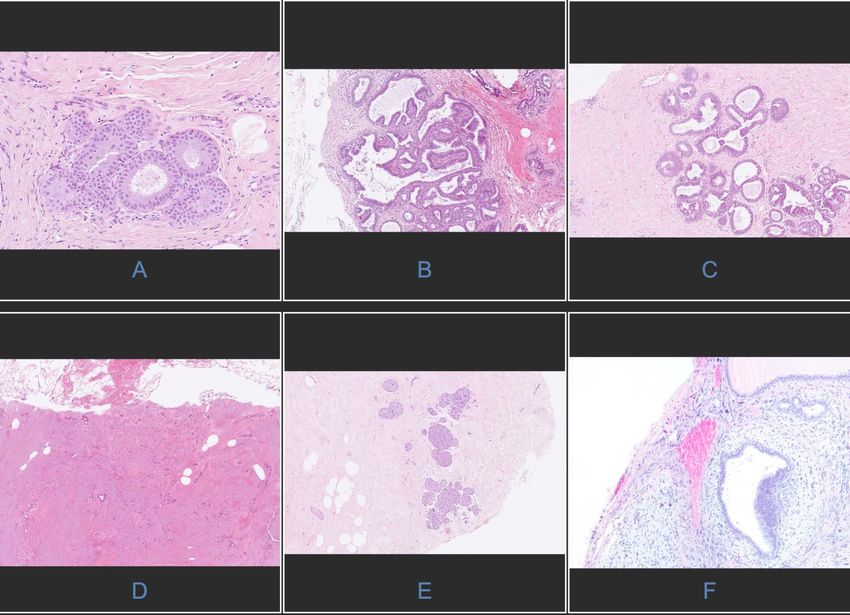
Figure 1. Six mammographic pattern
http://pharmacologyonline.silae.it
ISSN: 1827-8620PhOL Crivelli, et al. 45 (pag 39-45)
Figure 2. Six histological pattern
http://pharmacologyonline.silae.it
ISSN: 1827-8620You can also read