Local Heating with Lithographically Fabricated Plasmonic Titanium Nitride Nanoparticles
←
→
Page content transcription
If your browser does not render page correctly, please read the page content below
Letter

                                                                                                                                  pubs.acs.org/NanoLett

Local Heating with Lithographically Fabricated Plasmonic Titanium
Nitride Nanoparticles
Urcan Guler,† Justus C. Ndukaife,†,‡ Gururaj V. Naik,† A. G. Agwu Nnanna,‡ Alexander V. Kildishev,†
Vladimir M. Shalaev,† and Alexandra Boltasseva*,†,§
†
 School of Electrical and Computer Engineering and Birck Nanotechnology Center, Purdue University, West Lafayette, Indiana
 47907, United States
‡
 Water Institute, Purdue University Calumet, Hammond, Indiana 46323, United States
§
 DTU Fotonik, Department of Photonics Engineering, Technical University of Denmark, Lyngby DK-2800, Denmark
    *
    S Supporting Information

    ABSTRACT: Titanium nitride is considered a promising alternative
    plasmonic material and is known to exhibit localized surface plasmon
    resonances within the near-infrared biological transparency window.
    Here, local heating efficiencies of disk-shaped nanoparticles made of
    titanium nitride and gold are compared in the visible and near-infrared
    regions numerically and experimentally with samples fabricated using
    e-beam lithography. Results show that plasmonic titanium nitride
    nanodisks are efficient local heat sources and outperform gold
    nanodisks in the biological transparency window, dispensing the need
    for complex particle geometries.
    KEYWORDS: LSPR, plasmonic heating, TiN nanoparticle, photothermal effect, photodynamic therapy, biomaterials

P    lasmonics has been a hot topic for the last two decades
     with innumerable promising results in several different
application areas.1,2 Although the field has been investigated
                                                                             regions.17 The LSPR spectral position of small, spherical TiN
                                                                             nanoparticles matches very well with the biological trans-
                                                                             parency window where plasmonics-based biological research is
widely by many researchers from all around the world, recent                 carried out.18,19 Currently, these applications are dominated by
research trends still follow the early examples of this field. For            Au nanoparticles because of their biocompatibility and
instance, gold (Au) and silver (Ag) are the most popular                     relatively good plasmonic properties in the visible region.20
choices as plasmonic materials. However, it was shown that an                However, gold’s LSPR peaks occur at shorter wavelengths and
ancient example of plasmonics, the Lycurgus Cup, contains                    researchers have worked to redshift the resonance into the
silver−gold alloyed nanoparticles. This reveals the fact that the            biological window by using complex structures.21,22
search for alternative plasmonic materials is as old as the history             Beyond the biological applications, local heating with
of plasmonics itself.3 With the significant improvement of                    plasmonic nanoparticles is considered to be a promising
nanoscale fabrication and characterization techniques, the initial           method for several other applications ranging from catalysis to
tendency of the scientific community was to examine noble                     heat-assisted magnetic recording.23−27 TiN nanoparticles are
metals as plasmonic materials due to their suitable dielectric               promising alternatives to replace noble metals due to their
functions in the visible region of the electromagnetic                       increased heating efficiencies over Au in the red and near-
spectrum.4−6 As the understanding of physical mechanisms                     infrared regions and provide additional exciting properties such
has progressed, researchers are now working to find, or                       as high melting temperature, chemical stability, mechanical
synthesize, the optimum materials for individual application                 durability, and compatibility to CMOS technology. In addition,
areas and related spectral windows.7−9                                       plasmonic TiN nanoparticles with simple shapes would be
   Titanium nitride (TiN) is known to exhibit plasmonic                      favored when compared to complex metal nanostructures.
behavior.10−14 Owing to the similarities of its optical properties           Shape-tuned metal nanostructures may bring additional
to those of Au with additional significant advantages such as                 difficulties due to their structural complexities. For example,
low cost, thermal stability, and compatibility with silicon                  using nanorods with large aspect ratios results in a polarization
nanofabrication techniques, TiN draws attention as a promising               dependent response of particles and would reduce the total
alternative plasmonic material.15,16 Recently, we have shown                 efficiency due to lack of orientation control over the particles in
that near-field enhancement due to the localized surface
plasmon resonance (LSPR) in nitride plasmonic materials is                   Received: September 7, 2013
comparable to Au, and the dipolar resonance peak naturally                   Revised: November 23, 2013
occurs at the border of the visible and the near-infrared                    Published: November 26, 2013

                              © 2013 American Chemical Society        6078                        dx.doi.org/10.1021/nl4033457 | Nano Lett. 2013, 13, 6078−6083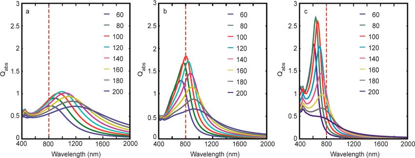
Nano Letters                                                                                                                                        Letter

Figure 1. Absorption efficiencies of (a) Au and (b) TiN spherical nanoparticles obtained from Mie calculations. Peak values for both materials are
equal but the spectral position is at 520 nm for Au and 700 nm for TiN. A broad resonance with equivalent strength located very close to the
biological transparency window, makes TiN a promising plasmonic material for biological applications.

many applications. Sharp edges in complex-shaped particles                  camera, FLIR A320, by measuring the temperature change in
would be exposed to higher field intensities and temperatures,               the substrate. Details of the experimental methods are given in
resulting in shape deformations, which will eventually degrade              the Supporting Information.
the performance.                                                               A simple and efficient method for comparing different
   Local heating with plasmonic nanoparticles is based on the               materials for local heating applications is to calculate absorption
idea of delivering energy with electromagnetic waves to the                 efficiency of spherical nanoparticles of varying sizes through the
region of interest, where plasmonic nanoparticles concentrate               Mie scattering formulation.32 Au and TiN optical constants
light and generate heat within a confined volume through                     retrieved from thin films are employed in the calculations where
ohmic losses at the resonance wavelengths.18,28 The heat                    parameter sweep over the particle radius and excitation
generated by a nanoparticle is known to be proportional to the              wavelength are performed. Size effects on the permittivities
intensity of the illuminating light where the proportionality               are neglected since the dimensions of the particles examined in
constant is the absorption efficiency of the particle, defined as              this work are relatively large. The absorption efficiencies
the absorption cross-section normalized by the geometrical                  obtained from Mie calculations are plotted in Figure 1. The
cross-sectional area of the particle under illumination.29 Thus,            well-known behavior of the reference material, Au, is evident in
assuming a constant illumination intensity, the absorption                  Figure 1a, where the LSPR peak is located around 520 nm with
efficiency of the particle can be used as a figure of merit (FOM)              an optimal particle radius around 55 nm. The peak efficiency at
for comparison purposes. Among the parameters affecting the                  these parameters is around 2.5. As expected, Au nanoparticles
absorption efficiency of a plasmonic particle, size and shape                 exhibit a narrow peak that for most of the applications dictate
have been extensively studied.30,31 Other important parameters              precise control on the size dispersion of particles.33 In order to
that affect the absorption efficiency are the optical constants of             obtain a strong response from Au nanoparticles in the
the nanoparticle that are dependent on the plasmonic material               biological transparency window, it is a common practice to
and the illumination wavelength.                                            use particles with large aspect ratios and complex shapes.21,34
   In this Letter, we compare TiN and Au nanoparticles within               However, as the size of the particle increases, higher order
the perspectives of local heating for biological and other                  modes become supported and interference effects between
important applications and show that TiN performs better than               multiple modes reduce the efficiency. TiN nanoparticles, on the
Au in the spectral range that includes the biological                       other hand, exhibit a broad resonance peak located around 700
transparency window. Electron beam lithography (EBL) is                     nm with maximum absorption efficiency around 2.5, identical
used for the fabrication of Au and TiN nanoparticle arrays                  to the Au peak value. A broad, dipolar peak of comparable
ranging in size from 60 to 180 nm in diameter with varying                  strength occurring at wavelengths closer to the biological
lattice constants. They are deposited on sapphire substrates,               transparency window is a significant advantage of TiN
which have a thermal conductivity of (κ = 42 W/m × K).36 Au                 nanoparticles over Au. From the initial comparison based on
samples are deposited with electron beam deposition while TiN               Mie calculations, one can claim that TiN nanoparticles of
samples are deposited with dc reactive magnetron sputtering at              simple geometries with reduced restrictions on the size-
400 and 800 °C. Spectroscopic ellipsometry (V-VASE, J. A.                   dispersion can provide better efficiencies for heating
Woollam Co.) is used for the transmittance measurements of                  applications when compared to Au.
the nanoparticle arrays and for the dielectric constant retrieval              In order to verify these calculations, Au and TiN nanodisk
from thin films grown simultaneously with nanoparticles.                     arrays with identical shapes are fabricated using the EBL
Retrieved optical constants are employed in numerical                       technique. Au samples are prepared by electron beam
simulations with the finite element method (FEM) based                       deposition while TiN samples are deposited using dc reactive
commercial software (COMSOL Multiphysics). Nanodisk                         magnetron sputtering at substrate temperatures of 400 and 800
arrays are illuminated with a laser beam at an 800 nm                       °C.16 TiN nanodisks grown at different temperatures are
wavelength and heat generation is observed with a thermal                   examined due to the nonstoichiometric nature of the material.35
                                                                     6079                         dx.doi.org/10.1021/nl4033457 | Nano Lett. 2013, 13, 6078−6083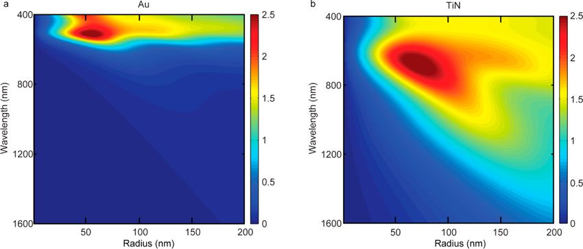
Nano Letters Letter
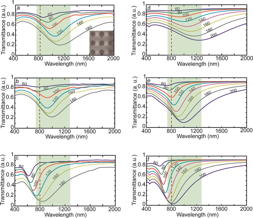
Figure 2. Transmittance spectra for 30 nm thick nanodisk arrays of TiN grown at (a) 400 °C, (b) 800 °C, and (c) Au. The spectral position of the
transmittance dips due to the extinction of particles indicates that TiN nanodisks exhibit plasmonic resonances throughout the biological
transparency window (green regions). The plasmonic peaks of Au nanoparticles with smaller sizes are located at relatively short wavelengths, and
larger particles are required to reach the spectral region of interest. (d−f) The simulation results of the transmittance through the arrays of the
particles match well with the experimental data allowing the following simulation based discussions on absorption efficiencies. The dashed line shows
the excitation wavelength used for the heating experiments in this work. The inset shows EBL patterned TiN nanoparticles deposited at 400 °C.
Figure 3. Simulated absorption efficiencies of nanodisks for TiN grown at (a) 400 °C, (b) 800 °C, and (c) Au. Broad resonance peaks throughout
the biological transparency window obtained from nanodisks of TiN suggest that high local heating efficiencies can be reached. The dashed line
shows the excitation wavelength used for the heating experiments in this work.
Samples grown at higher temperatures are known to be more shows the experimental transmittance data of the nanoparticle
metallic (larger magnitude of the real part of permittivity) when arrays of TiN grown at two different temperatures, 400 °C (a)
compared to the samples grown at lower temperatures. The and 800 °C (b), and Au as the reference material. A
nanodisks range in size from 60 to 180 nm with varying lattice comparison between TiN nanoparticles grown at different
constants on sapphire substrates, which has a thermal temperatures shows that due to the nonstoichiometric nature of
conductivity of (κ = 42 W/m × K).36 The LSPR peak the material the resonance behavior of these particles can be
positions of these particles are determined from the extinction tuned by varying the growth parameters. It is evident that the
dips in the optical transmittance data obtained from biological transparency window, highlighted with green in the
spectroscopic ellipsometry. Thin films grown under the same figure, can be covered by the LSPR peaks of TiN nanoparticles
deposition conditions as with the nanoparticles were with varying sizes and growth conditions. On the other hand,
characterized with ellipsometry and retrieved optical constants transmittance data from Au nanodisks have dips starting at 600
were used in subsequent 3D FEM simulations. Figure 2a−c nm with the smallest particles and increasing up to 850 nm with
6080 dx.doi.org/10.1021/nl4033457 | Nano Lett. 2013, 13, 6078−6083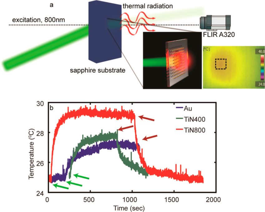
Nano Letters                                                                                                                                         Letter

larger particles. Figure 2d−f shows the simulated transmittance            efficiencies at the nanometer scale when compared to Au due to
data of the nanoparticle arrays that agree quite well with the             their LSPR being intrinsically located near the biological
experimental results. Although the spectral position and                   window.
strength of the extinction peaks provides important details                  In this work, heating efficiencies of nanoparticles are also
about the performance of nanoparticles, the crucial FOM for                examined experimentally. Sapphire substrates with arrays of Au
local heating applications is the absorption efficiency of the               and TiN nanoparticles are imaged with a FLIR infrared camera
particles as mentioned earlier. It should be noted that a                  with a pitch size of 25 μm and illuminated with a laser at 800
stronger resonance dip in the transmittance spectra of Au                  nm. Figure 4a provides an illustration of the experiment while
sample is due to the larger extinction efficiency of these
particles, which is the sum of absorption and scattering.
However, it is not easy to experimentally compare the
absorption efficiencies of small nanoparticles with high
precision. Since there is good agreement between numerically
simulated and experimentally measured transmittance data
presented in Figure 2, the absorption efficiencies of the
nanoparticles are determined via numerical simulations.
   Absorption efficiencies calculated from FEM simulations of
nanodisk arrays are presented in Figure 3 for TiN and Au.
Figure 3a,b shows the absorption efficiencies of TiN nanodisks
of varying sizes grown at 400 and 800 °C, respectively. As
mentioned, due to the nonstoichiometric nature of TiN, both
spectral position and strength of the resonance depends on the
growth temperature, and their efficiencies vary accordingly. For
TiN nanodisks grown at 400 °C, the smallest particles with a
diameter of 60 nm have their peak absorption efficiency at a
wavelength of 800 nm, while the largest particles with a
diameter of 200 nm have their peak absorption efficiency at a
wavelength of 1200 nm. These wavelengths are the limits of the             Figure 4. (a) Illustration of nanodisk arrays illuminated with laser light
biological transparency window. Nanodisks with a size of 120               at 800 nm. (Inset) Steady-state thermal image of the sapphire substrate
nm provide the best absorption efficiency at a wavelength of                 with illuminated TiN nanoparticles. The dashed rectangle shows the
1000 nm. Although the efficiency decreases with increasing                   position of the EBL fabricated nanodisk array. (b) Time-dependent
particle size after the optimum size of 120 nm, one can see that           temperature of the sapphire substrate heated by the plasmonic
                                                                           nanodisks excited with 800 nm laser light. TiN nanodisks grown at 800
200 nm particles provide a broad efficiency peak, which covers               °C clearly perform better than Au nanoparticles. Green and red arrows
the entire window and weakly varies with wavelength when                   indicate the time the laser is turned on and off for each sample,
compared to smaller particles. Particles grown at 800 °C have              respectively.
stronger and sharper efficiency peaks varying between 700 to
1000 nm with increasing dimensions. The highest absorption
efficiency is obtained with 100 nm disks at the wavelength of                the inset shows an image obtained in the steady state of the
800 nm. These particles provide relatively high absorption                 system where the location of the nanodisk array heat source is
efficiencies up to 1000 nm, which makes them favorable for                   designated by a dashed rectangle. As an excitation source in the
biological applications. Optical constants retrieved from the              biological transparency window, a Ti:sapphire regenerative
reference thin film samples for each material are provided in               amplifier with emission at 800 nm is chosen. Only the largest
Figure S1 of the Supporting Information. The TiN sample                    particles are examined at this step for two reasons: (1) in this
grown at 800 °C exhibits a more metallic behavior that results             wavelength region, the resonance from Au particles is observed
in a blue-shifted plasmonic peak when compared to TiN                      only for large particles (180 nm) and (2) the larger surface
deposited at 400 °C. Figure 3c shows the absorption                        coverage provides increased heating that can be observed with
efficiencies for Au nanoparticles where strong but spectrally                the thermal camera. Substrates are heated by the optically
narrow peaks are observed. Not surprisingly, the spectral                  excited nanodisks and the time dependent average temperature
positions of these particles are at shorter wavelengths and can            change of the substrate was recorded. Figure 4b shows the
hardly match the lower limit of biological transparency window,            temperature averaged over the area of nanodisk arrays located
even with large diameter particles. However, large particles have          on sapphire substrate. Owing to their high thermal con-
poor absorption efficiencies lending to the use of more                      ductivities (42W/m × K), sapphire substrates act as strong heat
complicated geometries, such as nanoshells, to obtain the                  sinks and prevent reaching high temperatures. Using a low
desired performance.37 In the case of Au, the efficiency of 200              thermal conductivity substrate such as glass would yield higher
nm particles is severely degraded and the resulting peak is                temperature readings. However, a glass substrate was not used
hardly visible. A comparison of the absorption efficiencies for              in this work because of its low melting point when compared to
nanodisks shows that although Au particles provide high peak               the high temperatures used for TiN deposition. When the laser
values for small wavelengths, they are not as efficient as TiN               is turned on, the temperature increase observed from the
nanoparticles of similar sizes in the red and near-infrared                samples follows the previously reported experimental and
regions, including the biological transparency window.                     theoretical results for a system consisting of plasmonic
Increasing the particle size redshifts the resonance position              nanoparticles as heat sources and the host medium acting as
but reduces the absorption efficiency. Consequently, TiN                     an external reservoir.20 Although the excitation wavelength
particles have the potential to provide higher energy conversion           corresponds to the extinction peak of the Au nanodisks, the
                                                                    6081                           dx.doi.org/10.1021/nl4033457 | Nano Lett. 2013, 13, 6078−6083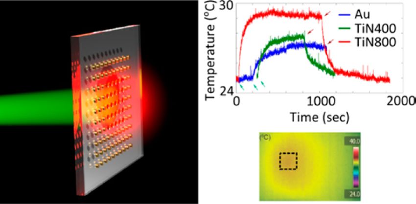
Nano Letters                                                                                                                                      Letter

steady-state temperature increase obtained from Au arrays is              sccm N2 flow. Two growth temperatures were used for TiN
low when compared to that of TiN nanoparticles grown at 400               samples, 400 and 800 °C. The deposition rate is approximated
and 800 °C. Extinction dips observed in the transmittance data            as 21 Å/min and the final thickness of the structures were
presented in Figure 2 reveal that TiN particles of 180 nm size            measured as 30 nm with atomic force microscopy (Veeco
grown at 400 and 800 °C are expected to provide better heating            Dimension 3100 AFM). Because of the high-temperature
efficiencies when excited at wavelengths around 1100 and 1000               growth of TiN samples, an 80 nm thick Cr lift-off layer was
nm, respectively. Also, the numerical data for the absorption             added to the standard lithography process. The nanoparticle
efficiencies predict that TiN grown at 800 °C should give better            pattern was transferred from ZEP 520A resist to Cr layer by use
heating performance at the excitation wavelength of 800 nm.               of reactive ion etching (Panasonic E620 Etcher). After TiN
This is in agreement with the larger experimentally observed              deposition, the Cr lift-off layer was removed with wet etching
temperature increase from the TiN sample grown at 800 °C.                 (Cyantek CR-7). The same procedure is followed for Au
Although the illuminating light is off the resonance peak for              samples in order to ensure shape consistency. During each
TiN nanodisks grown at 800 °C, these particles still provide a            deposition, a thin film sample was grown as a reference for
temperature increase of 4 °C, which is 2 times the heating                dielectric constant retrieval.
obtained from Au nanoparticles of same size. Densely packed                  Optical Measurements. The dielectric functions of each
TiN nanoparticles of smaller sizes excited with 800 nm light              material used in this work were retrieved from thin films by use
will provide a much higher temperature increase due to the                of a variable angle spectroscopic ellipsometer (V-VASE, J. A.
spectral match of the resonance peak wavelength with the                  Woollam Co.). The Drude-Lorentz model was used for data
excitation source.                                                        retrieval; details of data fit are given in the Supporting
   In this work, we have experimentally demonstrated the                  Information. The same ellipsometer setup was employed for
superior performance of TiN over Au for local heating                     transmittance measurements with optional focusing probes,
applications with lithographically fabricated nanodisks. How-             which allow examination of lithographic samples with
ever, large-scale EBL is not feasible method to generate TiN              dimensions down to 500 μm × 500 μm.
nanoparticles. Therefore, obtaining plasmonic TiN nano-                      Thermal Measurements. Samples were excited with 650
particles through chemical methods would be of high interest              mW of weakly focused laser light from a Ti:sapphire
to researchers in the field of plasmonics. Although synthesis of           regenerative amplifier at a wavelength of 800 nm. Illumination
TiN powders has been a hot topic due to above-mentioned                   was performed from backside and at a slight angle in order to
superior physical properties of the material, plasmonic                   prevent damage to the infrared camera. Heated samples were
properties have not been reported so far. A wide range of                 imaged with a thermal camera (FLIR A320). The substrate
methods to generate TiN powders have been investigated                    temperature was averaged over the area of the nanoparticle
including ammonolysis of titanium dioxide nanoparticles,38                arrays for each sample, resulting in a time-dependent
vapor synthesis,39 benzene-thermal route,40 urea route,41                 temperature line plot. The laser source was turned off after
mechanical ball milling,42 and laser ablation.43 Our current              reaching a steady state for each sample. All samples were placed
research is focused on fabricating plasmonic TiN powders and              on a fixed holder on the optical axis in order to ensure constant
promising initial results have been obtained. The development             laser intensity for each sample.
of a chemical synthesis method for plasmonic TiN nano-
particles would also stimulate the studies on surface chemistry
and potential toxicity of nanostructured TiN, which is already a
                                                                          ■
                                                                          *
                                                                              ASSOCIATED CONTENT
                                                                          S Supporting Information
widely used material in biomedical applications.44−46
                                                                          The dielectric functions retrieved from thin film samples and
   In conclusion, TiN nanodisks were studied in the scope of
                                                                          included in FEM simulations are presented; oscillator model
local heating applications and their heating efficiency was
                                                                          and fit parameters are provided. The absorption efficiencies for
compared to the widely used Au nanodisks. Both Mie
                                                                          spherical nanoparticles of TiN and Au located in water are
calculations and FEM simulations suggest that TiN nano-
                                                                          given. SEM images of Au and TiN samples are given. The
particles can perform better than Au in the biological
                                                                          thermal measurement presented in Figure 4 of main text was
transparency window due to the spectral match of the dipolar
                                                                          simulated for TiN nanoparticles grown at 800 °C and
resonances to the region of interest. Particles with identical
                                                                          compared to experimental result in Figure S4. This material
geometries fabricated via EBL are compared through trans-
                                                                          is available free of charge via the Internet at http://pubs.acs.org.

                                                                          ■
mittance and heating experiments in order to verify the
numerical findings. Experiments reveal that TiN nanodisks can
outperform Au nanodisks of same geometrical parameters and                    AUTHOR INFORMATION
eliminate the need for complex-shaped metal nanostructures in             Corresponding Author
practical applications. Considering the fact that TiN is a                *E-mail: aeb@purdue.edu.
biocompatible, CMOS compatible, and durable material with a               Notes
high melting point, our results prove that TiN is a promising
                                                                          The authors declare no competing financial interest.

                                                                          ■
material candidate for many areas such as biological and
medical applications, energy harvesting, heat-assisted magnetic
recording, and others.                                                        ACKNOWLEDGMENTS
   Materials and Methods. Sample Fabrication. EBL is used                 The authors acknowledge support from the following grants:
for nanodisk fabrication due to the high accuracy of the                  ONR-MURI Grant N00014-10-1-0942, ARO Grant 57981-PH
technique on the geometry of the features. Au samples were                (W911NF-11-1-0359), and NSF MRSEC Grant DMR-
deposited on a sapphire substrate with electron beam                      1120923. J.N. also acknowledges financial assistance from
evaporation and TiN samples were deposited with DC reactive               Purdue Calumet Water Institute. We are thankful to Nathaniel
magnetron sputtering from a Ti target under 4 sccm Ar and 6               Kinsey for the help in preparation of this manuscript.
                                                                   6082                         dx.doi.org/10.1021/nl4033457 | Nano Lett. 2013, 13, 6078−6083
Nano Letters
■
Letter
REFERENCES (35) Patsalas, P.; Logothetidis, S. J. Appl. Phys. 2001, 90 (9), 4725−
4734.
(1) Barnes, W. L.; Dereux, A.; Ebbesen, T. W. Nature 2003, 424 (36) CrysTec GmbH. http://www.crystec.de/daten/al2o3.pdf (ac-
(6950), 824−830. cessed February 2013).
(2) Atwater, H. A. Sci. Am. 2007, 296 (4), 56−63. (37) Hirsch, L. R.; Stafford, R. J.; Bankson, J. A.; Sershen, S. R.;
(3) Freestone, I.; Meeks, N.; Sax, M.; Higgitt, C. Gold Bull. 2007, 40 Rivera, B.; Price, R. E.; Hazle, J. D.; Halas, N. J.; West, J. L. Proc. Natl.
(4), 270−277. Acad. Sci. U.S.A. 2003, 100 (23), 13549−13554.
(4) Lal, S.; Link, S.; Halas, N. J. Nat. Photonics 2007, 1 (11), 641− (38) Li, J.; Gao, L.; Sun, J.; Zhang, Q.; Guo, J.; Yan, D. J. Am. Ceram.
648. Soc. 2001, 84 (12), 3045−3047.
(5) Maier, S. A. Plasmonics: Fundamentals and Applications: (39) Dekker, J. P.; van der Put, P. J.; Veringa, H. J.; Schoonman, J. J.
Fundamentals and Applications; Springer: New York, 2007. Mater. Chem. 1994, 4 (5), 689−694.
(6) Schuller, J. A.; Barnard, E. S.; Cai, W.; Jun, Y. C.; White, J. S.; (40) Hu, J.; Lu, Q.; Tang, K.; Yu, S.; Qian, Y.; Zhou, G.; Liu, X. J. Am.
Brongersma, M. L. Nat. Mater. 2010, 9 (3), 193−204. Ceram. Soc. 2000, 83 (2), 430−432.
(7) Blaber, M. G.; Arnold, M. D.; Ford, M. J. J. Phys. Chem. C 2009, (41) Giordano, C.; Erpen, C.; Yao, W.; Milke, B.; Antonietti, M.
113 (8), 3041−3045. Chem. Mater. 2009, 21 (21), 5136−5144.
(8) Boltasseva, A.; Atwater, H. A. Science 2011, 331 (6015), 290− (42) Calka, A. Appl. Phys. Lett. 1991, 59 (13), 1568−1569.
291. (43) Takada, N.; Sasaki, T.; Sasaki, K. Appl. Phys. A: Mater. Sci.
(9) Naik, G. V.; Shalaev, V. M.; Boltasseva, A. Adv. Mater. 2013, 25 Process. 2008, 93 (4), 833−836.
(24), 3264−3294. (44) Wisbey, A.; Gregson, P. J.; Tuke, M. Biomaterials 1987, 8 (6),
(10) Steinmüller-Nethl, D.; Kovacs, R.; Gornik, E.; Rödhammer, P. 477−480.
Thin Solid Films 1994, 237 (1), 277−281. (45) Hyde, G. K.; McCullen, S. D.; Jeon, S.; Stewart, S. M.; Jeon, H.;
(11) Hibbins, A. P.; Sambles, J. R.; Lawrence, C. R. J. Modern Opt. Loboa, E. G.; Parsons, G. N. Biomed. Mater. 2009, 4 (2), 025001.
1998, 45 (10), 2051−2062. (46) Williams, D. F. Biomaterials 2008, 29 (20), 2941−2953.
(12) Reinholdt, A.; Pecenka, R.; Pinchuk, A.; Runte, S.; Stepanov, A.
L.; Weirich, T. E.; Kreibig, U. Eur. Phys. J. D 2004, 31 (1), 69−76.
(13) Cortie, M. B.; Giddings, J.; Dowd, A. Nanotechnology 2010, 21
(11), 115201.
(14) Chen, N. C.; Lien, W. C.; Liu, C. R.; Huang, Y. L.; Lin, Y. R.;
Chou, C.; Chang, S. Y.; Ho, C. W. J. Appl. Phys. 2011, 109 (4),
043104.
(15) Naik, G. V.; Kim, J.; Boltasseva, A. Opt. Mater. Express 2011, 1
(6), 1090−1099.
(16) Naik, G. V.; Schroeder, J. L.; Ni, X.; Kildishev, A. V.; Sands, T.
D.; Boltasseva, A. Opt. Mater. Express 2012, 2 (4), 478−489.
(17) Guler, U.; Naik, G.; Boltasseva, A.; Shalaev, V.; Kildishev, A.
Appl. Phys. B: Lasers Opt. 2012, 107 (2), 285−291.
(18) Cole, J. R.; Mirin, N. A.; Knight, M. W.; Goodrich, G. P.; Halas,
N. J. J. Phys. Chem. C 2009, 113 (28), 12090−12094.
(19) Smith, A. M.; Mancini, M. C.; Nie, S. Nat Nanotechnol. 2009, 4
(11), 710−711.
(20) Richardson, H. H.; Carlson, M. T.; Tandler, P. J.; Hernandez,
P.; Govorov, A. O. Nano Lett 2009, 9 (3), 1139−1146.
(21) Baffou, G.; Quidant, R. Laser Photonics Rev. 2013, 7 (2), 171−
187.
(22) Halas, N. MRS Bull. 2005, 30 (05), 362−367.
(23) Adleman, J. R.; Boyd, D. A.; Goodwin, D. G.; Psaltis, D. Nano
Lett 2009, 9 (12), 4417−4423.
(24) Hung, W. H.; Aykol, M.; Valley, D.; Hou, W.; Cronin, S. B.
Nano Lett 2010, 10 (4), 1314−1318.
(25) Boyd, D. A.; Greengard, L.; Brongersma, M.; El-Naggar, M. Y.;
Goodwin, D. G. Nano Lett 2006, 6 (11), 2592−2597.
(26) Neumann, O.; Urban, A. S.; Day, J.; Lal, S.; Nordlander, P.;
Halas, N. J. ACS Nano 2012, 7 (1), 42−49.
(27) Kryder, M. H.; Gage, E. C.; McDaniel, T. W.; Challener, W. A.;
Rottmayer, R. E.; Ganping, J.; Hsia, Y.-T.; Erden, M. F. Proc. IEEE
2008, 96 (11), 1810−1835.
(28) Baffou, G.; Quidant, R.; Garcia de Abajo, F. J. ACS Nano 2010,
4 (2), 709−716.
(29) Bohren, C.; Huffman, D. Absorption and Scattering of Light by
Small Particles; Wiley-VCH: New York, 1998.
(30) Govorov, A. O.; Richardson, H. H. Nano Today 2007, 2 (1),
30−38.
(31) Baffou, G.; Girard, C.; Quidant, R. Phys. Rev. Lett. 2010, 104
(13), 136805.
(32) Mie, G. Ann. Phys. 1908, 330 (3), 377−445.
(33) Ye, X.; Jin, L.; Caglayan, H.; Chen, J.; Xing, G.; Zheng, C.;
Doan-Nguyen, V.; Kang, Y.; Engheta, N.; Kagan, C. R.; Murray, C. B.
ACS Nano 2012, 6 (3), 2804−2817.
(34) Loo, C.; Lin, A.; Hirsch, L.; Lee, M. H.; Barton, J.; Halas, N.;
West, J.; Drezek, R. Technol. Cancer Res. Treat. 2004, 3 (1), 33−40.
6083 dx.doi.org/10.1021/nl4033457 | Nano Lett. 2013, 13, 6078−6083You can also read