Lumbar Multifidus Muscle Thickness During Graded Quadruped and Prone Exercises
←
→
Page content transcription
If your browser does not render page correctly, please read the page content below
Original Research

Lumbar Multifidus Muscle Thickness During Graded Quadruped and Prone
Exercises
KONSTANTINOS DAFKOU†1, ELEFTHERIOS KELLIS‡1, ATHANASIOS ELLINOUDIS‡1, and
CHRYSOSTOMOS SAHINIS†1
1Laboratory of Neuromechanics, Department of Physical Education and Sport Sciences at Serres, Aristotle

University of Thessaloniki, GRC
†Denotes   graduate student author, ‡Denotes professional author

                                         ABSTRACT
International Journal of Exercise Science 14(7): 101-112, 2021. Exercises for lumbar multifidus (LM)
muscle are important for injury and low back pain prevention and treatment. This study examined the differences
in LM contraction thickness between variations of the superman and bird dog exercises. Twenty-one recreational
athletes performed the superman exercise from the prone position with the following grading: rest, right upper
extremity lift (RU), right lower extremity (RL) and upper and lower extremities lift (UL). They also performed the
following bird dog variations from the quadruped position: rest, RU, RL and left upper - right lower extremity lift
(LURL). LM muscle thickness of both sides was recorded using two ultrasonography (US) devices. LM thickness
during superman-UL, was significantly greater compared with the other exercises and significantly lower during
upper extremity exercises compared with lower extremity exercises (p < 0.05). No significant differences in LM
thickness between sides was found (p > 0.05). The US measurements of LM thickness displayed good to excellent
intrarrater reliability for both muscle sides. It appears that superman-UL is the most effective exercise for a greater
contraction thickness of LM. Further, in order to progressively increase LM muscle thickness, upper extremity tasks
should be performed prior to lower extremity tasks and combined upper and lower lifting tasks.

KEY WORDS: Trunk stabilization; ultrasonography; rehabilitation

INTRODUCTION

Core stability exercises are an integral part of training programs (12, 28, 37) which are used for
injury prevention (28, 34) or sport performance increase (28). Lumbar multifidus (LM) is a deep
muscle of the spine and an important stabilizer of the trunk (1, 10, 26). When the LM muscle is
well developed and functional it controls the magnitude of spinal motion and provides stiffness
to maintain mechanical stability (1). On the other hand, atrophy in the LM muscle is closely
related to chronic low back pain (8). However, due to the fact that this muscle is located in the
deepest layers of the lumbar musculature, specific exercises should be performed for selective
LM training. Therefore, exercises that selectively recruit the LM are considered effective for
improving lumbar stabilization (12, 28, 33).
Int J Exerc Sci 14(7): 101-112, 2021
Exercises that increase LM activity can be performed from different body positions such as
sitting, standing, prone, supine or quadruped (3, 4, 19, 38). Exercises from the quadruped and
prone positions, implementing upper and lower extremities raises, such as the “bird dog” and
the “superman”, have been proposed as ideal for selective training of the LM muscle, without
increasing lumbar spine stress (12, 13, 17, 27, 38). The superman exercise is performed from the
prone position and involves simultaneous upper and lower extremities raises (13, 17, 23). The
Bird dog exercise is performed from the quadruped position and it involves simultaneous raises
of the contralateral upper and lower extremities (17, 27, 38). During the superman exercise the
LM thickness shows an increase of 34% compared to rest (13) while bird dog exercises shows
lower values (20-30%) (15, 16). This provides an indication that bird dog exercise provides a
lower intensity training stimulus than the superman exercise. This is supported by
electromyography (EMG) analysis (6) which has shown a greater activation of the superman
exercise compared to the bird dog exercise. However, given the deep location of the LM, the use
of surface EMG to monitor LM activation may be influenced by crosstalk from surrounding
muscles (36). To our knowledge, ultrasound (US) contraction thickness has not been directly
compared between those exercises.
Variation or grades of either Superman or Bird dog exercise include lifting of upper or lower
extremities in various combinations. Grading these exercise variations can assist in setting
exercise progression during the training period. Studies have shown (13, 17, 23) that the
superman exercise performed with both upper and lower extremities lifted elicits greater
thickness than that performed with either the upper or the lower extremity alone. Bird dog
exercise with diagonal upper and lower extremity raises also shows greater thickness than
upper or lower extremity lifts alone (27). Only two studies (6, 17) compared surface EMG
patterns between various exercises (superman, bird dog, dead-bug, curl-up). Ekstrom et al. (6)
found that superman lifting both upper and lower extremities was more effective in activating
the LM muscle compared to only upper extremities or diagonal upper and lower extremities
lifting during superman exercise. EMG studies (17, 38) reported that the bird dog exercise with
diagonal upper and lower extremity lift shows higher EMG LM activation compared with the
same exercise with lifting only the upper or lower extremity. However, Kim et al., (17) did not
observe any differences in EMG between variations of superman exercise. Hence, the efficiency
of variations of superman and bird-dog exercises to recruit the LM muscle, needs further
investigation.
Asymmetries in LM muscle between the two sides of the body are often encountered and have
been proposed as possible indicators for lumbar pathology (8, 30). Such asymmetries may
appear even in healthy individuals and they may require specific training (8, 30). Research (27,
38) has shown that during leg raise from the quadruped position the ipsilateral side of LM EMG
activity is greater than the contralateral side, while arm raises affect more the contralateral than
the ipsilateral side. Masaki et al. (27) reported that shoulder and hip abduction from the
quadruped position is more effective for activating the LM of the contralateral side rather than
the same side. However, whether upper and lower extremity lifting from the prone or
quadruped positions affect differently the contraction thickness of left and right LM muscle has
not been examined.
International Journal of Exercise Science http://www.intjexersci.com
102Int J Exerc Sci 14(7): 101-112, 2021
To develop optimal training programs, it is essential to understand which exercises can
selectively strengthen a specific muscle (35). Most studies implemented EMG for the assessment
of LM muscle activation (19, 27, 31, 38). This technique displays some limitations such as the
influence of crosstalk activity from adjacent muscles (35) and the inconvenience of using
invasive needle electrodes (16, 35). US imaging offers a non-invasive evaluation of changes in
LM thickness during exercise relative to rest (“contraction thickness ratio”) (11, 16) which is
strongly correlated with LM EMG activation (16, 18, 21). US thickness has also a linear
relationship with cross sectional area determined using magnetic resonance imaging (MRI) (2).
Further, US measurements display good to excellent interrater (intraclass correlation coefficient
(ICC) > 0.75) and intra-rater reliability (ICC > 0.90) for both experienced and novice raters,
during rest and contraction of the LM (5,15).
Understanding the influence of various tasks on LM thickness may assist in ranking exercises
based on their intensity and hence, to set the exercise progression during a given intervention
period. Further, identifying exercises that specifically target one side of the body over the other
may be particularly useful when asymmetries in LM function between sides are detected.
Therefore, the purpose of the present study was, first, to examine differences in LM thickness
between superman and bird dog exercises, second, to compare LM thickness between different
grades of the superman and bird dog exercises, and finally, to examine differences in LM
thickness during exercise between the two sides of the body.
METHODS
The experimental protocol included the measurement of LM muscle thickness on left and right
side simultaneously, using US technology, during eight different conditions. Four of these were
performed from the prone position and included graded superman exercises : 1) rest , 2) right
upper extremity (RU), 3) right lower extremity lift (RL), 4) upper and lower extremities lift (UL)
and the remaining four were bird dog exercises after assuming quadruped position: 5) rest, 6)
RU, 7) RL and 8) left upper and right lower extremities lift (LURL). Ten participants returned to
the laboratory for a re-test measurement in a follow-up session 2 days later. Intra-rater reliability
of US measurements was examined across the 2 repeated measures of all 8 conditions.
Participants
Twenty-one recreational athletes (mean ± standard deviation (SD): age: 21.5 ± 2.11 years; mass
81.4 ± 5.17 kg; height 181 ± 6.31 cm) participated in this study voluntarily. The participants were
all healthy males, free from musculoskeletal injuries and they had not undergone a surgery in
the lumbar area. Also, participants were excluded if they reported a recent history (within a
year) of LBP. The participants gave their informed written consent after receiving information
regarding the goals and procedures of the study. This research was carried out fully in
accordance to the ethical standards of the International Journal of Exercise Science (29) and was
approved by the Aristotle University Ethics Committee.
International Journal of Exercise Science http://www.intjexersci.com
103Int J Exerc Sci 14(7): 101-112, 2021
Protocol
LM muscle thickness was acquired, bilaterally at the L5 vertebrae level, with the use of two
synchronized US devices (SSD-3500, ALOKA, Japan and GE LOGIQ 400 CL PRO, GE Medical
Systems, U.K) provided with linear array probes of 10 MHz wave frequency and a length of 6
cm. Two investigators operated each ultrasound unit and did all the scanning for this study, for
all participants simultaneously. One of them had 7-year experience in the use of US and the
other had finished 1-year practice with the specific protocol prior to commencement of this
study.
Prior to measurements each participant was familiarized to the procedures by being instructed
in and practicing the exercises to be performed, until they could correctly execute each exercise.
For the measurements from the prone position, the participant was positioned on a therapy bed
in a relaxed position with both hands lying next to the body. For the superman-RU exercise, the
participants were asked to lift the right upper extremity, with the elbow fully extended and the
shoulder abducted at 180°. During the superman-RL only the right lower extremity was lifted,
by extending the hip, knee and ankle joints. Finally, for the superman-UL exercise the
participants were instructed to lift both upper and lower extremities at the same time, with
elbows extended and shoulders abducted at 180° and legs extended at the knee and ankle joints.
The quadruped position was assumed by placing the hands shoulder-width and the knees on
the bed, right below the shoulder and hip joints, respectively. The bird dog-RU was performed
by lifting the right arm to the horizontal level, at 180° shoulder abduction. The bird dog-RL
exercise performed with right leg raise to 0° hip extension. The last exercise (bird dog-LURL)
was executed by lifting, the left upper extremity to 180° shoulder abduction and the right lower
extremity to 0° hip extension, at the same time. The duration of the contractions was
approximately 10 s, in order US images to be captured. Within this contraction period the
participants were asked to take a breath and hold it and then, US images for both sides were
frozen, simultaneously. A resting interval of 1 minute was included between each exercise trial.
During data collection, all the testing conditions were completed without pain or discomfort.
The experimental exercises were performed in a randomized order.
For the acquisition of the US images the two transducers were initially placed longitudinally
along the spine with the mid-point over the L4 spinous process. They were, then, moved
laterally and turned slightly medially until the L4/5 zygapophyseal joint could be identified
(16). At this point the probe is directly over LM muscle and after ensuring a good visualization
at the US screen, marks were drawn on the skin with a surgical marker, for consistency during
measurements. LM muscle thickness measurements were obtained via the electronic on-screen
calipers of the US software. Muscle thickness was expressed as the distance between the facet
joint and the plane between the subcutaneous tissue and LM multifidus muscle (Figure 2).
In each testing position, the LM thickness measurements which were acquired during each
exercise were normalized to the corresponding thickness at rest. The contraction thickness ratio
(CTR), was calculated as the percentage change from rest to exercise using the following
equation: thickness contraction – thickness rest / thickness rest. To reduce variability by
approximately 50% (22), the mean of three trials was used.
International Journal of Exercise Science http://www.intjexersci.com
104Int J Exerc Sci 14(7): 101-112, 2021
Statistical Analysis
Data were checked for normality using the Kolmogorov-Smirnov test. A two-way mixed
analysis of variance (ANOVA) design was used to determine the effect of condition (8 levels)
and side (left and right) on LM muscle thickness. If significant, a post-hoc analysis Tukey test
was applied to determine significant differences between various pairs of means. A separate
two-way mixed ANOVA was applied to examine the differences in contraction thickness ratio
(CTR) between six exercises and two sides of LM muscle. Post-hoc Tukey test was applied to
determine significant differences between various pairs of means. The generalized eta squared
values (η2) were calculated as a measure of effect sizes for each independent variable and their
interaction. The level of significance was set at α = 0.05.
Ten of the recruited participants returned to the laboratory for a re-test measurement two days
after the first session. Re-testing was performed exactly as the first testing session. Each of the
two investigators operated the same US unit and tested the same muscle side. They both
remained blind to each other’s assessment of muscle thickness during the testing process. The
generated data from these 10 participants were used for the reliability analysis of the study.
An ICC was calculated to assess intra-examiner reliability (ICC2,1) with a 95% confidence
interval (CI: 95%) based on the average of 3 measurements per session. An ICC value ≤ 0.50 was
considered low, 0.50 to 0.75 moderate, ≥ 0.75 good and ≥ 0.90 excellent (20). Agreement between
the measurements was examined using Bland-Altman analysis (Bias ± limits of agreement (LoA)
(25). Bias was calculated as the absolute difference in thickness (mm) between test and retest
sessions; values closer to 0 indicated greater agreement. The LoA was calculated as 1.96*SD
representing a measure of random error between measurement sessions. In addition, the
standard error of measurement (SEM) was calculated using the following formula:
SEM = SD * √1 – ICC.
RESULTS
Mean (± SD) values for both sides of LM muscle thickness at all 8 conditions are presented in
Table 1. The ANOVA showed a non-statistically significant Condition by Side effect on muscle
thickness (F7, 280 = 0.13, p > 0.05). However, there was a significant main effect of Condition (F7,
280 = 145.45, p < 0.05, η = 0.78). Post-hoc Tukey tests revealed that muscle thickness was
2
significantly lower at rest (both prone and quadruped) compared with all exercise conditions (p
< 0.05) except from the bird dog-RU exercise. Moreover, muscle thickness during superman-UL
was significantly greater compared with the other exercises (p < 0.05). Further, compared to the
exercises that implemented lower extremity lift (superman-RL, bird dog-RL and bird dog-
LURL) muscle thickness was significantly lower during upper extremity lifting exercises
(superman-RU and bird dog-RU) (p < 0.05). Finally, no significant differences in thickness
between sides (F1, 40 = 0.006, p > 0.05) were found.
International Journal of Exercise Science http://www.intjexersci.com
105Int J Exerc Sci 14(7): 101-112, 2021
Table 1. Mean (± SD) thickness of left and right lumbar multifidus (LM) in each testing condition. Mean percentage
(%) differences (± standard error of measurement) between each exercise and resting condition are also reported.
Condition Left LM (mm) CTR (%) Right LM (mm) CTR (%)
Rest Prone 30.03 ± 3.51 30.26 ± 3.13
Superman-RU 37.39 ± 4.88 24.80 ± 11.52 37.03 ± 5.30 22.69 ± 15.29
Superman-RL 40.83 ± 4.56 33.43 ± 12.66 40.89 ± 3.39 33.18 ± 10.14
Superman-UL 42.70 ± 5.44 42.84 ± 15.58 43.05 ± 4.64 42.95 ± 14.54
Rest Quadruped 29.86 ± 4.05 29.98 ± 3.18
Bird dog-RU 31.68 ± 4.25 6.09 ± 13.79 31.24 ± 4.05 3.62 ± 11.89
Bird dog-RL 39.25 ± 3.94 31.48 ± 12.47 39.61 ± 3.72 31.42 ± 10.04
Bird dog-LURL 39.89 ± 4.51 36.54 ± 11.74 40.21 ± 4.32 35.85 ± 11.15
Note. n = 21, CTR = contraction thickness ratio; LURL = Left Upper and Right Lower extremity lift; RL = Right
Lower Extremity lift; RU = Right Upper Extremity lift; UL = upper and lower extremities lift.
Group CTR values in each of 6 exercise conditions are presented in Figure 1. The ANOVA
showed a non-statistically significant Condition by Side effect on CTR (F5, 200 = 0.16, p > 0.05).
However, there was a significant main effect of Condition (F5, 200 = 96.40, p < 0.05, η2 = 0.70). Post-
hoc Tukey tests revealed that superman and bird dog exercises were significantly different in
terms of CTR, only in the superman-UL variation, which yielded the greatest CTR compared to
the other exercises (p < 0.05). Further, the CTR of the exercises that implemented lower extremity
lift (superman-RL, bird dog-RL and bird dog-LURL) was significantly greater compared with
the upper extremity lifting exercises (superman-RU and bird dog-RU) (p < 0.05). Finally, no
significant differences in CTR between sides (F1.40 = 0.08, p > 0.05) were observed.
Figure 1. Mean group values of the contraction thickness ratio (CTR) of the LM muscle left and right side in each
exercise condition (error bars indicate standard deviation). * significantly different compared with each exercise
condition, ^ significantly different compared with Superman-RU and bird dog-RU, p < 0.05.
International Journal of Exercise Science http://www.intjexersci.com
106Int J Exerc Sci 14(7): 101-112, 2021
Reliability results for muscle thickness are presented in Table 2. The ICC2,1 values ranged from
0.86 to 0.98 for the left side of LM muscle and from 0.87 to 0.98 for the right side of LM muscle.
In absolute terms, the SEM values ranged from 0.01 mm to 0.78 mm and from 0.06 mm to 0.57
mm for left and right side of LM muscle, respectively. The systematic error was low, ranging
from -0.33 to 0.93 mm for left side and from -0.50 to 1.55 mm for right side. The random error
ranged between -1.34 – 2.29 mm for left side and between -1.37 – 3.79 mm for right side of LM
muscle.
Table 2. Reliability values for LM muscle thickness in different exercises.
Exercise Side Test R-test ICC2,1 SEM Bias Lower LoA Upper LoA
Left 30.74 ± 2.55 31.07 ± 2.37 0.89 0.01 -0.33 -1.12 0.46
Rest Prone
Right 31.36 ± 2.71 31.86 ± 2.76 0.90 0.06 -0.50 -1.37 0.37
Left 36.62 ± 3.17 36.60 ± 3.22 0.98 0.07 0.02 -0.34 0.38
Superman-RU
Right 36.79 ± 3.73 36.92 ± 3.74 0.98 0.08 -0.13 -0.55 0.29
Left 39.97 ± 4.37 39.70 ± 3.98 0.95 0.27 0.27 -0.61 1.15
Superman-RL
Right 41.48 ± 3.06 41.20 ± 3.00 0.87 0.55 0.38 -0.71 1.47
Left 41.37 ± 4.98 40.68 ± 4.70 0.97 0.26 0.69 -0.06 1.44
Superman-UL
Right 42.28 ± 3.78 41.94 ± 3.61 0.98 0.08 0.34 -0.07 0.75
Left 29.13 ± 3.15 29.18 ± 3.84 0.86 0.67 -0.05 -1.34 1.24
Rest Quadruped
Right 30.68 ± 3.53 30.49 ± 3.64 0.97 0.12 0.19 -0.33 0.71
Left 31.55 ± 3.89 30.77 ± 4.24 0.86 0.78 0.78 -0.73 2.29
Bird dog-RU
Right 31.72 ± 4.12 30.92 ± 4.12 0.90 0.57 0.80 -0.51 2.11
Left 38.75 ± 4.55 37.82 ± 4.68 0.95 0.30 0.93 -0.04 1.90
Bird dog-RL
Right 39.37 ± 4.33 39.22 ± 4.20 0.98 0.44 1.55 -0.69 3.79
Left 38.69 ± 4.91 38.88 ± 4.68 0.97 0.23 -0.19 -0.93 0.55
Bird dog-LURL
Right 39.65 ± 4.55 39.54 ± 4.77 0.98 0.14 -0.19 -0.93 0.55
Note. Measures of reliability: ICC = intraclass correlation coefficient; SEM = standard error of measurement; Bias ±
LoA = 95% limits of agreement; LURL= Left Upper and Right Lower extremity lift; RL = Right Lower Extremity
lift; RU = Right Upper Extremity lift; UL = upper and lower extremities lift.
DISCUSSION
The main findings were that: a) the LM thickness and CTR were significantly greater during
superman-UL compared with all the other exercises, b) LM thickness and CTR were greater
when the lower extremity was lifted compared with upper extremity lifting tasks and, c) no
bilateral differences in thickness were observed during all exercise conditions.
Of all exercises, the superman-UL showed the highest LM thickness and CTR (Table 1). Hwang
and Park (13) reported that during the superman-UL, LM thickness increased approximately by
34% compared to rest, which is line with the present study. Previous EMG studies also
demonstrated that the superman exercise shows activity levels over 60% of maximum voluntary
contraction (MVC) which is greater than that observed in the bird dog exercises (6, 17, 32). EMG
studies have shown that superman-UL displays EMG activation in the range of 62-82% MVC (6,
32) which is greater than that observed during bird dog-LURL (6, 32, 38). The greater CTR
observed during the superman-UL could be attributed to a greater lumbar extension while the
International Journal of Exercise Science http://www.intjexersci.com
107Int J Exerc Sci 14(7): 101-112, 2021
bird dog exercise mostly requires spinal alignment. Paraspinal muscles like the LM, apart from
controlling spinal motion in the transverse and sagittal planes contribute to lumbar extension
movements (14, 23, 24). It is possible that superman-UL exercise yielded a greater CTR
compared to the bird dog exercise since it challenges both the ability of LM muscle to maintain
the orientation of the lumbar spine, while acting as an extensor. In contrast, during bird dog
exercise, LM muscle is activated in order to maintain the alignment of the spine , by resisting
torsional forces created from the opposing upper and lower extremity lift (27). Based on this
finding, it could be suggested that the superman-UL exercise can be incorporated in the final
stages of performance difficulty, in stabilization training programs with unloaded isometric
exercises.
The results of this study indicated that the exercises with lower extremity lifting showed greater
LM CTR than those incorporating upper extremity lifting (Figure 2 and Figure 3). More
specifically, CTR during superman-RU and bird dog-RU ranged from 3 to 24%, while during
the superman-RL and bird dog-RL exercises from 31 to 34% (Table 1). This is in agreement with
previous studies which examined upper and lower extremity lifts either from the quadruped or
the prone position (13, 17). The differences in measured thickness between exercises may be the
result of a difference in stability between them. By lifting the upper or lower extremities of the
ground, the base of support decreases, increasing the instability and thereby, it triggers the trunk
stabilization muscles to contract in order to maintain an aligned spine (17). Further, there is
evidence that during arm lifting tasks, global/local (erector spinae/LM) activation ratio is
higher compared to leg lifting tasks (17, 27, 38) which may provide an additional explanation of
our findings. Therefore, taking into account that the exercises we applied had a different impact
on LM thickness, upper extremity lifting exercises (superman-RU and bird dog-RU) could be
performed first at early stages of rehabilitation or in the case of well-trained individuals
disregarded, followed by lower extremity raises (superman-RL and bird dog-RL), opposing
upper and lower extremity lifting exercises (bird dog-LURL) and last, the superman-UL, where
all the extremities are lifted, as the most effective of these exercises, to recruit this muscle.
Even though our exercises required unilateral upper or lower extremity movements, there were
no significant differences in thickness between left and right LM muscle thickness (Table 1). In
fact, both sides of the LM displayed excellent symmetry in size, during rest and contraction
(Figure 2 and Figure 3). This was the first study that examined bilateral differences in LM muscle
thickness, during graded exercises from prone and quadruped position, using two synchronized
US devices. Our results are in contrast to previous studies which reported significant
asymmetries in LM size and recruitment between sides (6, 27, 30, 38). For example, Niemeläinen
et al., (30) observed that many healthy adults had asymmetry in LM cross-sectional area between
sides greater than 10%. In addition, it has been reported that during bird dog-LURL, LM
activation is greater on the side of the lifted leg compared to the activity of the contralateral side.
(27, 38). In advance, Masaki et al., (27) found that opposing shoulder and hip abduction
movements from the quadruped position resulted in higher LM activation on the side of the
lower extremity lift as opposed to the contralateral side. However, these studies (7, 27, 38) used
EMG electrodes and, hence, direct comparison with our findings is difficult. It is also possible
that the ability of individuals to trigger and affect LM unilaterally, depends on the presence of
International Journal of Exercise Science http://www.intjexersci.com
108Int J Exerc Sci 14(7): 101-112, 2021
asymmetry between sides (3). Therefore, based on our observations, unilateral
activation/contraction patterns of LM muscle might not be apparent and hence, segmental
training is not feasible, at least in healthy individuals with no asymmetries in LM muscle
thickness.
Figure 2. Illustration of the ultrasound measurement technique of left (L) and right (R) LM muscle at (A) prone rest,
(B) superman- right upper extremity lift (RU), (C) superman-right lower extremity lift (RL) and (D) superman-
upper and lower extremities. Thickness measurements were made between the superficial and deep borders of LM
muscle (drawn with line).
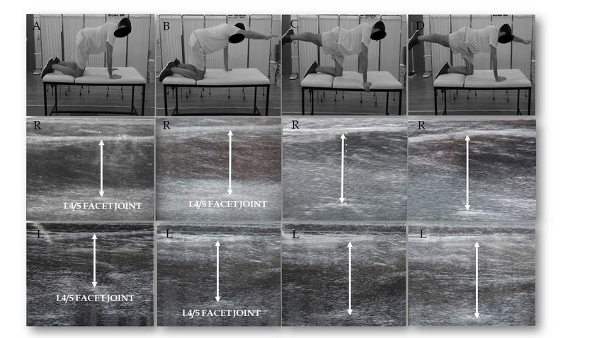
Figure 3. Illustration of the ultrasound measurement technique of left (L) and right (R) LM muscle at (A) quadruped
rest, (B) bird dog- right upper extremity lift (RU), (C) bird dog-right lower extremity lift (RL) and (D) bird dog-left
upper right lower. Thickness measurements were made between the superficial and deep borders of LM muscle
(drawn with line).
International Journal of Exercise Science http://www.intjexersci.com
109Int J Exerc Sci 14(7): 101-112, 2021
The present study has several limitations. First, the research was conducted with recreationally
active males with no musculoskeletal injuries. This restricts the results from representing all
ages, both genders and patients with LBP problems, who may have altered lumbar muscle
activity and contraction ability. Second, the amount of loading in intervertebral joints during
these graded exercises is not clear, even though they have been proposed as safe for athletes and
patients (9, 12). Moreover, the examination of other surrounding muscles, when performing
these exercises, such as the erector spinae and gluteal muscles would give further information,
for the contribution of each muscle to the movement. Our design did not include performance
of the superman exercise with contralateral upper and lower extremity raises. This would allow
a better comparison of superman and bird-dog exercises; instead, we chose to examine the
superman-UL exercise, as it was examined in previous studies (6, 13, 17). Finally, US technology,
although is established as reliable and valid in measuring changes in muscle morphology,
remains an indirect method for the assessment of muscle activity compared with EMG. This
could be a possible explanation for not detecting bilateral differences in LM muscle.
The results of this study indicated that LM CTR was greater during superman compared with
bird dog exercises. Further, LM CTR increased progressively when superman or bird dog
exercises were performed in the following order: only upper extremity raised, only lower
extremities raised and finally, combined upper and lower extremities raised. No side to side
differences in LM muscle thickness were observed in any testing condition. These findings can
be used to set exercise program progression when designing exercise interventions for the LM.
REFERENCES
1. Bakkum BW, Cramer GD. Chapter 4 - Muscles that influence the spine [Internet]. In: Clinical anatomy of the
spine, spinal cord, and Ans (Third Edition). Cramer GD, Darby SA, eds. Saint Louis: Mosby; 2014.
2. Belavý DL, Armbrecht G, Felsenberg D. Real-time ultrasound measures of lumbar erector spinae and multifidus:
Reliability and comparison to magnetic resonance imaging. Physiol Meas 36(11), 2015.
3. Berglund L, Aasa B, Michaelson P, Aasa U. Effects of low-load motor control exercises and a high-load lifting
exercise on lumbar multifidus thickness. Spine (Phila Pa 1976) 42(15): E876–82, 2017.
4. Danneels LA, Vanderstraeten GG, Cambier DC, Witvrouw EE, Bourgois J, Dankaerts W, De Cuyper HJ. Effects
of three different training modalities on the cross sectional area of the lumbar multifidus muscle in patients with
chronic low back pain. Br J Sports Med 35(3): 186–91, 2001.
5. Djordjevic O, Djordjevic A, Konstantinovic L. Interrater and intrarater reliability of transverse abdominal and
lumbar multifidus muscle thickness in subjects with and without low back pain. J Orthop Sports Phys Ther 44(12):
979–88, 2014.
6. Ekstrom RA, Osborn RW, Hauer PL. Surface electromyographic analysis of the low back muscles during
rehabilitation exercises. J Orthop Sports Phys Ther 38(12): 736–45, 2008.
7. Feldwieser FM, Sheeran L, Meana-Esteban A, Sparkes V. Electromyographic analysis of trunk-muscle activity
during stable, unstable and unilateral bridging exercises in healthy individuals. Eur Spine J 21(SUPPL. 2), 2012.
8. Hides J, Gilmore C, Stanton W, Bohlscheid E. Multifidus size and symmetry among chronic LBP and healthy
asymptomatic subjects. Man Ther 13(1): 43–9, 2008.
International Journal of Exercise Science http://www.intjexersci.com
110Int J Exerc Sci 14(7): 101-112, 2021
9. Hides JA, Jull GA, Richardson CA. Long-term effects of specific stabilizing exercises for first-episode low back
pain. Spine (Phila Pa 1976) 26(11), 2001.
10. Hodges PW. Core stability exercise in chronic low back pain. Orthop Clin North Am 34(2): 245–54, 2003.
11. Hodges PW, Pengel LHM, Herbert RD, Gandevia SC. Measurement of muscle contraction with ultrasound
imaging. Muscle Nerve 27(6): 682–92, 2003.
12. Huxel Bliven KC, Anderson BE. Core stability training for injury prevention. Sports Health 5(6): 514–22, 2013.
13. Hwang YI, Park DJ. Comparison of lumbar multifidus thickness and perceived exertion during graded
superman exercises with or without an abdominal drawing-in maneuver in young adults. J Exerc Rehabil 14(4):
628–32, 2018.
14. Jemmett RS, MacDonald DA, Agur AMR. Anatomical relationships between selected segmental muscles of the
lumbar spine in the context of multi-planar segmental motion: A preliminary investigation. Man Ther 9(4): 203–10,
2004.
15. Kellis E, Ellinoudis A, Intziegianni K, Kofotolis N. Muscle thickness during core stability exercises in children
and adults. J Hum Kinet 71(1): 131–44, 2020.
16. Kiesel KB, Uhl TL, Underwood FB, Rodd DW, Nitz AJ. Measurement of lumbar multifidus muscle contraction
with rehabilitative ultrasound imaging. Man Ther 12(2): 161–6, 2007.
17. Kim CR, Park DK, Lee ST, Ryu JS. Electromyographic changes in trunk muscles during graded lumbar
stabilization exercises. PM R 8(10): 979–89, 2016.
18. Kim CY, Choi JD, Kim SY, Oh DW, Kim JK, Park JW. Comparison between muscle activation measured by
electromyography and muscle thickness measured using ultrasonography for effective muscle assessment. J
Electromyogr Kinesiol 24(5): 614–20, 2014.
19. Kim MJ, Oh DW, Park HJ. Integrating arm movement into bridge exercise: Effect on EMG activity of selected
trunk muscles. J Electromyogr Kinesiol 23(5): 1119–23, 2013.
20. Koo TK, Li MY. A Guideline of selecting and reporting intraclass correlation coefficients for reliability research.
J Chiropr Med 15(2): 155–63, 2016.
21. Koppenhaver SL, Hebert JJ, Parent EC, Fritz JM. Rehabilitative ultrasound imaging is a valid measure of trunk
muscle size and activation during most isometric sub-maximal contractions: A systematic review. Aust J Physiother
55(3): 153–69, 2009.
22. Koppenhaver SL, Parent EC, Teyhen DS, Hebert JJ, Fritz JM. The effect of averaging multiple trials on
measurement error during ultrasound imaging of transversus abdominis and lumbar multifidus muscles in
individuals with low back pain. J Orthop Sport Phys Ther 39(8): 604–11, 2009.
23. Macdonald DA, Dawson AP, Hodges PW. Behavior of the lumbar multifidus during lower extremity
movements in people with recurrent low back pain during symptom remission. J Orthop Sports Phys Ther 41(3):
155–64, 2011.
24. Macintosh JE, Valencia F, Bogduk N, Munro RR. The morphology of the human lumbar multifidus. Clin
Biomech 1(4): 196–204, 1986.
International Journal of Exercise Science http://www.intjexersci.com
111Int J Exerc Sci 14(7): 101-112, 2021
25. Martin Bland J, Altman DG. Statistical methods for assessing agreement between two methods of clinical
measurement. Lancet 327(8476): 307–10, 1986.
26. Martuscello JM, Nuzzo JL, Ashley CD, Campbell BI, Orriola JJ, Mayer JM. Systematic review of core muscle
activity during physical fitness exercises. J Strength Cond Res 27(6): 1684–98, 2013.
27. Masaki M, Tateuchi H, Tsukagoshi R, Ibuki S, Ichihashi N. Electromyographic analysis of training to selectively
strengthen the lumbar multifidus muscle: Effects of different lifting directions and weight loading of the extremities
during quadruped upper and lower extremity lifts. J Manipulative Physiol Ther 38(2): 138–44, 2015.
28. McGill S. Core training: Evidence translating to better performance and injury prevention. Strength Cond J 32(3):
33–46, 2010.
29. Navalta J, Stone W, Lyons S. Ethical issues relating to scientific discovery in exercise science. Int J Exerc Sci 12(1):
1, 2019.
30. Niemeläinen R, Briand MM, Battié MC. Substantial asymmetry in paraspinal muscle cross-sectional area in
healthy adults questions its value as a marker of low back pain and pathology. Spine (Phila Pa 1976) 36(25): 2152–
7, 2011.
31. Okubo Y, Kaneoka K, Imai A, Shiina I, Tatsumura M, Izumi S, Miyakawa S. Electromyographic analysis of
transversus abdominis and lumbar multifidus using wire electrodes during lumbar stabilization exercises. J Orthop
Sport Phys Ther 40(11): 743–50, 2010.
32. Oliver GD, Stone AJ, Plummer H. Electromyographic examination of selected muscle activation during
isometric core exercises. Clin J Sport Med 20(6): 452–7, 2010.
33. Pillastrini P, Ferrari S, Rattin S, Cupello A, Villafañe JH, Vanti C. Exercise and tropism of the multifidus muscle
in low back pain: A short review. J Phys Ther Sci 27(3): 943–5, 2015.
34. Schuermans J, Danneels L, Van Tiggelen D, Palmans T, Witvrouw E. Proximal neuromuscular control protects
against hamstring injuries in male soccer players: A prospective study with electromyography time-series analysis
during maximal sprinting. Am J Sports Med 45(6): 1315–25, 2017.
35. ShahAli S, Shanbehzadeh S, ShahAli S, Ebrahimi Takamjani I. Application of ultrasonography in the assessment
of abdominal and lumbar trunk muscle activity in participants with and without low back pain: A systematic
review. J. Manipulative Physiol. Ther. 42(7): 541–50, 2019.
36. Stokes IAF, Henry SM, Single RM. Surface EMG electrodes do not accurately record from lumbar multifidus
muscles. Clin Biomech 18(1): 9–13, 2003.
37. Wirth K, Hartmann H, Mickel C, Szilvas E, Keiner M, Sander A. Core stability in athletes: A critical analysis of
current guidelines. Sport Med 47(3): 401–14, 2017.
38. Yoon TL, Cynn HS, Choi SA, Choi WJ, Jeong HJ, Lee JH, Choi BS. Trunk muscle activation during different
quadruped stabilization exercises in individuals with chronic low back pain. Physiother Res Int 20(2): 126–32, 2015.
International Journal of Exercise Science http://www.intjexersci.com
112You can also read