Metabolic alterations in the critically ill child - Pediatric Medicine
←
→
Page content transcription
If your browser does not render page correctly, please read the page content below

Review Article
                                                                                                                                      Page 1 of 17

Metabolic alterations in the critically ill child
Renán A. Orellana, Jorge A. Coss-Bu

Department of Pediatrics, Baylor College of Medicine, Texas Children’s Hospital, Houston, Texas, USA
Contributions: (I) Conception and design: All authors; (II) Administrative support: None; (III) Provision of study materials or patients: None; (IV)
Collection and assembly of data: None; (V) Data analysis and interpretation: None; (VI) Manuscript writing: All authors; (VII) Final Approval of
manuscript: All authors.
Correspondence to: Jorge A. Coss-Bu, MD, FAAP. Professor, Department of Pediatrics, Baylor College of Medicine, Texas Children’s Hospital, 6651
Main St. E1420, Houston, Texas 77030, USA. Email: jacossbu@texaschildrens.org.

                Abstract: The metabolic response to critical illness is characterized by changes in energy metabolism
                and distribution of substrates and nutrients to sustain cellular function, repair, and to support the immune
                response and growth. Such response is regulated by simultaneous neurohormonal and inflammatory
                responses mediated by neurotransmitters, cytokines, hormones, and metabolic signals. The stress-mediated
                changes in energy expenditure (EE) are characterized by an initial phase of decreased followed by an
                increased EE. Inflammation triggered by cytokines is divided in several categories: interleukins, chemokines,
                interferons, tumor necrosis factor, and growth factors. The interleukins are divided into pro-inflammatory
                (responsible for cell activation, tissue damage and necrosis) and anti-inflammatory interleukins (involved in
                dampening and reversing the inflammatory process. The counter-regulatory response is characterized by
                increased concentrations of catecholamines, glucagon, and cortisol and oppose the effects of insulin. Those
                same elements that regulate stress, inflammation and energetic failure also induce an increase in whole-body
                protein synthesis, intensification of muscle protein catabolism, and a reduction in muscle protein synthesis to
                support a surge of amino acids (AA) released into the systemic circulation to serve as biochemical precursors,
                signals and structural substrates and oxidation for energy if needed. Both energetic and macronutrient
                processes interact and regulate themselves to establish a new homeostatic situation suited to fight infections,
                sustain life, and promote recovery. Due to current advances in intensive care, and modern life sustaining
                therapies, a prolongation of the inflammatory and catabolic state and nitrogen loss occurs, rendering the
                individual resistant to traditional nutrition support therapies.

                Keywords: Energy metabolism; critical illness; child; catabolism; metabolic stress

                Received: 30 June 2020; Accepted: 17 December 2020; Published: 28 February 2021.
                doi: 10.21037/pm-20-64
                View this article at: http://dx.doi.org/10.21037/pm-20-64

Introduction                                                                 chronic conditions) (5,6). This response includes changes
                                                                             in energy expenditure (EE), metabolic changes mediated
The acute metabolic response that follows an acute
                                                                             by proinflammatory cytokines such as interleukin (IL)-1β,
illness, trauma, and surgery is characterized by increased
catabolism, release of increased amounts of glucose, amino                   IL-6, IL-12, IL-18, tumor necrosis factor alfa (TNF-α),
acids (AA), and fatty acids from the body’s stores (1,2). Sir                and interferon gamma (IFN-γ), hormonal responses with
David P. Cuthbertson described the fundamental aspects of                    changes in levels of growth hormone (GH), thyroid-
this metabolic response to injury more than half-a-century                   stimulating hormone (TSH), insulin growth factor binding
ago (3,4). This response varies depending of the nature                      proteins (IGFBP); and several metabolic reactions including
and severity of the insult, as well as, factors related to the               increased gluconeogenesis, increased fatty acid and
host (i.e., age, metabolic reserve capacity, and presence of                 carbohydrate oxidation and increased loss of muscle mass

© Pediatric Medicine. All rights reserved.                                             Pediatr Med 2021;4:8 | http://dx.doi.org/10.21037/pm-20-64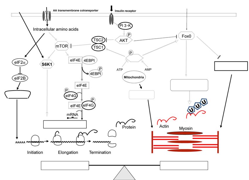
Page 2 of 17                                                                                                                                 Pediatric Medicine, 2021

Figure 1 The Metabolic response to injury. Reproduced with permission from Sparks EA, Fisher JG, Khan F, et al. Chapter 62: The
critically ill child. In: Duggan C, Watkins JB, Koletzko B, et al. editors. Nutrition in pediatrics (5th ed.), 2016:957-69. New Haven, CT and
Cary, NC: PMPH USA, LTD.

  100%
                                            Adaptive Energy 0-10%                                    metabolic rate (BMR), thermogenesis, physical activity, and
                                                                                                     adaptive energy. BMR accounts for 60–70%, thermogenesis
   80%                                      TEE 15-30%
                                                                                                     for a 10% and physical activity represents 20-30% of
   60%                                      DIT 5-10%                                                TEE; adaptive energy is the energy expended to adapt to
                                                                                                     environmental conditions, particularly changes in ambient
   40%
                                                                                                     temperature; throughout circumstances of stress or injury
   20%
                                            BMR 60%                                                  these percentages varies depending on the degree of insult,
                                                                                                     substrate intake and amount of physical activity (15-19).
    0%                                                                                                  Energy needs are related to age and represents up to 3
         TEE: Total energy expenditure; DIT: Diet induced thermogenesis; BMR: Basal metabolic rate
                                                                                                     to 4 times higher per body weight for infants compared to
Figure 2 Components of energy expenditure.                                                           adults (20), and are also dependent on changes on metabolic
                                                                                                     rate and body nutrients reserve. In the presence of an insult
                                                                                                     the response will be proportional to the magnitude, nature,
(7-12) (Figure 1).                                                                                   and duration of the injury (21). Increased levels of counter-
                                                                                                     regulatory hormones will result in opposition to the actions
                                                                                                     of insulin and GH. This increased resistance to the action
Changes in EE
                                                                                                     of insulin and GH will result in a catabolic response with
Energy metabolism is the most important function of                                                  breakdown of glycogen, protein, and fat to provide enough
the body and it regulates basal metabolism, growth, and                                              substrate to support the metabolic response (22,23).
physical activity; it is controlled at the cellular level by                                            The main reason of the augmented energy dissipation
complex reactions of the neurohormonal system and                                                    in younger children is credited to the energy expense for
regulates the utilization of substrate. The total objective of                                       growth. At 6 months of age the growth velocity is maximal
these controlling processes is to preserve energy stability                                          representing up to 6–8% of energy utilized for growth,
and the central nervous system (CNS) acts an essential                                               this process slows down at the age of 12 months once
function in achieving this balance by triggering functions at                                        the BMR is 55 kcal/kg/day, when 2% of the EE is used
the hormonal, neural, and metabolic level (13,14).                                                   for growth (16). During periods of acute stress, however,
   To be able to understand the concept of energy balance                                            somatic growth is very difficult to achieve and cannot
is essential to examine the elements of total energy                                                 occur. Second, critically ill children are usually sedated or
expenditure (TEE) (Figure 2). TEE has four elements: basal                                           treated with muscle relaxants, therefore their activity level

© Pediatric Medicine. All rights reserved.                                                                   Pediatr Med 2021;4:8 | http://dx.doi.org/10.21037/pm-20-64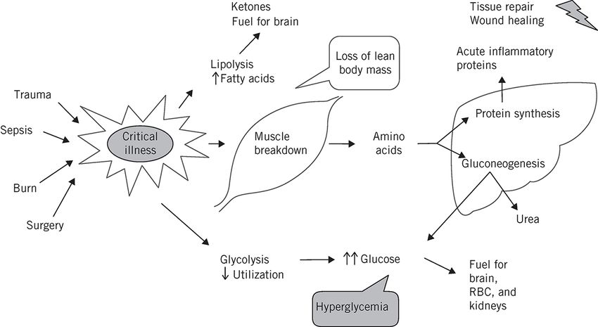
Pediatric Medicine, 2021                                                                                              Page 3 of 17

is reduced significantly lowering their energy needs. Third,         The role of cytokines including TNF-α, IL-1β,
the insensible losses are significantly reduced, particularly     IL-6, IL-8 as important mediators of infection and tissue
for patients on mechanical ventilatory support. Therefore,        injury, and INF-γ as metabolic mediator have been
is very important to take into consideration these changes        identified in many experimental studies. In subjects with
while implementing a nutrition support plan.                      sepsis who died, IL-1β levels were higher compared to
    The traditional concept has been that acutely ill patients    subjects that survived, implying a link between elevated
present a hypermetabolic condition known as “flow phase”,         levels of IL-1β and outcome in patients with sepsis (57).
preceded by a phase of reduced EE aimed to preserve               Several studies have been performed to provide evidence
energy known as “ebb phase” (17,24-32). Many studies              for the role of endogenously produced TNF in the
have reported measurements of EE by indirect calorimetry          development of cachexia, muscle wasting, and decreased
in children admitted to the pediatric intensive care unit         albumin synthesis (51,52,58). The most important
(PICU) (15,17,24-46), the aggregate result of all these           supplier of IL-6 is macrophages at the tissue with elevated
measurements yields an average metabolic index [measured          concentrations noted in several inflammatory conditions
energy expenditure (MEE)/predicted BMR] of 1.02±0.10              such as cardiovascular and autoimmune diseases, or
(SD), indicating an average metabolic condition. Of note          neoplasia (52). Higher levels of IL-6 have been reported
are the studies that reported decreased MEE in postsurgical       in pediatric patients with sepsis compared to subjects
infants and neonates using indirect calorimetry and tracer        with systemic inflammation not associated with infection
methodology indicating a hypometabolic state (47-49),             (59,60). Additionally, in children with sepsis a higher level
therefore, the importance of adjusting the caloric intake in      of IL-6 is linked to more severity (61), making IL-6 a useful
this population of infants and neonates to avoid overfeeding.     tool to predict outcome in sepsis. IL-8 functions include
    In summary, the metabolic response is characterized           chemotaxis and neutrophil stimulation and several studies
by dysregulation of the energy metabolism, therefore, is          have found to be a good discriminator for survival. Wong
important to understand and identify these changes during         et al. reported in two studies (62,63), higher levels of IL-8
the acute phase of the injury in order to implement timely        in deceased pediatric patients with septic shock compared
and appropriate interventions to support metabolically            to subjects that survived, also found that a level of IL-8 ≤
the acutely ill patient, while avoiding underfeeding and          to 220 pg/mL on admission to the intensive care unit was
overfeeding (16,25-27,50).                                        a good predictor of survival. Two reports in children with
                                                                  cancer found that a low level of IL-8 was a good predictor
                                                                  of a low risk of bacteremia (64), and that levels >300 pg/mL
Inflammatory response and cytokines
                                                                  in conjunction with high C-reactive protein (CRP) in
The metabolic response to tissue injury is initiated by           children >12 years of age were associated with worse
activation of the cytokine cascade. Cytokines are a group         outcome in this population (65).
of proteins (
Page 4 of 17 Pediatric Medicine, 2021
given its sensitivity and specificity to distinguish among injury-induced stimuli that triggers the CNS, resulting in
benign vs. severe bacterial infection or the presence of a non- alterations at the hypothalamic-anterior pituitary axes, these
bacterial infection process. A systematic review evaluating include the adrenal gland (increased cortisol secretion), the
CRP to diagnose bacterial infection accurately in ambulatory somatotrophic (increased GH secretion), the thyrotrophic
pediatric patients with fever, reported a sensitivity of 77% [decreased triiodothyronine (T3) and increased reverse T3
and specificity of 79%; this low sensitivity value suggests that (rT3) secretion], and the gonado-/lactotrophic (decreased
CRP cannot be used to exclude all bacterial infection (72). testosterone, increased prolactin) axes (79,80). In addition,
Additionally, it is useful to monitor response to treatment the CNS also acts through the peripheral sympathetic
after a diagnosis of infection has been done, with serial nervous system to increase catecholamine secretion.
persistent high CRP levels or higher levels after 48 hours After an insult, a condition of increased resistance to
indicate inadequate treatment (67). The more recent studies the actions of the GH at the peripheral tissues is developed
in the literature of the use of CRP as a diagnostic tool has (9,81), this response is in part a result of the secreted
focused on the comparison of its diagnostic accuracy with the cytokines. The increase in circulating amounts of GH (82)
use of PCT (68). is heralded by a reduction in concentrations of GH-binding
Procalcitonin is made by the thyroid to control serum protein, indicating a reduction on the expression of the
calcium concentrations and constitutes a precursor of GH receptor at the level of peripheral tissues (83,84). The
calcitonin and it is produced by the parafollicular cells (C cells) reduction in negative feedback inhibition explains the
of the thyroid and by the neuroendocrine cells of the lung ample availability of GH during the initial stages of the
and the intestine, and thyroid C cells are the only ones that stress response; this answer of the GH axis is instrumental
express the enzymes that produce the mature calcitonin (73). in the fight for existence, where indirect insulin-like growth
The thyroid gland produces PCT and this production factor-1 (IGF-1) mediated somatotrophic effects of GH are
occurs under normal conditions with low levels detected. attenuated, resulting in increased levels in the circulation
Under an infection challenge the production of PCT by of glucose and fatty acids (80). The decreased level of
non-thyroidal tissue is increased significantly, suggesting somatotropism, because of a lack of pulsatile GH secretion
that initial inflammatory stimulation from TNF-α, IL- might add to the etiology of the wasting syndrome that
1β and IL-6 is important (68). Multiple studies have distinguishes by a protracted course of a severe condition
evaluated the diagnostic use and advantage of using PCT (79,80,85).
to discriminate sepsis from systemic inflammatory response Shortly after the onset of severe stress there is a rapid
syndrome (SIRS) (74-78), and reported that children with decline of circulating levels of T 3 with a concomitant
established infection had elevated values of PCT compared increase in rT3 as a result of disrupted peripheral conversion
to children without infection (SIRS only) (78), serum PCT of T4 (86,87). The instant reduction in levels of T3 might
concentration was significantly elevated in children with be seen as an answer to preserve energy while the substrate
sepsis compared to children without infection with SIRS intake is markedly reduced (80). The persistence of low
after cardiopulmonary bypass, (74), and in a cohort of T3 after normalization of TSH levels is known as the low
children admitted to the PICU, PCT was better than CRP euthyroid syndrome. The reduction in T3 levels throughout
in discriminating subjects with SIRS and sepsis with PCT the initial period following the insult is a reflection of
elevated concentrations associated with higher severity the severity of the disease process (88) and this has been
of illness (77). The level of evidence published to date in shown in clinical studies where a low T3 is associated with
children with infection and sepsis, preclude the routine use increased mortality (79,89,90).
of PCT as a biomarker in clinical practice, as a prognostic Cortisol levels increase during the acute phase
tool and risk stratification, or to help with the decision of of the response to injury as a result of the increased
antibiotic treatment duration (56). release of corticotropin-releasing hormone (CRH) and
adrenocorticotropic hormone (ACTH), this is explained either
by a direct mechanism or inhibition of the negative feedback
Hormonal response
by cortisol (91,92). The majority of the suppressive effects of
The stress responses to injury, trauma or sepsis are mediated cortisol on immune and inflammatory reactions appear to be
by a number of different hormones, protein messengers, a consequence of the modulation of production or activity
and the development of a complex system of neural of cytokines (i.e., IL-1, IL-2, IL-3, IL-6, interferon-γ,
© Pediatric Medicine. All rights reserved. Pediatr Med 2021;4:8 | http://dx.doi.org/10.21037/pm-20-64Pediatric Medicine, 2021 Page 5 of 17
TNF-α), chemokines, eicosanoids, complement activation, (103,104).
and other inflammatory mediators (i.e., bradykinin, The fat tissue represents the main supplier of fuel and
histamine, macrophage migration inhibitory factor) (93). it is deposited as triacylglycerides and the breakdown or
The most necessary and dynamic hypercortisolism induced lipolysis produces non-esterified free fatty acids (FFA) (105).
by stress in critically ill patients results in energy provision Lipolysis at the extracellular and intracellular space is
by shifting carbohydrate, fat, and protein metabolism, regulated by lipoprotein and hormone-sensitive lipase (HSL).
suppress inflammation, and boost hemodynamics by During conditions of inflammation there is a significant
augmented sensitization of the vasopressor response to increase in serum levels of triacylglycerides and FFA and
catecholamines (80,93,94). Plausible disadvantages of decrease in high-density lipoprotein concentration (106).
prolonged hypercortisolism include impaired wound A d d i t i o n a l l y, i n c r e a s e d l e v e l s o f c y t o k i n e s a n d
healing and myopathy, complications often seen during catecholamines induce blockage of lipoprotein lipase and
lengthy course of critical illness (79,80,90,94). reduced extracellular lipolysis, simultaneously, upregulation
of HSL results in lipolysis at the fat tissue (105). An
increased triacylglycerides synthesis results in reduced
Carbohydrate and lipid metabolism
clearance of triglycerides, resulting in hepatic steatosis (107),
During conditions of stress, hyperglycemia is a consequence and storage of triacylglycerides and FFA at the muscle
of a mixture of enhanced gluconeogenesis and enhanced (108,109), heart (110), and kidney (111), this is the result
insulin resistance resulting in decreased glucose uptake of a disrupted uptake FFA and its oxidation. No reported
by the cells (95) (Figure 1). These two mechanisms are definite evidence that an alteration of the FFA oxidation
potentially mediated by increases in counter regulatory is present at the mitochondrial and peroxisomal level in
hormones and proinflammatory cytokines and potentially the acutely ill patient, but in subjects with type 2 diabetes
these cytokines prevent insulin to be secreted by the and metabolic syndrome, these alterations modify the
pancreas via activation of α adrenergic receptors (96-98). insulin signaling and enhances insulin resistance (112,113).
Increased concentrations of counterregulatory hormones Hypertriglyceridemia is an important concern because of
and proinflammatory cytokines participate in the the altered endothelial function, lipotoxicity, and increased
regulation of glycogenolysis and gluconeogenesis with inflammation and often lipid infusion during acute injury
resultant hyperglycemia (95), and glycogen stores are conditions worsens hypertriglyceridemia (105).
rapidly depleted with glycogenolysis resulting in limited
glucose production (96). The elevated concentrations of
AA and protein and metabolism in the critically
catecholamines during this acute response to injury results
ill
in elevated glucagon levels with gluconeogenesis being
maintained despite elevated levels of insulin (99). Other The complex interrelation of protein metabolism and
hormonal changes including increased GH and decreased metabolic partitioning during critically illness across the
IGF-1 concentrations enable the destruction of muscle human lifespan emphasizes the need to individualize protein
releasing alanine to enhance gluconeogenesis (100). Acute support therapy towards achievement of proteostasis
injury is distinguished by insulin resistance either central or (protein metabolic homeostasis) and organ support rather
peripheral (101), and insulin resistance at the hepatic level than simply balancing nitrogen expenditure (114-116).
being central and mediated by glucagon, epinephrine, and To sustain tissue integrity and organ function in healthy
cortisol (99). The Insulin resistance at the muscle and fat conditions, body protein is continuously degraded and
tissue is classified as peripheral and is explained by changes resynthesized in all tissues and cells, a process known
in the insulin-signaling pathway regulated by inflammatory as protein turnover. Tissue proteins in different organs
cytokines and counter regulatory hormones (101). This are constituted by AA, which normally can be either
peripheral insulin resistance might continue for a protracted incorporated into tissue protein or undergo oxidation for
time after recovery from an acute injury, as described in energy production when energy intake is inadequate to
pediatric patients (102). Several studies have reported satisfy the metabolic demands. Normally, tissue protein
defects in beta-cell function of the pancreas with reduction breakdown releases AA to the peripheral circulation, and
in its ability to produce insulin in critically ill children those circulating AA may be reutilized for accretion of
© Pediatric Medicine. All rights reserved. Pediatr Med 2021;4:8 | http://dx.doi.org/10.21037/pm-20-64Page 6 of 17 Pediatric Medicine, 2021
tissue protein or may perform intracellular or physiologic Nitrogen shuttle and metabolic partitioning
functions. Adult individuals are less efficient than children
During critical illness, in contrast to normal states, injury
and neonates to convert dietary protein into net accretion
and inflammation induce protein breakdown release AA and
and maintenance of body protein, but youngsters also
nitrogen to the systemic circulation to provide substrate for
require additional protein per unit of mass to include
whole body protein metabolism (128-130) (Figure 3A,B).
fractional needs required to sustain growth in their
Such metabolic response is not reversed by provision of
maintenance protein and AA requirements (115).
exogenous protein as is innately driven and regulated by
Skeletal muscle mass accounts for a major component
stress hormones, neural mediators, and cytokines. Systemic
of the lean body mass (LBM) as the largest protein reserve
inflammation enhances protein synthesis in the liver and
in the body. During illness, muscle and LBM correlates
immune cells displaying as increased whole body protein
with severity of illness, systemic inflammation, impairment
synthesis rates. Circulating plasma AA released from
of the respiratory function and clinical outcomes in both
body protein are preferentially used for gluconeogenesis,
pediatric and adult patients (117-119). Other components
of the body protein reserve include circulating proteins, oxidation to produce energy, as substrate for immune cells
such as visceral proteins, acute phase reactants, hemoglobin, and enterocyte metabolism, and to supply nitrogen to the
leucocytes, and immunoglobulins. In normal conditions, the liver for synthesis of acute phase reactants. Therefore,
balance between protein and AA intake, protein turnover, circulating plasma AA concentrations are extracted from the
and nitrogen loss is aimed to maintain LBM, sustain protein systemic circulation and thus achieve lower levels in patients
compartments and homeostasis, and, in the case of children with critical illness when compared to healthy subjects
and neonates, also for lean mass growth (120). In critically (128,130). Intestinal epithelial breakdown and a decrease in
ill states, pre-albumin and retinol-binding protein are more visceral protein synthesis (i.e., albumin, and pre-albumin)
accurate to evaluate the response of the de novo plasma ensues when protein or AA are not provided in the enteral
protein pool to dietary protein intake because of their lumen for its absorption and release to the splanchnic bed
shorter half-life, when compared to circulating proteins (131,132). In healthy conditions, portal rather than arterial
with a longer half-life, such as albumin (121,122). AA is preferentially used for hepatic protein synthesis
For infants and pediatric patients, rapid growth of visceral protein after enteral feeding (132). Protein
occurs because of efficient protein accretion of skeletal metabolic partitioning occurs based on specific organ needs,
muscle mass, mediated in large part by very high protein as different organ systems may require and uptake specific
synthesis rates in skeletal muscle and extreme sensitivity to AA or when a particular AA may serve as a precursor or as
anabolic stimulation triggered by post prandial elevation a physiologic signal during critical illness (115). Thirty to
of circulating insulin and AA. Such robust post prandial fifty percent of essential AA in the diet may be catabolized
response to insulin and AA stimulation in the young does by the small intestine in first-pass metabolism for enteral
not affect muscle protein degradation and it declines as the utilization by the enterocyte and splanchnic extraction
young individual becomes an adult (123-126). Therefore, (133,134).
normal infants and children have a more efficient use of As opposed to the increase in whole body protein
dietary protein and AA released from endogenous proteins synthesis during systemic inflammatory states, in skeletal
breakdown to conserve and grow LBM. muscle protein synthesis decreases and protein degradation
Critical illness induces loss and catabolism of body increases, to decrease uptake and utilization of AA by
protein by the presence of starvation, immobility, stress, muscle tissue and to release and shuttle AA and nitrogen
and inflammation. With current advances in intensive care to the immune cells and visceral tissues (135,136). This
and life sustaining support with extracorporeal therapies, preference on protein degradation over protein synthesis in
dialysis, mechanical ventilation, medications (such as skeletal muscle leads to muscle atrophy and loss of LBM,
steroids, sedatives, and immunosuppressors) and the and it is also associated with growth failure in children
presence of organ dysfunction can cause prolongation of the (137,138). In this regard, critically ill children have a
inflammatory and catabolic state and add to the promotion higher protein turnover than adults, due to relatively
of nitrogen loss. Such prolongation of the catabolic state amplified baseline higher whole-body protein synthesis and
creates a chronic cumulative nitrogen deficit (127). breakdown, limiting loss of LBM by their protective robust
© Pediatric Medicine. All rights reserved. Pediatr Med 2021;4:8 | http://dx.doi.org/10.21037/pm-20-64Pediatric Medicine, 2021 Page 7 of 17
A B
NH3 and urea excretion AA oxidation NH3 and urea
AA oxidation
excretion
Plasma free Lean mass
Muscle tissues
AA pool Muscle Plasma free Injured
AA pool tissues
Cellular immune
response splanchnic
Visceral Liver and splanchnic
protein viscera viscera Liver
Dietary Dietary Intestine Acute phase Visceral
protein protein reactants protein
Intestine
Figure 3 Compartmental model of whole-body protein kinetics. Diagram of the compartmental model representing whole body protein
kinetics in normal conditions (A), and during fasting in the presence of critical illness and inflammation (B). Arrows towards the Plasma
free amino acid (AA) pool compartment represent pathways towards catabolism and release of AA to the systemic circulation, while arrows
towards the organ compartments indicate AA intake and tissue attrition. Circulating AA in the free AA pool may undergo oxidation for
energy production and nitrogen waste products. Muscle protein turnover is high at baseline and their anabolic drive towards synthesis is very
sensitive to anabolic stimulation in young animals. This response dampens as the organism matures, and muscle protein breakdown may not
be worsened by a catabolic insult with maturity.
baseline anabolic rates (135,139). Critically ill adults can translational machinery while simultaneously impairing
achieve maximal rate of protein loss in the first 10 days, and the efficiency of translation of mRNA into protein in
loose more than 14% of total body protein over 3 weeks muscle (144,145). Protein degradation in skeletal muscle is
(140,141). regulated by molecular signals involved in translation (146).
From studying fast or slow proteins in animal models and Protein kinase B (PKB, also known as Akt) an insulin
humans, it appears that is the rapid increase and variation signaling protein, appears to link translation and protein
in the plasma AA concentrations what leads to protein degradation signal activation. Translation comprises
synthesis in muscle, not the absolute AA concentrations. activation of the mammalian target of rapamycin (mTOR)
In neonatal animal studies, intermittent boluses of protein through PKB and intracellular AA. Intracellular AA also
have improved feeding efficiency, by inducing a greater activates translation initiation via stimulation of mRNA
stimulatory effect on skeletal muscle protein synthesis than binding to the 43S ribosomal complex; and through eIF2B,
continuous enteral feedings (126,142,143). which stimulates the binding of the initiator methionyl-
tRNA (met-tRNAi) to form the 43S pre-initiation complex;
and dephosphorylation of the eukaryotic elongation factor
Intracellular protein turnover in critical illness
2 (eEF2) for peptide chain elongation (144,147). Systemic
In skeletal muscle and in most organs, cellular protein mass circulating AA require active transmembrane transport to
or function are maintained by regulation of the protein become intracellular. PKB activation also inhibits Caspase-3
synthesis and degradation balance (Figure 4). Protein activity and restrains activation of the Fox0 group of
synthesis in all organs occurs by triggering of a signaling proteins. Proteases such as caspase-3 facilitate intact muscle
pathway that stimulates translation of mRNA into protein fiber decomposition to release monomeric contractile
and it can be regulated differently in different organs proteins, such as actin and myosin, for further disintegration
during critical illness. In this regard, systemic inflammation into AA by the ubiquitin-proteasome system (148).
increases hepatic protein synthesis by activating the PKB and mTOR inhibition increase E3 ubiquitin ligase
© Pediatric Medicine. All rights reserved. Pediatr Med 2021;4:8 | http://dx.doi.org/10.21037/pm-20-64Page 8 of 17 Pediatric Medicine, 2021
Contractility Circulating amino Acids Insulin Stress, Cortisol/TNF-α/IL6
Stretch
AMPK
Autophagy
Ubiquitin Ligases
Met-tRNA Caspases
Translation
SYNTHESIS DEGRADATION
Figure 4 Molecular regulation of protein synthesis and degradation in skeletal muscle. Representation of the signal activation sequence that
leads to protein balance in tissues, cells and organs. In muscle, protein synthesis occurs when amino acids, insulin and contractility promote
mRNA translation into protein. The regulation of protein degradation involves myofibrillar degradation by the ubiquitin-proteasome
system and autophagy. AMPK, an intracellular energy sensor, modulates the balance between the activation of signaling pathways for protein
synthesis and degradation in the presence of inflammation and stress. AA degraded from muscle are either released into the circulating AA
pool, or are reutilized by skeletal muscle through translation.
expression of muscle atrophy F-box (MAFbx, atrogin1) and immobility leads to enhanced catabolic processes and
and muscle RING finger 1 (MuRF1), which have been decreased protein synthesis (153).
associated with activation of the ubiquitin-proteosomal Alteration in energy metabolism during systemic
system (149,150). High protein synthesis rates in young inflammatory states leads to decreased translation and
animals are due to an enhanced translational process that enhanced degradation signal activation in skeletal muscle.
declines as the animal matures (125,151). In contrast, Inflammation may cause mitochondrial dysfunction and
animal studies suggest that the more intense activation energy failure causing enhanced catabolic signals and
of degradation signaling at baseline in skeletal muscle of decreased protein synthesis. 5’-AMP-activated protein
young animals cannot be enhanced by inflammation, and kinase (AMPK), an intracellular energy sensor, is activated
that catabolic signal activation in skeletal manifests its in the presence of energy starvation, inhibits mTOR
severity as maturation advances (150). Autophagy appears and protein synthesis signal activation and activates the
to be an innate process that is activated by inflammation ubiquitin-proteosomal system (149,150,154). In neonatal
and antagonized by the presence of intracellular AA, which animals, insulin has shown to antagonize AMPK activation
can antagonize autophagy signal activation (152). Moreover, and thus, appears to stimulate protein synthesis and
protein synthesis and degradation in skeletal muscle can decrease muscle protein degradation signal activation in
be regulated by the presence or absence of fiber stretch, skeletal muscle during inflammation, suggesting that insulin
© Pediatric Medicine. All rights reserved. Pediatr Med 2021;4:8 | http://dx.doi.org/10.21037/pm-20-64Pediatric Medicine, 2021 Page 9 of 17
resistance plays a role in skeletal muscle catabolism during While the epinephrine and norepinephrine are usually
critical illness (136,154). associated with catabolic processes on energy metabolic
rate, they may have an anabolic effect on skeletal muscle
protein metabolism (165). Critical illness is associated with
Protein catabolism and anabolic resistance
transitory reduced levels of IGF-1, acquired GH resistance,
Critical illness is a rapidly changing physiologic state, in and a decreased anabolic response to GH (141).
which protein requirements, utilization and balance is Branched-chain AA (leucine, isoleucine, valine),
evolving in accordance to the progression of the acute threonine, and lysine supply close to the 75% of the body’s
physiologic alterations. Critical illness may induce nitrogen requirement (166). Even though certain AA may
a catabolic response and a loss of LBM that may be directly exert physiologic or cellular effects, AA imbalances
unresponsive to exogenous nutrient support, in contrast to may also be negative for metabolic homeostasis, and during
simple starvation (137). During critical illness, the effects of critical illness they may become conditionally essential.
the autonomic stress response, insulin resistance, cortisol, That is because all 20 protein AA and their metabolites are
cytokines, and the dysfunction of anabolic hormones required for normal cell physiology and function, and their
may decrease the expected response to adequate protein single deficiency or oversupply may blunt their intrinsic
provision. Both injury and inflammation lessen the response beneficial effects (167). AA are intrinsically anabolic and
to anabolic hormones and nutrients that enhance protein can stimulate a marked rise in muscle protein synthesis
deposition in skeletal muscle and maintenance of the LBM independent of insulin stimulation. AA requirements are
(141,144). also influenced by age because of increased requirements in
To preserve LBM, circulating insulin and its response the presence of active growth in the young individual (157).
are crucial for skeletal muscle protein deposition, as In critical illness, Alanine, Glutamine, Glycine, and Aspartic
they stimulate protein synthesis, inhibit muscle protein acid can act as gluconeogenic substrates, shuttling nitrogen
degradation, and improves energy homeostasis in skeletal from peripheral skeletal muscle to the circulating AA
muscle (142,154). In this regard, insulin continues to pool. Glutamine is a major constituent in muscle protein,
stimulate skeletal muscle protein synthesis and inhibits shuttling about one-third of all AA nitrogen and serves as
muscle protein degradation during critical illness but fuel for enterocytes and cellular immune response (168).
does not attenuate whole body proteolysis when provided Arginine, and its precursor citrulline, are precursors of
at higher than physiological concentrations (155-158), nitric oxide, creatine, agmatine and other polyamines, and
possibly due to the antagonism of circulating cytokines modulates protein anabolism (133,167,169). Parenteral
(105,159). As we explained previously, assessment of the BCAA have been used to improved outcomes in critical
response of protein metabolism to insulin at the whole- illness without success (128). Leucine, and its metabolite
body level may not reflect the favorable effects of insulin beta-hydroxy-beta-methylbutyrate, have a direct anabolic
in skeletal muscle during critical illness, since insulin does effect in skeletal muscle, and have been used to stimulate
not affect the elevated protein synthesis rates in liver during nitrogen maintenance (152,170,171).
systemic inflammation (160). Thus, due to such metabolic The hypercatabolic state of injury or sepsis has
partitioning during critical illness, the advantageous effects been characterized a marked negative nitrogen balance
of insulin on whole body protein metabolism are permissive (25,27,50,136,172). Nitrogen excretion is linked to the
for protein synthesis and suppressive for protein breakdown metabolic expenditure because it is affected by severity
only if adequate AA are provided (105,136,157). In addition, of illness. Whole body nitrogen utilization is affected by
insulin has been reported to have intrinsic anti-inflammatory energetic deficits, and protein can also be oxidized for
properties and positive effects on reestablishing glucose and energy in catabolic states (25,27). During intensive care
energy homeostasis and stimulation of protein anabolism in support during critical illness, nitrogen can be lost in
skeletal muscle (157,161-163). urine, stool, skin, and in extracorporeal elements such as
In pediatric critical illness other important mediators dialysate, extracorporeal circuits and thoracic or abdominal
of the stress response such as corticosteroids cause insulin drainage (173-176). Therefore, even when provided with
resistance, hyperglycemia, net release of glutamine the appropriate estimated requirements, the critically ill
from muscle, and decrease in translation initiation and may lose more protein than that able to assimilate (173).
enhancement of protein degradation in muscle (130,164). Although aiming for a positive protein balance has been
© Pediatric Medicine. All rights reserved. Pediatr Med 2021;4:8 | http://dx.doi.org/10.21037/pm-20-64Page 10 of 17 Pediatric Medicine, 2021
used as a surrogate measure of LBM preservation, it does and plasticity, metabolomics and epigenetics (167,180).
not assess protein or AA utilization, quality of intake or During conditions of protein starvation, cells respond to the
protein reserves or metabolic partitioning. Moreover, stress of AA deprivation through sensing the AA deficiency,
sufficient amounts of energy are needed to efficiently utilize leading to modulation of global protein synthesis to save EE
the supplemented protein. When protein and energy are through translation reprogramming to maintain metabolic
supplied during critical illness, whole body protein synthesis homeostasis (180). Adequate understanding of energy and
rates are increased without affecting protein breakdown. macronutrient sustenance to the metabolic adaptation to
Therefore, improvement in protein balance at the expense prolonged survival in intensive care when supporting critical
of higher protein synthesis may occur despite resultant illness will allow improved survival and recovery, and in
ongoing losses of body protein and attaining protein children, restoration of growth potential.
balance may not prevent loss of LBM or skeletal muscle
mass (27,173,177).
Acknowledgments
Even when faced with a critical illness, infants and
children contrast from adults in their requirement for a Funding: Financial and material support from the Section
continuous supply of substrate and energy to maintain of Critical Care and the Department of Pediatrics, Baylor
growth and their protein needs. Acceptable quantities of College of Medicine and Texas Children’s Hospital.
energy are needed to efficiently use the supplemented
protein, since whole body nitrogen utilization is affected
Footnote
by energetic deficits, and protein is catabolized to loss
and oxidized for energy in catabolic conditions (25). It is Provenance and Peer Review: This article was commissioned
recommended to adjust the normal caloric partitioning (50– by the Guest Editors (Lyvonne Tume, Frederic Valla
60% of calories from carbohydrates, 25–35% from protein, and Sascha Verbruggen) for the series “Nutrition in the
and 10–25% from fat) to adjust to the increased protein Critically Ill Child” published in Pediatric Medicine. The
needs to prevent AA to be used for energy production article was sent for external peer review organized by the
during critical illness. The calculation of calorie-to-nitrogen Guest Editors and the editorial office.
ratio, whether total or non-protein calories, supports the
concept of providing adequate caloric intake when high Conflicts of Interest: Both authors have completed the
protein is provided (178). Traditionally, and based on ICMJE uniform disclosure form (available at http://dx.doi.
expert opinion, the recommended calorie-to-nitrogen ratio org/10.21037/pm-20-64). The series “Nutrition in the
requirement has been suggested around 130–150 kcal/gram Critically Ill Child” was commissioned by the editorial
of nitrogen (1 gram of protein =6.25 grams of nitrogen) office without any funding or sponsorship. JACB reports
during critical illness in adults. In contrast, an energy to personal fees from Nestle USA, outside the submitted work.
nitrogen (E/N) expenditure ratio of 382:1 kcal/gram of The authors have no other conflicts of interest to declare.
nitrogen has been described in healthy active young men
and was proposed to help design the adequate caloric Ethical Statement: The authors are accountable for all
partitioning for enteral nutrition or parenteral nutrition aspects of the work in ensuring that questions related
support (173). In that report, the E/N ratio decreases to the accuracy or integrity of any part of the work are
continuously with increasing protein loss and is not a appropriately investigated and resolved.
constant value (173). This evidence indicates the need for
studies that specifically match intake to expenditures in Open Access Statement: This is an Open Access article
critically ill individuals across the lifespan to encompass for distributed in accordance with the Creative Commons
the large variations in EE, protein loss, and E/N ratios in Attribution-NonCommercial-NoDerivs 4.0 International
diverse patient populations. License (CC BY-NC-ND 4.0), which permits the non-
Proteostasis in the critically ill child implies understanding commercial replication and distribution of the article with
protein metabolism and adaptation to stress (115). the strict proviso that no changes or edits are made and the
Studies on protein metabolism discovered that humans original work is properly cited (including links to both the
adapt to prolonged low protein intake and maintain of formal publication through the relevant DOI and the license).
health and LBM (179) by means of metabolic adaptation See: https://creativecommons.org/licenses/by-nc-nd/4.0/.
© Pediatric Medicine. All rights reserved. Pediatr Med 2021;4:8 | http://dx.doi.org/10.21037/pm-20-64Pediatric Medicine, 2021 Page 11 of 17
References 15. Chwals WJ, Lally KP, Woolley MM, et al. Measured
energy expenditure in critically ill infants and young
1. Cogo PE, Carnielli VP, Rosso F, et al. Protein turnover,
children. J Surg Res 1988;44:467-72.
lipolysis, and endogenous hormonal secretion in critically
16. Coss-Bu JA, Mehta NM. Energy Metabolism. In: Caballero
ill children. Crit Care Med 2002;30:65-70.
B, Finglas P, Toldrá F. editors. The Encyclopedia of Food
2. Shaw JH, Wildbore M, Wolfe RR. Whole body protein
and Health. Oxford: Academic Press, 2016:503-10.
kinetics in severely septic patients. The response to
17. Framson CM, LeLeiko NS, Dallal GE, et al. Energy
glucose infusion and total parenteral nutrition. Ann Surg
expenditure in critically ill children. Pediatr Crit Care
1987;205:288-94.
Med 2007;8:264-7.
3. Cuthbertson DP. Further observations on the disturbance
18. Schofield WN. Predicting basal metabolic rate, new
of metabolism caused by injury, with particular reference to
standards and review of previous work. Hum Nutr Clin
the dietary requirements of fracture cases. 1936;23:505-20.
Nutr 1985;39 Suppl 1:5-41.
4. Cuthbertson DP. Second annual Jonathan E. Rhoads
19. Talbot FB. Basal metabolism standards for children. Am J
Lecture. The metabolic response to injury and its
Dis Child 1938;55:455-9.
nutritional implications: retrospect and prospect. JPEN J
20. Holliday MA. Metabolic rate and organ size during growth
Parenter Enteral Nutr 1979;3:108-29. from infancy to maturity and during late gastation and
5. Hasselgren PO. Catabolic response to stress and injury: early infancy. Pediatrics 1971;47:Suppl 2:169+.
implications for regulation. World J Surg 2000;24:1452-9. 21. Chwals WJ. Overfeeding the critically ill child: fact or
6. Jeschke MG, Chinkes DL, Finnerty CC, et al. fantasy? New Horiz 1994;2:147-55.
Pathophysiologic response to severe burn injury. Ann Surg 22. Bier DM. Growth hormone and insulin-like growth factor
2008;248:387-401. I: nutritional pathophysiology and therapeutic potential.
7. Baumann H, Gauldie J. The acute phase response. Acta Paediatr Scand Suppl 1991;374:119-28.
Immunol Today 1994;15:74-80. 23. Chwals WJ, Bistrian BR. Role of exogenous growth
8. Dahn MS, Jacobs LA, Smith S, et al. The relationship of hormone and insulin-like growth factor I in malnutrition
insulin production to glucose metabolism in severe sepsis. and acute metabolic stress: a hypothesis. Crit Care Med
Arch Surg 1985;120:166-72. 1991;19:1317-22.
9. Ross R, Miell J, Freeman E, et al. Critically ill patients 24. Briassoulis G, Venkataraman S, Thompson AE. Energy
have high basal growth hormone levels with attenuated expenditure in critically ill children. Crit Care Med
oscillatory activity associated with low levels of insulin-like 2000;28:1166-72.
growth factor-I. Clin Endocrinol (Oxf) 1991;35:47-54. 25. Coss-Bu JA, Jefferson LS, Walding D, et al. Resting
10. Shaw JH, Wolfe RR. Determinations of glucose turnover energy expenditure and nitrogen balance in critically ill
and oxidation in normal volunteers and septic patients pediatric patients on mechanical ventilation. Nutrition
using stable and radio-isotopes: the response to glucose 1998;14:649-52.
infusion and total parenteral feeding. Aust N Z J Surg 26. Coss-Bu JA, Jefferson LS, Walding D, et al. Resting
1986;56:785-91. energy expenditure in children in a pediatric intensive
11. Wilmore DW. Catabolic illness. Strategies for enhancing care unit: comparison of Harris-Benedict and Talbot
recovery. N Engl J Med 1991;325:695-702. predictions with indirect calorimetry values. Am J Clin
12. Wolfe RR, Jahoor F, Herndon DN, et al. Isotopic Nutr 1998;67:74-80.
evaluation of the metabolism of pyruvate and related 27. Coss-Bu JA, Klish WJ, Walding D, et al. Energy
substrates in normal adult volunteers and severely burned metabolism, nitrogen balance, and substrate utilization in
children: effect of dichloroacetate and glucose infusion. critically ill children. Am J Clin Nutr 2001;74:664-9.
Surgery 1991;110:54-67. 28. Havalad S, Quaid MA, Sapiega V. Energy expenditure
13. Roberts SB, Rosenberg I: Nutrition and aging: changes in in children with severe head injury: lack of agreement
the regulation of energy metabolism with aging. Physiol between measured and estimated energy expenditure. Nutr
Rev 2006;86:651-67. Clin Pract 2006;21:175-81.
14. Sisley S, Sandoval D. Hypothalamic control of energy 29. Kerklaan D, Hulst JM, Verhoeven JJ, et al. Use of Indirect
and glucose metabolism. Rev Endocr Metab Disord Calorimetry to Detect Overfeeding in Critically Ill
2011;12:219-33. Children: Finding the Appropriate Definition. J Pediatr
© Pediatric Medicine. All rights reserved. Pediatr Med 2021;4:8 | http://dx.doi.org/10.21037/pm-20-64Page 12 of 17 Pediatric Medicine, 2021
Gastroenterol Nutr 2016;63:445-450. activity as a determinant of total energy expenditure in
30. Mehta NM, Bechard LJ, Leavitt K, et al. Cumulative critically ill children. Clinical nutrition 2007;26:744-51.
energy imbalance in the pediatric intensive care unit: role 43. Verhoeven JJ, Hazelzet JA, van der Voort E, et al.
of targeted indirect calorimetry. JPEN J Parenter Enteral Comparison of measured and predicted energy expenditure
Nutr 2009;33:336-44. in mechanically ventilated children. Intensive Care Med
31. Sy J, Gourishankar A, Gordon WE, et al. Bicarbonate 1998;24:464-8.
kinetics and predicted energy expenditure in critically ill 44. White MS, Shepherd RW, McEniery JA. Energy
children. Am J Clin Nutr 2008;88:340-7. expenditure measurements in ventilated critically ill
32. Vazquez Martinez JL, Martinez-Romillo PD, Diez children: within- and between-day variability. JPEN J
Sebastian J, et al. Predicted versus measured energy Parenter Enteral Nutr1999;23:300-4.
expenditure by continuous, online indirect calorimetry in 45. White MS, Shepherd RW, McEniery JA: Energy
ventilated, critically ill children during the early postinjury expenditure in 100 ventilated, critically ill children:
period. Pediatr Crit Care Med 2004;5:19-27. improving the accuracy of predictive equations. Crit Care
33. Chwals WJ, Bistrian BR. Predicted energy expenditure in Med 2000;28:2307-12.
critically ill children: problems associated with increased 46. Witte MK. Metabolic measurements during mechanical
variability. Crit Care Med 2000;28:2655-6. ventilation in the pediatric intensive care unit. Respir Care
34. Chwals WJ, Lally KP, Woolley MM. Indirect calorimetry Clin N Am 1996;2:573-86.
in mechanically ventilated infants and children: 47. Alaedeen DI, Queen AL, Leung E, et al. C-Reactive
measurement accuracy with absence of audible airleak. protein-determined injury severity: length of stay predictor
Crit Care Med 1992;20:768-70. in surgical infants. J Pediatr Surg 2004;39:1832-4.
35. de Klerk G, Hop WC, de Hoog M, et al. Serial 48. Jaksic T, Shew SB, Keshen TH, et al. Do critically ill
measurements of energy expenditure in critically ill surgical neonates have increased energy expenditure? J
children: useful in optimizing nutritional therapy? Pediatr Surg 2001;36:63-7.
Intensive Care Med 2002;28:1781-5. 49. Letton RW, Chwals WJ, Jamie A, et al. Early
36. Joosten KF, Verhoeven JJ, Hazelzet JA. Energy postoperative alterations in infant energy use increase
expenditure and substrate utilization in mechanically the risk of overfeeding. J Pediatr Surg 1995;30:988-92;
ventilated children. Nutrition 1999;15:444-8. discussion 992-3.
37. López-Herce Cid J, Sanchez Sanchez C, Mencia 50. Coss-Bu JA, Hamilton-Reeves J, Patel JJ, et al. Protein
Bartolome S, et al. Energy expenditure in critically ill Requirements of the Critically Ill Pediatric Patient. Nutr
children: correlation with clinical characteristics, caloric Clin Pract 2017;32:128S-141S.
intake, and predictive equations. An Pediatr (Barc) 51. Dinarello CA. Historical insights into cytokines. Eur J
2007;66:229-39. Immunol 2007;37 Suppl 1:S34-45.
38. Oosterveld MJ, Van Der Kuip M, De Meer K, et al. Energy 52. Chousterman BG, Swirski FK, Weber GF. Cytokine storm
expenditure and balance following pediatric intensive care and sepsis disease pathogenesis. Semin Immunopathol
unit admission: a longitudinal study of critically ill children. 2017;39:517-28.
Pediatr Crit Care Med 2006;7:147-53. 53. Behrens EM, Koretzky GA. Review. Cytokine Storm
39. Selby AM, McCauley JC, Schell DN, et al. Indirect Syndrome: Looking Toward the Precision Medicine Era.
calorimetry in mechanically ventilated children: a new Arthritis Rheumatol 2017;69:1135-43.
technique that overcomes the problem of endotracheal 54. Canna SW, Behrens EM. Making sense of the cytokine
tube leak. Crit Care Med 1995;23:365-70. storm: a conceptual framework for understanding,
40. Taylor RM, Cheeseman P, Preedy V, et al. Can energy diagnosing, and treating hemophagocytic syndromes.
expenditure be predicted in critically ill children? Pediatr Pediatr Clin North Am 2012;59:329-44.
Crit Care Med 2003;4:176-180. 55. Hall MW, Greathouse KC, Thakkar RK, et al.
41. van der Kuip M, de Meer K, Oosterveld MJ, et al. Immunoparalysis in Pediatric Critical Care. Pediatr Clin
Simple and accurate assessment of energy expenditure North Am 2017;64:1089-1102.
in ventilated paediatric intensive care patients. Clinical 56. Lanziotti VS, Póvoa P, Soares M, et al. Use of biomarkers
nutrition 2004;23:657-63. in pediatric sepsis: literature review. Rev Bras Ter Intensiva
42. van der Kuip M, de Meer K, Westerterp KR, et al. Physical 2016;28:472-82.
© Pediatric Medicine. All rights reserved. Pediatr Med 2021;4:8 | http://dx.doi.org/10.21037/pm-20-64Pediatric Medicine, 2021 Page 13 of 17
57. Mera S, Tatulescu D, Cismaru C, et al. Multiplex cytokine children with fever. J Pediatr 2008;153:570-4.
profiling in patients with sepsis. APMIS 2011;119:155-63. 73. Becker KL, Snider R, Nylen ES. Procalcitonin assay in
58. Chang HR, Bistrian B. The role of cytokines in the systemic inflammation, infection, and sepsis: clinical utility
catabolic consequences of infection and injury. JPEN J and limitations. Crit Care Med 2008;36:941-52.
Parenter Enteral Nutr 1998;22:156-66. 74. Arkader R, Troster EJ, Lopes MR, et al. Procalcitonin does
59. Huang SY, Tang RB, Chen SJ, et al. Serum interleukin-6 discriminate between sepsis and systemic inflammatory
level as a diagnostic test in children with sepsis. J Chin response syndrome. Arch Dis Child 2006;91:117-20.
Med Assoc 2003;66:523-7. 75. Casado-Flores J, Blanco-Quiros A, Asensio J, et al.
60. Zurek J, Vavrina M. Procalcitonin biomarker kinetics to Serum procalcitonin in children with suspected sepsis: a
predict multiorgan dysfunction syndrome in children with comparison with C-reactive protein and neutrophil count.
sepsis and systemic inflammatory response syndrome. Iran Pediatr Crit Care Med 2003;4:190-5.
J Pediatr 2015;25:e324. 76. Fioretto JR, Martin JG, Kurokawa CS, et al. Comparison
61. Pavare J, Grope I, Kalnins I, et al. High-mobility group between procalcitonin and C-reactive protein for early
box-1 protein, lipopolysaccharide-binding protein, diagnosis of children with sepsis or septic shock. Inflamm
interleukin-6 and C-reactive protein in children with Res 2010;59:581-6.
community acquired infections and bacteraemia: a 77. Rey C, Los Arcos M, Concha A, et al. Procalcitonin and
prospective study. BMC Infect Dis 2010;10:28. C-reactive protein as markers of systemic inflammatory
62. Wong HR. Pediatric septic shock treatment: new clues from response syndrome severity in critically ill children.
genomic profiling. Pharmacogenomics 2007;8:1287-90. Intensive Care Med 2007;33:477-84.
63. Wong HR, Cvijanovich N, Wheeler DS, et al. 78. Simon L, Saint-Louis P, Amre DK, et al. Procalcitonin
Interleukin-8 as a stratification tool for interventional trials and C-reactive protein as markers of bacterial infection
involving pediatric septic shock. Am J Respir Crit Care in critically ill children at onset of systemic inflammatory
Med 2008;178:276-82. response syndrome. Pediatr Crit Care Med 2008;9:407-13.
64. Cost CR, Stegner MM, Leonard D, et al. IL-8 predicts 79. Langouche L, Van den Berghe G. The dynamic
pediatric oncology patients with febrile neutropenia neuroendocrine response to critical illness. Endocrinol
at low risk for bacteremia. J Pediatr Hematol Oncol Metab Clin North Am 2006;35:777-791, ix.
2013;35:206-11. 80. Vanhorebeek I, Van den Berghe G. The neuroendocrine
65. Santolaya ME, Alvarez AM, Aviles CL, et al. Prospective response to critical illness is a dynamic process. Crit Care
validation of a risk prediction model for severe sepsis in Clin 2006;22:1-15, v.
children with cancer and high-risk febrile neutropenia. 81. Baxter RC, Hawker FH, To C, et al. Thirty-day
Pediatr Infect Dis J 2013;32:1318-23. monitoring of insulin-like growth factors and their binding
66. Tillett WS, Francis T. Serological reactions in penumonia proteins in intensive care unit patients. Growth Horm IGF
with a non-protein somatic fraction of penumococcus. J Res 1998;8:455-63.
Exp Med 1930;52:561-71. 82. Voerman HJ, Strack van Schijndel RJ, de Boer H, et
67. McWilliam S, Riordan A. How to use: C-reactive protein. al. Growth hormone: secretion and administration in
Arch Dis Child Educ Pract Ed 2010;95:55-8. catabolic adult patients, with emphasis on the critically ill
68. Standage SW, Wong HR. Biomarkers for pediatric sepsis patient. Neth J Med 1992;41:229-44.
and septic shock. Expert Rev Anti Infect Ther 2011;9:71-9. 83. Defalque D, Brandt N, Ketelslegers JM, et al. GH
69. Ballou SP, Kushner I. C-reactive protein and the acute insensitivity induced by endotoxin injection is associated
phase response. Adv Intern Med 1992;37:313-36. with decreased liver GH receptors. Am J Physiol
70. Gabay C, Kushner I. Acute-phase proteins and other 1999;276:E565-72.
systemic responses to inflammation. N Engl J Med 84. Hermansson M, Wickelgren RB, Hammarqvist F, et
1999;340:448-54. al. Measurement of human growth hormone receptor
71. Kushner I. C-reactive protein and the acute-phase messenger ribonucleic acid by a quantitative polymerase
response. Hosp Pract (Off Ed) 1990;25:13, 16, 21-8. chain reaction-based assay: demonstration of reduced
72. Sanders S, Barnett A, Correa-Velez I, et al. Systematic expression after elective surgery. J Clin Endocrinol Metab
review of the diagnostic accuracy of C-reactive protein to 1997;82:421-8.
detect bacterial infection in nonhospitalized infants and 85. Van den Berghe G, Wouters P, Weekers F, et al.
© Pediatric Medicine. All rights reserved. Pediatr Med 2021;4:8 | http://dx.doi.org/10.21037/pm-20-64You can also read