Microevolution of Puumala hantavirus in its host, the bank vole (Myodes glareolus) - Core
←
→
Page content transcription
If your browser does not render page correctly, please read the page content below
Microevolution of Puumala hantavirus in its host,
              the bank vole (Myodes glareolus)

                              Maria Razzauti Sanfeliu

                               Research Programs Unit
                         Infection Biology Research Program
                    Department of Virology, Haartman Institute
                     Faculty of Medicine, University of Helsinki
                      and the Finnish Forest Research Institute

                              Academic dissertation
                                     Helsinki 2012

 To be presented for public examination, with the permission of the Faculty of Medicine,
                                                                                 th
 University of Helsinki, in Lecture Hall 2, Haartman Institute on February the 24 at noon
Supervisors                    Professor, docent Alexander Plyusnin
                               Department of Virology, Haartman Institute
                               P.O.Box 21, FI-00014 University of Helsinki, Finland
                               e-mail: alexander.plyusnin@helsinki.fi

                               Professor Heikki Henttonen
                               Finnish Forest Research Institute (Metla)
                               P.O.Box 18, FI-01301 Vantaa, Finland
                               e-mail: heikki.henttonen@metla.fi

Reviewers                      Docent Petri Susi
                               Biosciences and Business
                               Turku University of Applied Sciences,
                               Lemminkäisenkatu 30, FI-20520 Turku, Finland
                               e-mail: petri.susi@turkuamk.fi

                               Professor Dennis Bamford
                               Department of Biological and Environmental Sciences
                               Division of General Microbiology
                               P.O.Box 56, FI-00014 University of Helsinki, Finland
                               e-mail: dennis.bamford@helsinki.fi

Opponent                       Professor Herwig Leirs
                               Dept. Biology, Evolutionary Ecology group
                               Groenenborgercampus, room G.V323a
                               Groenenborgerlaan 171, B-2020
                               University of Antwerpen, Belgium
                               e-mail: herwig.leirs@ua.ac.be

ISBN 978-952-10-7687-9 (paperback)
ISBN 978-952-10-7688-6 (PDF)
Helsinki University Print. http://ethesis.helsinki.fi
© Maria Razzauti Sanfeliu, 2012

                                              2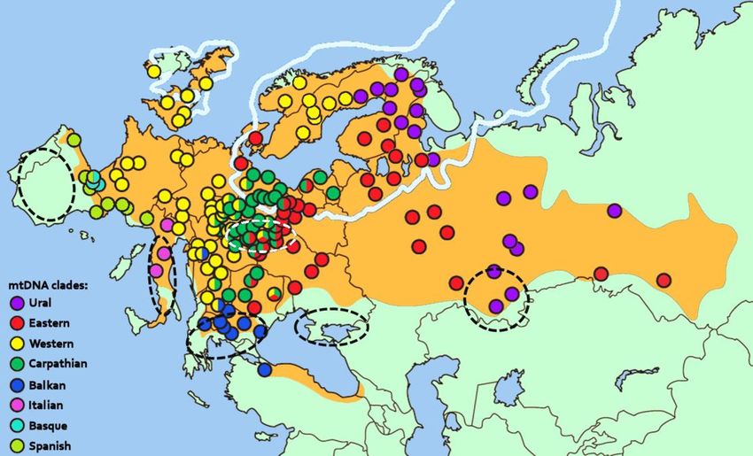
Contents

LIST OF ORIGINAL PUBLICATIONS .................................................................................. 5
ABBREVIATIONS .............................................................................................................. 6

SUMMARY ........................................................................................................................ 9

REVIEW OF THE LITERATURE .........................................................................................11

    History of hantaviruses................................................................................................11
    Taxonomy of hantaviruses ......................................................................................... 13
    Genome organization and virion structure of hantaviruses......................................... 18
    Life cycle of hantaviruses ........................................................................................... 20
    Transmission of hantaviruses ..................................................................................... 23
    Hantaviral infections .................................................................................................. 26
       Clinical features of hantaviral diseases ..................................................................... 26
       Risk factors of acquiring hantaviral diseases ............................................................ 27
       Diagnosis of hantaviral infections ............................................................................ 27
       Incidence and seroprevalence of hantaviral infections ............................................. 28
       Prophylaxes for hantaviral diseases ......................................................................... 30
       Prevention and control of hantaviruses.................................................................... 31

    Evolution of hantaviruses ........................................................................................... 32
    Hantaviruses and their hosts ...................................................................................... 35
    Puumala virus and its host .......................................................................................... 38
    Geographic distribution of Puumala virus and the post-glacial history of bank voles .. 40
AIM OF THE STUDY ........................................................................................................ 43

MATERIALS AND METHODS .......................................................................................... 44

    Rodent trapping ......................................................................................................... 44
    Processing of rodent samples ..................................................................................... 46
    Immunoblotting ......................................................................................................... 46
    RNA/DNA extraction .................................................................................................. 47
    Recovery of viral genome sequences .......................................................................... 47

                                                                3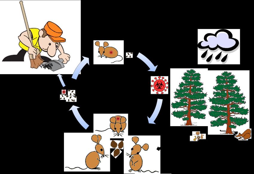
Mitochondrial DNA amplification ............................................................................... 48
    Genetic variation and phylogenetic analyses .............................................................. 48
RESULTS AND DISCUSSION ........................................................................................... 49
    Microevolution of PUUV related to host population dynamics.................................... 49
    PUUV lineages diversity and their post-glacial history ................................................ 59
CONCLUDING REMARKS AND FUTURE PROSPECTS .................................................... 63
ACKNOWLEDGEMENTS ................................................................................................. 65
REFERENCES .................................................................................................................. 67
REPRINTS OF ORIGINAL PUBLICATIONS ....................................................................... 88

                                                               4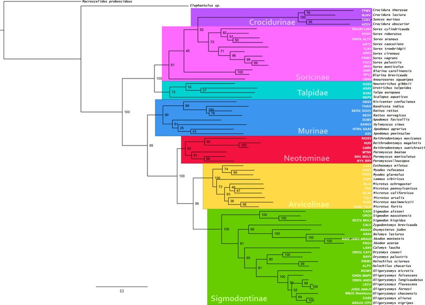
List of publications

LIST OF ORIGINAL PUBLICATIONS

   The present thesis is based on the following publications, which are referred to in the
text by roman numerals.

 I.     Maria Razzauti, Angelina Plyusnina, Heikki Henttonen & Alexander Plyusnin.
        2008. Accumulation of point mutations and reassortment of genomic RNA
        segments are involved in the microevolution of Puumala hantavirus in a bank
        vole (Myodes glareolus) population. The Journal of General Virology, 89(7):1649-
        1660.

 II.    Maria Razzauti, Angelina Plyusnina, Tarja Sironen, Heikki Henttonen & Alexander
        Plyusnin. 2009. Analysis of Puumala hantavirus in a bank vole population in
        Fennoscandia: Evidence for co-circulation of two genetic lineages and
        frequent reassortment between wild-type strains. The Journal of General
        Virology, 90(8):1923-1931.

 III.   Maria Razzauti, Angelina Plyusnina, Jukka Niemimaa, Heikki Henttonen &
        Alexander Plyusnin. 2012. Co-circulation of two Puumala hantavirus lineages in
        Latvia: A Russian lineage described previously and a novel Latvian lineage.
        Journal of Medical Virology, 84(2):314-318.

 IV.    Maria Razzauti, Angelina Plyusnina, Heikki Henttonen & Alexander Plyusnin.
        Microevolution of Puumala hantavirus during a complete population cycle of
        its host, the bank vole (Myodes glareolus). Manuscript.

   The published articles have been reprinted with the permission of the respective
copyright holders.

                                              5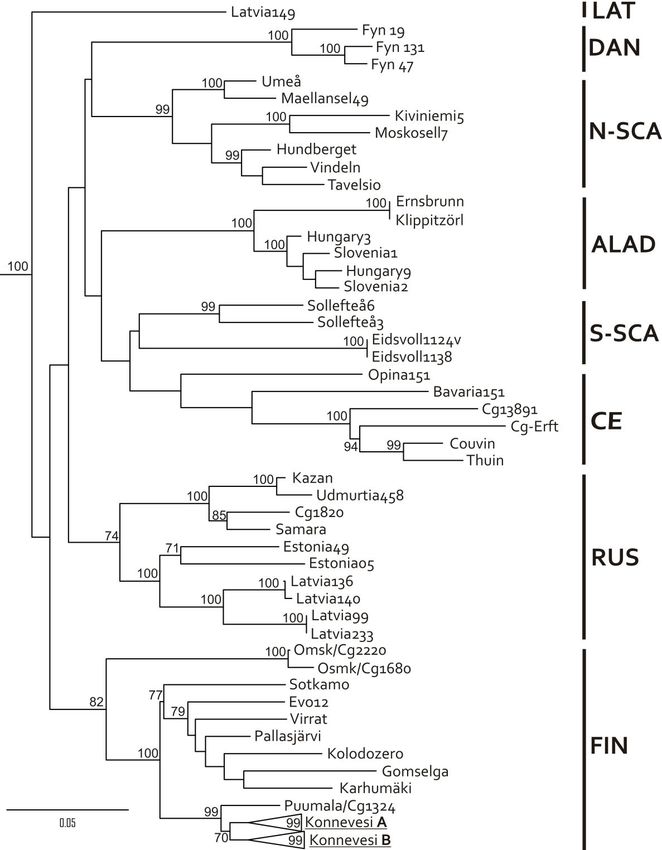
Abbreviations

ABBREVIATIONS

aa              amino acid

Ab              antibody

AD              Anno Domini

Ag              antigen

ALAD            Alpe-Adrian genetic lineage of PUUV

cDNA            complementary DNA

CE              Central European genetic lineage of PUUV

ca.             circa

CR              coding region

Cyt b           cytochrome b

DAN             Danish genetic lineage of PUUV

DM              distance matrix

EDTA            ethylene diamine tetra-acetic acid

ELISA           enzyme-linked immunosorbent assay

ER              endoplasic reticulum

ERGIC           endoplasic reticulum-Golgi intermediate compartments

FIN             Finnish genetic lineage of PUUV

FITC            fluorescein isothiocyanate

FM              Fitch-Margoliash

FRNT            focus reduction neutralization test

GTR             general time reversible

HCPS            hantavirus cardiopulmonary syndrome

HFRS            haemorrhagic fever with renal syndrome

HLA             human leukocyte antigen

ICTV            International Committee on Taxonomy of Viruses

IFA             immunofluorescence assay

IFN-β           interferon beta

                                6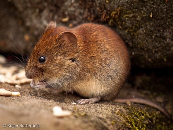
Abbreviations

IgG        immunoglobulin G

IgM        immunoglobulin M

IRF-3      interferon regulatory factor-3

Ka         kiloannum

kb         kilobase

kDa        kiloDalton

L          large genome segment of Bunyaviridae

LAT        Latvian genetic lineage of PUUV

LGM        Last Glacial Maximum

M          medium genome segment of Bunyaviridae

Ma         mega-annum

MCMC       Markov chain Monte Carlo

MHC        major histocompatibility complex

MJ         median-joining

ML         maximum likelihood

MP         maximum parsimony

mRNA       messenger RNA

mtDNA      mitochondrial DNA

NCR        non-coding region

NE         nephropathia epidemica

NF-κB      nuclear factor kappa B

NJ         neighbour-joining

nt         nucleotide(s)

N-SCA      North-Scandinavian genetic lineage of PUUV

OD         optical density

ORF        open reading frame

P bodies   cytoplasmic processing bodies

PBS        phosphate buffered saline

                           7
Abbreviations

PCR                            polymerase chain reaction

pNPP                           p-nitrophenyl phosphate

PUUV                           Puumala virus

RdRp                           RNA-dependent RNA polymerase

rN                             recombinant nucleocapsid protein

RT                             reverse transcription

S                              small genome segment of Bunyaviridae

SD                             standard deviation

SDS-PAGE                       sodium dodecyl sulfate polyacrylamide gel electrophoresis

S-SCA                          South-Scandinavian genetic lineage of PUUV

TNF                            tumor necrosis factor

RUS                            Russian genetic lineage of PUUV

vRNA                           viral RNA

WB                             western blot

A list of hantaviruses and their abbreviations can be found in Table 1 on page 13.

                                               8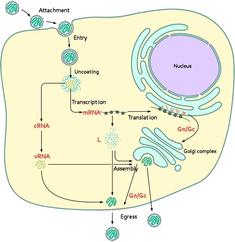
Summary

SUMMARY

    Puumala hantavirus (PUUV) is a zoonotic virus that causes nephropathia epidemica
(NE) in humans, a mild form of haemorrhagic fever with renal syndrome. An average of
10 000 cases are reported annually in Europe, many of which occur in Fennoscandia.
The incidence of NE is connected to the distribution and population density of the the
bank vole (Myodes glareolus), the main host of the virus. In Fennoscandia, high
incidences of NE occur at 3-4 year intervals due to the characteristic population cycles
of this woodland rodent.
   This study aimed to improve our understanding of PUUV microevolution by
examining genetic features of the virus in several bank vole populations of Finland and
Latvia.
    Genetic variation in PUUV circulating in a bank vole population at Konnevesi in
Central Finland was examined and monitored over five-years throughout a complete
bank vole cycle, including two peak-phases in 2005 and 2008 and two population
declines in 2006 and 2009 (i.e., viral bottlenecks). Altogether, 1369 bank voles were
captured and 26.3% were detected PUUV-infected. Partial sequences of the three viral
genome segments (Small, Medium and Large) were inspected from 365 PUUV
genomes. Genetic diversity was 6.2% for the S segment, 4.8% for the M segment, and a
surprisingly high 10.1% for the L segment. Each genome segment had accumulated
mutations as a separate gene pool. The majority of nucleotide substitutions were
synonymous and most of the deduced amino acid substitutions were conservative,
suggesting a strong stabilizing selection operating at the protein level. Genetic markers
found along the genome segments allowed for the recognition of two PUUV
genogroups co-circulating in the host population. Even though one of the genogroups
presented a higher genetic diversity, no signs of competition were observed between
them. Nearly 80% the variants exhibited a transient existence, and frequently occurring
variants were integrated by the most abundant segment genotypes suggesting a viral
mutational robustness. A substantial portion (19.1%) of genomes appeared to be
reassortants, with S and M typically being exchanged. Reassortant variants did not
outcompete parental variants and were commonly transient. Reassortment was
seasonal, occurring more frequently in autumn when infection risk increases. An
imperceptible intra-genogroup reassortment could contribute to the steady state of the
viral population, counteracting the effects of Muller’s ratchet.
   Co-circulation and interaction of two distinct PUUV lineages (Finnish and North-
Scandinavian) was monitored in a bank vole population at Pallasjärvi in Northern
Finland. To date, seven genetic lineages have been detected, and they exhibit
geographic structure within the host distribution. Here, we present new evidence of

                                          9
Summary

two lineages circulating in the same bank vole phylogroup (Ural clade). Genetic
diversity within each PUUV lineage was modest (up to 1.7%) and most substitutions
were synonymous. However, genetic differences between the two lineages were as
high as 18.9%. Phylogenetic analysis revealed that these distinct lineages naturally
reassort with a frequency to that genogroups circulating at Konnevesi, i.e., 32%. In
contrast to Konnevesi, only M segment was exchanged between PUUV lineages at
Pallasjärvi.
    Two distinct PUUV lineages were also found to co-circulate in Latvia. One
(Russian) has been previously described and the other awaits formal description. The
novel Latvian lineage is considerably divergent from other PUUV lineages and several
amino acid markers made it easily distinguishable. Phylogenetic analysis suggested
an independent evolutionary history for the segments of the Latvian lineage. Similar
to Pallasjärvi, both Russian and Latvian lineages were found in a single bank vole
phylogroup (Carpathian clade), confirming earlier observations that PUUV lineages
are not limited to a single host phylogroup.

                                         10
Review of the literature: History of hantaviruses
REVIEW OF THE LITERATURE
History of hantaviruses
Haemorrhagic fever with renal syndrome (HFRS) is a major public health problem and
has been occurring throughout Eurasia for hundreds of years [WHO]. HFRS encompasses
a group of clinically similar illnesses previously known as ”Korean hemorrhagic fever”,
”epidemic nephritis”, “war nephritis”, ”trench nephritis”, “field nephritis”, ”hemorrhagic
nephrosonephritis”, ”virus glomerulonephritis”, ”epidemic hemorrhagic fever” and
”nephropathia epidemica”. In 1982, the WHO recommended fixing “HFRS-like diseases”
as the name in widespread use. Hantavirus infections have a long history; ancient Chinese
writings dating back to 960 AD describe the symptoms of a haemorrhagic fever
syndrome. More recently, outbreaks of “field nephritis” were reported in over 60 000
combatants during wars of the past 150 years: 14 187 participants of the American Civil
War (1861-56); ~12 000 soldiers in World War I (1914-18); ~12500 soldiers of Japanese
troops in Manchuria, ~8 000 of Soviet troops in the Far East, ~10 000 of German and
Finnish troops in Lapland (1942), and ~6 000 German prisoners of war in Yugoslavia during
the World War II (1939-45); and 3 256 members of the United Nation Command in the
Korean War (1951-54) [Casals et al., 1969; Lee, 1996]. The continuous occurrence of HFRS
in the field provided an early indication that the aetiological agent was a zoonotic
microbe.
Clinical manifestations of the disease were studied during 1951-54 [Traub & Wisseman,
1978]. In 1976, Dr. Ho-Wang Lee and his colleagues suspected that the etiological agent
was a virus borne in rodents. Subsequently, they found an antigen in the lungs and
kidneys of the striped field mouse (Apodemus agrarius) collected in endemic foci. The
newly discovered virus was named Hantaan virus (HTNV) after the Hantan River in South
Korea, where the strain originated [Lee & Lee, 1976]. Lung tissues from Apodemus
agrarius coreae presented a specific immunofluorescent reaction with sera from patients
recovering from ”Korean hemorrhagic fever” [Lee et al., 1978].
Early attempts to infect wild rodents or establish a cell culture system for replication of
the presumptive virus failed. In 1981, a prototype virus strain was adapted for growth in
cultured human lung carcinoma cells (A-549) [French et al., 1981]. Later, monkey
epithelial kidney cells (Vero E6) were shown to support viral growth better than A-549
cells [Schmaljohn et al., 1985], an soon after, the first electron micrographs of the virus
were obtained by two groups working independently [McCormick et al., 1982; White et
al., 1982]. Although the morphology resembled that of a bunyavirus, Hantavirus was not
accepted as a new genus of the Bunyaviridae until Hantaan-like viruses were shown to
possess three RNA genomic segments that circularize in vivo as a result of terminal
complementary nucleotides that help to fold them into a “hairpin” structure [Schmaljohn
& Dalrymple, 1983].
11Review of the literature: History of hantaviruses
In 1979 Dr. Markus Brummer-Korvenkontio and colleagues demonstrated that lung
tissue of bank voles (Myodes glareolus) reacts with sera of Finnish HFRS-like patients
[Brummer-Korvenkontio et al., 1980, 1982]. Immunofluorescent assays revealed a virus
related to but distinct from HTNV. The newly recognized aetiological agent was called
Puumala virus (PUUV) named after the small town in South-eastern Finland where it was
first detected. PUUV causes a mild form of HFRS, called nephropathia epidemica (NE)
[Brummer-Korvenkontio et al., 1982]. Antibodies reacting with aetiological agents of
HFRS were found in sera of patients in Europe and Asia [Lee, 1982] and led to the
discovery of other hantaviruses. Seoul virus (SEOV) was found in rats lungs (Rattus rattus
and R. norvegicus) from urban areas of Seoul, and viral antigen was established in
laboratory rats [Lee et al., 1982]. In Maryland, another virus named Prospect Hill (PHV)
was recovered from the meadow vole (Microtus pennsylvanicus), although no association
with acute human disease was observed [Lee et al., 1985]. Previously considered
arbovirus, Thottapalayam virus (TPMV) was found in the Asian musk shrew (Suncus
murinus) in India and subsequently reclassified [Carey et al., 1971; Song et al., 2007]. And
Dobrava-Belgrade virus (DOBV) was isolated from the yellow-necked mouse (Apodemus
flavicollis) from Slovenia [Avšić-Županc et al., 1992].
In 1993, hantaviruses became a concern in the Americas after an acute respiratory
distress outbreak occurred among the Navajo Nation in "The Four Corners", an area
including adjacent parts of Arizona, New Mexico, Colorado and Utah [Nichol et al., 1993].
The newly-described disease was referred as "unexplained adult respiratory distress
syndrome" (ARDS). It was ultimately identified as a virus of the Hantaviridae family. The
Four Corners outbreak occurred during unusual environmental conditions due to the El
Niño in 1991-1992 that created a warm winter and a rainy spring in 1993. This contributed
to the explosive growth of vegetation, providing food and cover for a burgeoning rodent
population. The South-western USA experienced a tenfold increase in the deer mouse
(Peromyscus maniculatus) population, the species recognized as the virus reservoir [Childs
et al., 1994]. The virus was given the name of Muerto Canyon virus but, at the request of
the Navajo community, its name was eventually changed to Sin Nombre virus (SNV) (in
Spanish, "nameless virus"), and the linked disease became known as hantavirus
cardiopulmonary syndrome (HCPS).
Numerous viruses in the Old and New World have since been classified within
Hantavirus, but only some have been shown to cause human diseases. A detailed list of
known hantaviruses is presented in Table 1.
12Review of the literature: Taxonomy of hantaviruses
Taxonomy of hantaviruses
Bunyaviridae consists of five genera: Tospovirus, Nairovirus, Phlebovirus,
Orthobunyavirus and Hantavirus. Hantaviruses present a tri-segmented single-stranded
RNA genome of negative-polarity. Even though the other genera within Bunyaviridae are
mainly arthropod-borne hantaviruses are transmitted to humans through inhalation of
aerosolized rodent excretia. Thus far, no human infections have been associated with
hantaviruses borne by insectivores.
Rodent-borne hantaviruses are known as “RoBo-viruses” and insectivore-borne
hantaviruses as “InBo-viruses”, and the inclusive “RaInBo-viruses” has recently been
adopted. Hantaviruses are also commonly known as Old and New World viruses
depending on the geographic distribution of their hosts. Hantaviruses present a firmly
established host association, yet host-switching events have been reported in the
literature [Morzunov et al., 1998; Vapalahti et al., 1999; Nemirov et al., 2003].
Hantaviruses have been detected in murid and cricetid rodents of subfamilies Murinae
(Old World rats and mice), Arvicolinae (voles and lemmings), Neotominae and
Sigmodontinae (New World rats and mice). Inbo-viruses have been detected in soricine
and crocidurine soridics (shrews), as well as moles (Talpidae) (Table 1 and Fig. 8).
Table 1. List of hantaviruses. Hantavirus species currently recognized by the International
Committee on Taxonomy of Viruses are shown in bold. HFRS stands for haemorrhagic fever
with renal syndrome, NE stands for nephropathia epidemica, and HCPS stands for hantavirus
cardiopulmonary syndrome.
Human Geographic
Virus Abbreviation Host Reference
disease range
Hantaviruses carried by rodents in the family Muridae, subfamily Murinae
Bandicota indica
Thailand THAIV ? S Asia Xiao et al., 1994
(great bandicoot rat)
Rattus rattus (black rat),
Seoul SEOV HFRS Global Elwell et al., 1985
Rattus norvegicus (brown rat)
Rattus rattus
Gou GOUV ? Global Wang et al., 2000
(black rat)
Rattus tanezumi
Serang SERV ? SE Asia Plyusnina et al., 2009
(Asian house rat)
Niviventer confucianus
Da Bie Shan DBSV ? E Asia Wang et al., 2000
(Chinese white-bellied rat)
Apodemus agrarius
Hantaan HTNV HFRS E Eurasia Lee & Lee, 1978
(striped field mouse)
Apodemus agrarius Plyusnin et al., 1997
Saaremaa SAAV HFRS W and C Eurasia
(striped field mouse) Nemirov et al., 1999
13Review of the literature: Taxonomy of hantaviruses
Dobrava- Apodemus flavicollis Avšić-Županc et al.,
DOBV HFRS Europe
Belgrade (yellow-necked mouse) 1992, 1995
Liang et al., 1994
Amur/ Apodemus peninsulae Yashina et al., 2001
ASV HFRS E Asia
Soochong (Korean field mouse) Lokugamage et al., 2002
Baek et al., 2006
Hylomyscus simus Côte d'Ivoire,
Sangassou SANGV ? Klempa et al., 2006
(African wood mouse) Africa
Hantaviruses carried by rodents in the family Cricetidae, subfamily Arvicolinae
HFRS Myodes glareolus Europe to W Brummer-Korvenkontio
Puumala PUUV
(NE) (bank vole) Siberia et al., 1980
Myodes rufocanus
Hokkaido HOKV ? N Eurasia Kariwa et al., 1995
(grey-sided vole)
Myodes regulus
Muju MUJV ? Korea Song et al., 2007
(royal vole)
Microtus pennsylvanicus
Prospect Hill PHV - North America Lee et al., 1982, 1985
(meadow vole)
Microtus arvalis
Tula TULV - Europe to C Asia Plyusnin et al., 1994a
(European common vole)
Lemmus sibiricus
Topografov TOPV ? Palaearctic tundra Plyusnin et al., 1996a
(Siberian brown lemming)
Microtus californicus
Isla Vista ISLAV ? W North America Song et al., 1995
(Californian vole)
Bloodland Microtus ochrogaster
BLLV ? C North America Song et al., 1995
Lake (prairie vole)
Microtus maximowiczii
Khabarovsk KHAV ? NE Asia Hörling et al., 1996
(Maximowicz’s vole)
Microtus fortis
Vladivostok VLAV ? NE Asia Kariwa et al., 1999
(reed vole)
Microtus fortis
Yuanjiang YUJV ? NE Asia Zou et al., 2008
(reed vole)
Eothenomys miletus
Luxi LUXV HFRS SW China Zhang et al., 2011
(Yunnan red-backed vole)
Hantaviruses carried by rodents in the family Cricetidae, subfamily Neotominae
Peromyscus maniculatus
Sin Nombre SNV HCPS North America Nichol et al., 1993
(deer mouse)
Peromyscus maniculatus
Monongahela MGLV HCPS North America Song et al., 1996
(deer mouse)
Peromyscus leucopus Hjelle et al. 1995a,
New York NYV HCPS E North America
(white-footed mouse) 1995b
Peromyscus leucopus
Blue River BRV ? E North America Morzunov et al., 1998
(white-footed mouse)
Peromyscus beatae
Montano MTNV ? Central America Kariwa et al., 2011
(Orizaba deermouse)
14Review of the literature: Taxonomy of hantaviruses
El Moro Reithrodontomys megalotis
ELMCV ? W North America Hjelle et al., 1994
Canyon (western harvest mouse)
Limestone Reithrodontomys megalotis
LSCV ? W North America Sanchez et al., 2001
Canyon (western harvest mouse)
Reithrodontomys megalotis
Huitzilac HUIV ? W North America Kariwa et al., 2011
(western harvest mouse)
Reithrodontomys mexicanus
Rio Segundo RIOSV ? Central America Hjelle et al., 1995c
(Mexican harvest mouse)
Reithrodontomys sumichrasti
Carrizal CARV ? Central America Kariwa et al., 2011
(Sumichrast's harvest mouse)
Hantaviruses carried by rodents in the family Cricetidae, subfamily Sigmodontinae
Black Creek Sigmodon hispidus Ravkov et al., 1995
BCCV HCPS S North America
Canal (hispid cotton rat) Rawlings et al., 1996
Sigmodon hispidus
Muleshoe MULV ? S North America Rawlings et al., 1996
(hispid cotton rat)
Sigmodon alstoni
Cano Delgadito CADV ? N South America Fulhorst et al., 1997
(cane mouse)
Sigmodon mascotensis
(Jaliscan cotton rat)
Playa de Oro OROV ? Central America Chu et al., 2008
Oryzomys couesi
(Coues' rice rat)
Oryzomys couesi
Catacamas CATV ? Central America Milazzo et al., 2006
(Coues' rice rat)
Oryzomys palustris
Bayou BAYV HCPS SE North America Morzunoz et al., 1995
(rice rat)
Oligoryzomys microtis
Rio Mamore RIOMV HCPS N South America Hjelle et al., 1995a
(small-eared pygmy rice rat)
Oligoryzomys longicaudatus
Andes ANDV HCPS S South America Lopez et al., 1996
(long-tailed pigmy rice rat)
Oligoryzomys chacoensis WC South
Bermejo BMJV HCPS Levis et al., 1997
(Chacoan pygmy rice rat) America
Oligoryzomys flavescens
Lechiguanas LECV HCPS NC SouthAmerica Levis et al., 1997
(yellow pygmy rice rat)
Oligoryzomys costaricensis Vicent et al., 2000
Choclo CHOV HCPS NC South America
(Costa Rican pygmy rice rat) Hanson et al., 2011
Oligoryzomys delicatus Fulhosrt et al., 2004
Maporal MAPV HCPS NC South America
(delicate pygmy rice rat) Hanson et al., 2011
Oligoryzomys longicaudatus Levis et al., 1997
Oran ORNV HCPS S South America
(long-tailed pygmy rice rat) Bohlman et al., 2002
Castelo dos Oligoryzomys eliurus
CASV HCPS EC South America Johnson et al. 1999
sonhos (Brazilian pygmy rice rat)
Oligoryzomys nigripes
Itapua ITPV HCPS N South America Chu et al., 2003
(black-footed pygmy rice rat)
Oligoryzomys chacoensis
Neembucu HCPS N South America Chu et al., 2003
(Chacoan pygmy rice rat)
15Review of the literature: Taxonomy of hantaviruses
Oligoryzomys fornesi
Juquitiba JUQV HCPS N South America Vasconcelos et al., 1997
(Fornes' colilargo)
Oligoryzomys fornesi
Anajatuba ANAJV HCPS N South America Rosa et al., 2005
(Fornes' colilargo)
Oligoryzomys nigripes
(black-footed pygmy rice rat)
Oxymycterus judex
Araucaria ARAUV HCPS NC South America Raboni et al., 2005
(hocicudo)
Akodon montensis
(montane grass mouse)
Akodon montensis
Jaborá JABV ? C South America Goodin et al., 2009
(montane grass mouse)
Ape Aime- Akodon montensis
AAIV ? C South America Chu et al., 2003
Itapua (montane grass mouse)
Akodon azarae Levis et al., 1998
Pergamino PRGV ? N South America
(Azara’s grass mouse) Bohlman et al., 2002
Calomys laucha
Laguna Negra LANV HCPS N South America Johnson et al., 1997
(vesper mouse)
Bolomys obscurus Levis et al., 1998
Maciel MCLV ? S South America
(dark bolo mouse) Bohlman et al., 2002
Bolomys lasiurus
Araraquara ARAV HCPS EC South America Johnson et al., 1999
(hairy-tailed bolo mouse)
Holochilus chacarius
Alto Paraguay ALPV ? S South America Chu et al., 2003
(Chacoan marsh rat)
Holochilus sciureus
Rio Mearim RIMEV ? N South America Rosa et al., 2005
(Amazonian marsh rat)
Zygodontomys brevicauda
Calabazo CALV ? N South America Vicent et al., 2000
(short-tailed cane mouse)
Paranoa HPCS ? N South America Melo-Silva et al., 2007
Hantaviruses carried by insectivores in the family Soridae, subfamily Soricinae
Sorex araneus
Seewis SWSV ? Eurasia Song et al., 2007
(Eurasian common shrew)
Sorex araneus
Altai ALTV ? Eurasia Yashina et al., 2008a
(Eurasian common shrew)
Taiga and tundra
Sorex caecutiens zones from N
Artybash ARTV ? Yashina et al., 2008b
(masked shrew) Europe to E
Siberia
Taiga and tundra
Sorex caecutiens zones from N
Lena River LNAV ? Arai et al., 2008
(masked shrew) Europe to E
Siberia
Sorex monticolus
Jemez Springs JMSV ? W North America Arai et al., 2008
(dusky shrew)
Sorex cinereus
Ash River ARRV ? North America Arai et al., 2008
(cinereus shrew)
16Review of the literature: Taxonomy of hantaviruses
Sorex roboratus Taiga and forest-
Kenkeme KKNV ? Kang et al., 2010
(flat-skulled shrew) tundra of E Asia
Provinces of
Sorex cylindricauda Yunnan, Sichuan,
Qiandao Lake ? Gansu, Shaanxi Zhuo et al., 2010
(stripe-backed shrew) and Ningxia of
China
Sorex palustris Boreal and
Fox Creek FXCV ? montane regions Kang et al., 2009
(American water shrew) of North America
Sorex vagrans
Powell Butte PWBV ? North America Kang et al., 2009
(vagrant shrew)
Sorex trowbridgii
Tualatin River TLNV ? E North America Kang et al., 2009
(Trowbridge's Shrew)
Anourosorex squamipes
Cao Bang CBNV ? NE South Asia Song et al., 2007
(Chinese mole shrew)
Blarina brevicauda
Camp Ripley RPLV ? North America Song et al., 2007
(northern short-tailed shrew)
Blarina carolinensis
Iamonia IAMV ? W North America Arai et al., 2007
(southern short-tailed shrew)
Hantaviruses carried by insectivores in the family Soridae, subfamily Crocidurinae
Suncus murinus
Thottapalayam TPMV ? S Asia and E Africa Carey et al., 1971
(Asian house shrew)
Crocidura lasiura Temperate zone
Imjin MJNV ? Song et al., 2009
(Ussuri white-toothed shrew) of E Asia
Crocidura theresae
Tanganya TGNV ? W Africa Klempa et al., 2007
(Therese shrew)
Crocidura obscurior
Azagny AZGV ? W Africa Kang et al., 2011b
(west African pygmy shrew)
Hantaviruses carried by insectivores in the family Talpidae
Talpa europaea
Nova NVAV ? Europe Kang et al., 2009a
(European common mole)
Urotrichus talpoides
Asama ASAV ? Japan Arai et al., 2008
(Japanese shrew mole)
Neurotrichus gibbsii NW North
Oxbow OXBV ? Kang et al., 2009b
(American shrew mole) America
Scalopus aquaticus
Rockport RKPV ? North America Kang et al., 2011a
(eastern mole)
The Bunyaviridae study group of the International Committee on Taxonomy of Viruses
(ICTV) [Plyusnin et al., 2011] suggests the following criteria define hantavirus species: i)
they engage a unique ecological niche, i.e., primary reservoir species; ii) the amino acid
(aa) difference of the complete glycoprotein precursor and the nucleocapsid proteins
must exceed 7%; iii) they manifest at least a four-fold difference in two-way cross-
17Review of the literature: Genome organization and virion structure of hantaviruses
neutralization test; and iv) they cannot naturally reassort with other hantavirus species.
The ICTV policy on defining species considers viruses as polythetic entities. Several
hantaviruses do not meet all four criteria, yet, they are recognized as distinct species
[Plyusnin et al., 2002]. The classification of known hantaviruses, their hosts and
geographic distribution is detailed in Table 1.
Genome organization and virion structure of hantaviruses
Hantaviruses possess a tri-segmented single-stranded genome of negative polarity.
Each of the three segments has a consensus 3’-terminal nucleotide sequence (3’-
AUCAUCAUCUG), which is complementary to the 5’-terminal sequence and distinct from
those of the other four genera in the Bunyaviridae. These sequences contribute to forming
a panhandle structure through imperfect hydrogen bonding that likely plays a role in
replication [Schmaljohn & Dalrymple, 1983] (Figure 1).
Figure 1. Panhandle-forming nucleotides of each genomic RNA segment of PUUV: small (S),
medium (M) and large (L). Vertical lines reflex base pairs and colons the non-canonical pairing.
The corkscrew model of Flick et al. [2002] is shown for the S viral RNA on the right.
The large (L) segment is approximately 6500 nucleotides (nt) in length and encodes
the L protein, which has viral RNA-dependent RNA polymerase (RdRp) function
[Schmaljohn, 1990]. The 3’-terminal non-coding region (NCR) is shorter (100 nt in
average) than the other two segments. The RdRp presents five conserved aa regions
[Poch et al., 1989]; these motifs have proven helpful in the detection of novel lineages.
The L segment is typically the most conserved part of the genome; the diversity for more
distantly related viruses is up to 40%. The medium (M) segment, approx. 3600 nt in
length, encodes the Gn and Gc glycoproteins in a single open reading frame (ORF)
[Schmaljohn et al., 1986, 1987 ]. Its 3’-NCR is 200-250 nt in length. The cleavage site for
18Review of the iterature: Genome organization and virion structure of hantaviruses
two glycoproteins Gn and Gc is highly conserved (WAASA motif), yet glycoproteins are
considerably variable (up to 60%). The small (S) segment from 1600 to 2004 nt in length
encodes the nucleocapsid (N) protein [Schmaljohn et al., 1986]. The length of the 3’-NCR
for this segment varies extensively between different hantaviruses (~220-800 nt). There is
a highly variable domain within 700-900 nt region with more conserved flanking regions.
N protein sequences diversity is up to 57% (Fig. 2). Cricetid-borne hantaviruses are
exceptional in that they contain an evolutionary conserved non-structural (NSs) protein in
an overlapping second ORF similar to those of other orthobunyaviruses [Spiropoulou et
al., 1994; Plyusnin, 2002]. The NSs protein in arvicoline-borne viruses is 88-95 aa residues
long, but shorter (~60 aa) in sigmodontine- and neotomine-borne hantaviruses [Plyusnin
& Elliott, 2011]. The expression of NSs inhibits interferon beta (IFN-β), nuclear factor
kappa B (NF-κB) and interferon regulatory factor-3 (IRF-3) [Jääskeläinen et al., 2007,
2008]. Each of the viral ribonucleoproteins (RNPs) is encapsidated with the N protein
[Dahlberg et al., 1977].
Figure 2. Structure and genome organization of hantaviruses. The hantavirus virion (ø 80-120
nm) is enveloped and the surface encompasses a glycoproteins (Gn and Gc) layer. Single-
stranded RNA molecules of negative-polarity of Puumala hantavirus are represented on the
right side. The Small genome segment encodes the nucleocapsid (N) protein that is
structurally associated with viral RNAs. The Medium genome segment encodes the Gn and
Gc glycoproteins associated in the viral envelope. The Large genome segment encodes the L
protein. [Modified from the Swiss Institute of Bioinformatics].
In nature, hantavirus virions are commonly spherical and vary in size from 80 to 120
nm [McCormick et al., 1982]. Pleiomorphism and size variation have been suggested to
result from encapsidation of additional genome segments into the nucleocapsid
[Rodriguez et al., 1998]. The two glycoproteins Gn and Gc are embedded in the outer lipid
membrane. Ultrastructural studies suggest that the surface has a grid-like pattern and
19Review of the literature: Life cycle of hantaviruses
glycoproteins appear as fuzzy surface projections, approx. 7 nm in length [Martin et al.,
1985]. Recently, biochemical studies resolved the hantaviral glycoprotein complex, and
showed that it consists of Gn tetramers interconnected with Gc dimers [Hepojoki et al.,
2010] (Fig. 3). In 1996, Hutchinson and colleagues estimated the amounts of L, M and S
genome segments in infected Vero E6 cells and the plateau values were 3.2x10⁸, 6.5x10⁸
and 1.24x10⁹ RNA copies/0.1 mg RNA, respectively. This equals a L:M:S ratio of 1 : 1.9 :
3.9; the inverse numbers are 1 : 0.52 : 0.26, which is very close to the size ratio of the
genome segments ( 1 : 0.56 : 0.31). This suggests that the viral messenger RNAs (mRNA)
are produced in amounts that are inversely proportional to their length and that
elongation is the rate-limiting step in mRNA transcription [Hutchinson et al., 1996].
Figure 3. Electron cryotomography of Tula hantavirus. Particles vary in shape from tubular to
spherical. Some spiked (white triangles) and naked (arrowheads) areas of the surface are
indicated in panel A. Small spherical particles devoid of any RNP density are indicated with
asterisks. Straight, rod-shaped densities distinct from the RNP densities are indicated with
arrows in panel B. Scale bar, 50 nm. [Huiskonen and co-workers, 2010; reproduced with
permission].
Life cycle of hantaviruses
Hantaviruses infect endothelial and epithelial cells, follicular dendritic cells as well as
macrophages and lymphocytes without causing any direct cytopathic effect [Pensiero et
al., 1992; Temonen et al., 1993; Zaki et al., 1995]. Surface glycoproteins mediate the
attachment to host-cell receptors [Mackow et al., 2001]. Receptors for cellular entry are
associated with viral pathogenecity. β1 integrins (or the ligand fibronectine) are
20Review of the literature: Life cycle of hantaviruses
implicated in the infection of non-pathogenic hantaviruses while pathogenic species bind
to β3 integrins (or the ligand vitronectine) [Gavrilovskaya et al., 1998].
Virus entry occurs via clathrine-mediated endocytosis, in which viruses move from
early to late endosomes and/or lysosomes. The conformational changes in the Gc
glycoprotein induced by a low pH lead to fusion of the viral membrane with the
endosomal membrane. The virus then decapsulates in endolysosomal compartments to
liberate the three RNPs into the cytoplasm [Jin et al., 2002].
Following penetration of the host cells, RNPs are transported to the perinuclear region
[Ravkov & Compans, 2001] where the viral L protein transcribes negative-sense RNA
segments into functional mRNAs of the S, M and L segments. Initiation of viral RNA
(vRNA) transcription relies on a “cap-snatching” mechanism, in which an endonuclease
encoded in the L segment generates 5’-capped oligonucleotides (10-14 nt long) from host
cell mRNA [Bouloy et al., 1978]. Initiation of genomic RNA synthesis of the L protein uses
a “prime-and-realign” mechanism. This consists of short-capped primers annealing via
single G-C base pairing a few nucleotides upstream of the 3′ terminus of the vRNA
template. After a brief elongation, the extended primer is realigned with the viral
template so that the 3’-terminal nucleotide of the capped primer is at position -1 [Garcin
et al., 1995]. N binds the vRNA panhandle (with higher affinity in trimeric than in
monomeric conformation) unwinding it, and remains attached to the 5′ terminus, which
leaves the 3′ terminus accessible to the L protein [Mir et al., 2006]. Degraded cellular
mRNAs accumulate in cytoplasmic processing bodies (P bodies) that serve as a pool of
primers during the initiation of viral mRNA synthesis. The N protein enhances
transcription by sequestering 5’ mRNA caps stored in P bodies [Mir et al., 2008].
The N is the first protein to be synthesized and accumulates rapidly following infection
[Severson et al., 2001]. Its main role is to protect the vRNA from degradation by
nucleases, but it also has several key functions in the life cycle, namely formation of RNPs,
encapsidation of genomic (negative-sense) and antigenomic (positive-sense) RNA
[Patterson & Kolakofsky, 1984], binding of panhandles during transcription initiation [Mir
& Panganiban, 2006], interaction with the ribosomal protein RPS19 to facilitate the
loading of the 40S ribosomal subunit onto the virus mRNA [Cheng et al., 2011], and in
virus assembly [Ravkov & Compans, 2001]. Furthermore, recent studies suggest that the
N protein can modulate the host immune response [Taylor et al., 2009ab]. The L protein
acts as an RNA transcriptase, replicase and endonuclease [Reguera et al., 2010].
During vRNA replication, antigenomic RNA segments are used as a template for the
production of new RNAs. These are full-length complements of the genomic vRNA and
can serve as a template for mRNA or as the genome precursor at later stages. The
mechanism by which the virus switches from transcription to replication remains unclear
but is thought to involve increasing concentrations of N protein leading to more efficient
RNA encapsidation [Johnsson et al., 2001].
21Review of the literature: Life cycle of hantaviruses
Translation of the S and L mRNAs occurs on free ribosomes. The M mRNA is translated
on membrane-bound ribosomes to generate the glycoprotein precursor (GPC), which is
proteolytically cleaved into Gn and Gc proteins at the conserved WAASA-motif during
import into the endoplasic reticulum (ER) [Lober et al., 2001]. Interaction of Gn and Gc
proteins in the ER is essential for their vesicular transport to the Golgi complex [Ruusala et
al., 1992]. Accumulation of glycoproteins at the Golgi complex is thought to be
responsible for viral maturation and subsequent assembly [Spiropoulu, 2001].
Figure 4. Hantavirus life cycle. Glycoproteins integrated in the viral envelope attach to host
cell surface receptors. Penetration occurs via receptor-mediated endocytosis and uncoating
prior to release of the viral genome. Transcription of complementary RNA (cRNA) from the
viral RNA (vRNA) genome uses host-derived primers. Translation of the S and L mRNAs into
viral proteins using host machinery occurs on free ribosomes, whereas translation of M mRNA
takes place on membrane-bound ribosomes. Replicated and amplified vRNA is transported to
the Golgi apparatus to be assembled with the N protein. Assembly of all components occurs at
the Golgi apparatus or at the plasma membrane in New World hantaviruses. Egress follows
the fusion of Golgi vesicles harbouring mature virion particles with the plasma membrane.
[Modified from Johnsson et al., 2010].
22Review of the literature: Transmission of hantaviruses
The RNP complexes travel to the ER-Golgi intermediate compartments (ERGIC) via
dynein microtubules [Ramanathan et al., 2007], and then to the Golgi complex containing
the embedded viral Gn and Gc glycoproteins. Unlike many other negative-strand RNA
viruses, hantaviruses do not encode a matrix protein. Thus, the cytoplasmic tail of the Gn
protein mediates the interaction of the glycoprotein multimers with the N protein during
viral assembly [Jonhsson et al., 2001]. The Gn cytoplasmic tail also contains a late domain
motif (YXXL) that in other viruses has been shown to interact with cellular factors that
facilitate virus budding from cells [Spiropoulu et al., 2003]. Recently, a direct interaction
of the Gn cytoplasmic tail with the N protein and RNA has been reported for Old World
hantaviruses [Wang et al., 2010; Strandin et al., 2011]. Hantavirus particles are
transported in vacuoles to the plasma membrane where they egress by exocytosis
[Pettersson & Melin, 1996]. In contrast, electron microscopy has suggested that the cell
surface may act as an alternative maturation and budding site for New World
hantaviruses [Ravkov et al., 1997; Spiropoulou, 2001]. A schematic of the hantavirus
infection cycle is shown in Figure 4.
Transmission of hantaviruses
Hantavirus outbreaks are promoted by environmental factors (e.g., weather and food
availability), host population fluctuations, woodland disturbance by humans and/or
domestic animals, anthropogenic factors (e.g., deforestation, agricultural development,
urbanization, etc.) and ecological changes [Brummer-Korvenkontio et al., 1982;
Engelthaler et al., 1999; Buceta et al., 2004; Yan, et al., 2007; Clement et al., 2009; Kallio
et al., 2009; Klempa, 2009; Tersago et al., 2009]. The main factor associated with human
epidemics appears to be the host population density, in that the number of cases
increases when rodents are abundant. A high host population density increases the
number of infected animals. However, although rodent density plays an important role,
viral prevalence often follows a seasonal cycle (Fig. 5) [Kallio et al., 2009]. Bank vole
population dynamics, and consequently NE epidemiology, differ between biomes in
Europe. In temperate forests, masting (heavy seed crops of deciduous trees) increases
bank vole densities [Tersago et al., 2009] while in boreal forests population cycles of bank
voles are more influenced by predation [Henttonen, 1985; Henttonen et al., 1985].
23Review of the literature: Transmission of hantaviruses
Figure 5. Monthly cases of human nephropathia epidemica in Central Finland (black line)
reported by the Finnish National Institute for Health and Welfare (http://www3.ktl.fi/) and the
density index of bank voles (grey line) at Konnevesi 1995–2008 (vole trapping index = captured
individuals / 100 trap nights, monthly data are interpolated from trappings carried out four
times per year, trappings are indicated with dots). Biological years are marked with alternating
shaded and unshaded bars [Kallio et al., 2009; reproduced with permission].
Hantaviruses infect their hosts chronically. The main route for transmission between
rodent/insectivore reservoirs is via aggressive behaviour and/or exposure to aerosolized
contaminated droppings (Fig. 6) [Glass et al., 1988; Hinson et al., 2004; Kallio et al,
2006a]. Under laboratory conditions, an infected rodent can transmit the virus
horizontally to another rodent within the same cage or through infected bedding [Lee et
al., 1981a; Kallio et al., 2006b; Hutchinson et al., 2000]. Chronically infected animals have
high levels of neutralizing antibodies (Ab). However, infected rodents excrete virus for a
limited time shortly after infection, the duration of which appears to be different for each
hantavirus/host species [Lee et al., 1981ab; Hutchinson et al., 2000; Kallio et al., 2006a;
Harderstam et al., 2009]. Hantaviruses are not transmitted vertically and maternal Abs
acquired through breast-feeding protect the offspring for up to 80 days [Kallio et al.,
2006b]. Such acquired immunity plays a role in the infection dynamics of natural
populations. Breast-feeding bank voles are abundant in October but when lactation ends,
naive individuals (≥15 g of weight) become susceptible to hantavirus infection. This
suggests that maternal Abs protection may affect the infection dynamics in bank vole
populations [Kallio et al., 2010].
24Review of the literature: Transmission of hantaviruses
Figure 6. Hantavirus transmission cycle. Predation and weather conditions modulate food and
water resources that regulate rodent population densities. These parameters among others
influence the prevalence of hantaviruses. Hantaviruses are horizontally transmitted among
rodents or insectivores through aggressive behaviour and/or exposure to aerosolized
contaminated droppings. Humans are dead-end hosts and infected by breathing aerosolized
rodent excreta containing the virus.
Humans become incidental hosts when they are exposed to hantavirus-containing
excreta of rodent reservoirs. Typically, humans are infected by inhaling aerosolized urine
or faeces released by an infected rodent. Infection may also occur if contaminated
material or dust gets into broken skin or onto a mucous membrane. Hantaviruses can also
be transmitted via a bite from an infected rodent. Person-to-person transmission has only
been demonstrated for ANDV in Argentina [Wells et al., 1997].
Hantaviruses are susceptible to drying conditions, but remain viable for several days
when protected by moist organic material. Moreover, hantaviruses can survive up to 14
days at room temperature and probably much longer in cold and moist conditions, such as
those experience during the Finnish winter [Kallio et al., 2006a]. Hantaviruses can be
chemically inactivated with methanol, paraformaldehyde, 1% sodium hypochlorite, 2%
glutaraldehyde, 70% ethanol, acetone, methanol, detergents containing lysis buffer, acids
lower than pH 5.0, as well as UV irradiation [Kraus et al., 2005].
25Review of the literature: Hantaviral infections
Hantaviral infections
Clinical features of hantaviral diseases
The incubation period for HFRS is typically 2-3 weeks long, although a 6-week
incubation period has recently been described for PUUV [Kramski et al., 2009]. Classically,
HFRS occurs in five indiscrete phases: (i) febrile, (ii) hypotensive, (iii) oliguric, (iv) polyuric
and (v) convalescent phase. The initial febrile (i) stage typically lasts 3-6 days and is
characterized by fever, chills, facial flashing, headache, photophobia, blurry vision,
hypotension and malaise. Haemoconcentration, thrombocytopenia and leukocytosis can
be observed in the laboratory. Clinical symptoms of the febrile stage are eventually
augmented in the (ii) hypotensive phase (2-3 days long) that includes thirst, restlessness,
dizziness, nausea, vomiting and petechial rash. One-third of patients during this stage
develop shock and mental confusion. Symptoms of oliguric (iii) phase (Review of the literature: Hantaviral infections
HCPS is a more severe disease than HFRS with an average case-fatality rate of 30-40%
[Hjelle & Torres-Pérez, 2010]. Haemorrhagic manifestations are unusual and most of the
pathogenic events occur in the thoracic cavity [Hjelle et al., 1995d; Schmaljohn & Hjelle,
1997]. The incubation period varies from 9 to 33 days. The onset of HCPS is abrupt with
prodromal symptoms of fever, myalgia, headache, nausea, vomiting, abdominal pain and
sometimes dizziness. After 3-6 days, the cardiopulmonary phase leads to coughing,
dyspnoea, tachycardia, fever and hypotension. Haemoconcentration, thrombocytopenia,
left-shift leukocytosis and proteinuria are frequently observed. A rapid development of
pulmonary oedema requires intubation and mechanical ventilation. Severe
cardiopulmonary dysfunctions correlate with a poor prognosis.
Risk factors of acquiring hantaviral diseases
Several human risk factors have been suggested to promote hantavirus infection: Sex
of the patient; the male-female ratio of NE cases varies from 2 to 5:1 [Hjertqvist et al.,
2010; Klein et al., 2011]. Age of the patient; case fatality rates were higher among women
between the ages of 20-39 and more than 50 years old [Klein et al., 2011]. Damage to the
respiratory tract due to smoking [Vapalahti et al., 2010], and genetic predisposition.
Besides biological factors, several behavioural and societal components are suspected to
be involved.
A genetic predisposition to suffer from a severe course of hantaviral diseases has been
described for the adaptive immune response involving the human leukocyte antigen
(HLA) system. Patients with haplotype HLA-B*8, -DRB1*03:01, -DRB1*13, -C4A*Q0 or
-DQ2 have been observed to have a significantly higher risk for a severe course of NE
[Mustonen et al., 1996] and HLA-B*35:01 and -DRB1*14:02 alleles have been associated
with a severe courses of HCPS [Kilpatrick et al., 2004]. In contrast, HLA-B*27 and HLA-
B*07 haplotypes have been associated with a more benign clinical course of NE
[Mustonen et al., 1998; Korva et al., 2011]. HLA-DRB1*15 alleles were significantly more
common in a group of patients with a mild course of ANDV [Ferrer et al., 2007].
Diagnosis of hantaviral infections
The diagnosis of HFRS and HCPS is based on clinical and epidemiological data.
Serological tests for viral antigens (Ag) are used to confirm a hantaviral infection.
Immunoglobulin M (IgM) antibodies are detected in the early phase of the disease and
may persist in sera for several months. Immunoglobulin G (IgG) antibodies appear slightly
later than IgM, remain detectable for life [Settergren et al., 1991], and may provide
immunity against secondary infections. Classically, serological tests such as
immunofluorescence assay (IFA) [Brummer-Korvenkontio et al., 1980; Settergren et al.,
1987; Hedman et al., 1991], enzyme-linked immunosorbent assay (ELISA) [Vapalahti et
al., 1996; Kallio-Kokko et al., 1998], or immunoblot assay [Hjelle et al., 1997; Schubert et
27Review of the literature: Hantaviral infections
al., 2001] were widely used and effective diagnostics tools for hantavirus. The most
reliable serological methods for the detection of PUUV-specific IgM Abs are based on
either native or recombinant full-length N-Ag [Brus Sjölander et al., 1997]. Commercial
ELISA methods, in which different hantaviral Ags are used, are available from Progen
Diagnostics (Heidelberg, Germany) for PUUV and HTNV, and from Focus Technologies
(Cypress, CA, USA) for SEOV and SNV. A commercial immunoblot assay is available from
Mikrogen (Martinsried, Germany) for PUUV, HTNV, DOBV and SEOV. A rapid
immunochromatographic test is available from Reagena (Erilab Ltd., Kuopio, Finland) for
DOBV, HTNV and PUUV. Successful Ag detection by immunohistochemical methods has
been reported in biopsies and post-mortem samples from patients with severe HFRS or
HCPS [Poljak et al., 1994; Zaki et al., 1995]. Nowadays, the use of RT-PCR for the
detection of vRNA in body fluids and tissues of patients is preferred for a rapid
differentiation of viral strains.
Incidence and seroprevalence of hantaviral infections
Hantaviruses are the etiological agents of two diseases in humans: the New World
hantavirus cardiopulmonary syndrome (HCPS) and the Old World haemorrhagic fever
with renal syndrome (HFRS) [Schmaljohn & Hjelle, 1997]. The geographic distribution of
hantaviruses and epidemiology of their associated infections are linked to the distribution
of the rodent hosts. The impact of hantavirus infection on human health worldwide is
significant; an average of 200 000 HFRS patients are hospitalized each year throughout
the world [Lee, 1996] and approx. 200 cases of HCPS are reported annually in the
Americas. Although cases of HCPS are less numerous than HFRS, the average case
fatality is higher (30-40%) [Lednicky, 2003].
In Asia, clinical cases of HFRS have been reported mainly in China, the Republic of
Korea, and the Far East Federal District of Russia. In the recent years, China has
accounted for 70 to 90% of HFRS cases worldwide with 40 000 to 60 000 cases being
reported annually [Zhang et al., 2004]. In Eastern Russia, up to 200 HFRS cases are
reported each year [Yashina et al., 2000], 3% of all cases in the Federation [Tkachenko et
al., 2007]. Serological studies have indicated that there is strong evidence for human
hantaviral infections in Thailand, Laos, Viet Nam, Hong Kong, Taiwan, Malaysia, Israel,
Philippines, Singapore, Indonesia, Kuwait, Mongolia, India and Sri Lanka [Rollin et al.,
1986; Shortridge et al., 1987; Wong et al., 1989; Kao et al., 1996; George et al., 1998;
Quelapio et al., 2000; Lam et al., 2001; Pacsa et al., 2002; Groen et al., 2002; Zhang et al.,
2009; Chandy et al., 2009; Gamage et al., 2011]. Hantaviral prevalence in rodents has also
been measured in Japan, Thailand and Cambodia [Arikawa et al., 1985; Reynes et al.,
2003; Chandy et al., 2009].
In Europe, NE was described in Sweden as early as 1934 [Myhrman, 1934; Zeetterholm,
1934] and is the most prevalent hantaviral disease in Western and Central Europe. In 1979,
PUUV, the causative agent of NE, was first isolated in Finland [Brummer-Korvenkontio et
28Review of the literature: Hantaviral infections
al., 1980]. By the end of 2006, 35 424 European cases of NE had been reported, 95% of
which were after 1990 [Heyman et al., 2008, 2009]. However, mild cases can be
misdiagnosed as a flu-like disease, meaning the actual number is likely higher. Nowadays,
around 10 000 cases are reported each year in Europe. In Fennoscandia, there is a peak of
NE every 3-4 years due to the population cycle of bank voles. During the population peaks
of 1999, 2002, 2005 and 2008, the number of cases diagnosed in Finland were 2300, 2603,
2526 and 3500, respectively [National Infectious Disease Registry (http://www3.ktl.fi/)].
Since the introduction of PUUV diagnostics in Finland laboratories, 35 000 cases have
been confirmed [Heyman et al., 2011]. It has been estimated that PUUV seroprevalence
among Finns is 5% [Brummer-Korvenkontio et al., 1999]. In Sweden, a large NE outbreak
occurred in 2006-2007 with an incidence of 313 cases per 100 000 inhabitants in the most
affected areas [Evander & Ahlm, 2009]. Prior to 2010, 1234 cases of NE-like disease were
reported in Norway, at an average rate of 50 cases per year [Lundkvist et al., 1998]. In
European Russia, 10 000 – 12 000 clinical cases of PUUV and DOBV infection are reported
annually [Klempa et al., 2008]. In southern Baltic countries, PUUV seroprevalence is 0.5%
in Lithuania [Sandmann et al., 2005], 1.5% in Latvia [Lundkvist et al., 2002] and 5.1% in
Estonia. Also, a neutralizing Ab specific reaction for SAAV has been observed at a rate of
3.4% in Estonia [Golovljova et al., 2002]. In the Netherlands, Belgium, Luxembourg,
France, Germany and Austria, NE outbreaks in 2005 and 2007 were the most significant
since 1990, with a total of 1114 and 2106 confirmed cases, respectively [Heyman et al.,
2007, 2011]. In the Czech Republic since 1992, antibody seroprevalence against
hantaviruses peaked up 1.4% [Pejcoch et al., 2003]. In the Balkans, particularly the former
Socialist Federal Republic of Yugoslavia, HFRS outbreaks have been recorded since the
early 1950’s with a case fatality rate of up to 10%, where DOBV and PUUV were the
predominant pathogens [Lukac et al., 1990]. In Croatia, 317 cases of HFRS were diagnosed
during an epidemic in 2002 [Mulić & Ropac, 2002]. Around the same time, a large
epidemic occurred in Serbia and Montenegro in 2002 with 128 laboratory-confirmed cases
[Papa et al., 2006]. In Greece, 210 HFRS cases have been diagnosed since the first
detection in 1984, with a case fatality rate of 9% [Papa et al., 2001]. In Hungary, 342
clinical cases of hantavirus were confirmed between 1992 and 2010 [Heyman et al., 2011].
In Bulgaria during 1954-1986, 399 cases of HFRS were registered with a fatality rate of
15.7% [Chumakov et al., 1998]. Clinical infections of hantaviruses have also been recorded
in Romania [Manasia et al., 1977], Albania [Eltari et al., 1978], Slovenia [Avšić-Županc et
al., 1992], the United Kingdom [Pether et al., 1993], Denmark [Asikainen et al., 2000;
Nemirov et al., 2004], Slovakia [Sibold et al., 2001], Poland [Knap et al., 2006],
Switzerland [Fontana-Binard et al., 2008], and Turkey [Ertek & Burgan, 2009]. Sero-
epidemiological surveys have demonstrated the presence of hantavirus Ab in humans in
the Republic of Moldova [Mikhailenko et al., 1994], Spain [Lledó et al., 2003], Portugal
[Vapalahti et al., 2003] and Italy [Kallio-Kokko et al., 2006] although no clinical cases have
been reported there.
29You can also read