Morphing of Ibogaine: A Successful Attempt into the Search for Sigma-2 Receptor Ligands - MDPI
←
→
Page content transcription
If your browser does not render page correctly, please read the page content below
Article

Morphing of Ibogaine: A Successful Attempt into the
Search for Sigma-2 Receptor Ligands
Giuseppe Floresta 1, Maria Dichiara 1, Davide Gentile 1, Orazio Prezzavento 1,
Agostino Marrazzo 1, Antonio Rescifina 1,2,* and Emanuele Amata 1,*
 1 Department of Drug Sciences, University of Catania, V.le A. Doria, 95125 Catania, Italy;
   giuseppe.floresta@unict.it (G.F.); maria.dichiar@unict.it (M.D.); davide.gentile@studium.unict.it (D.G.);
   prezzave@unict.it (O.P.); marrazzo@unict.it (A.M.)
 2 Consorzio Interuniversitario Nazionale di ricerca in Metodologie e Processi Innovativi di Sintesi

   (C.I.N.M.P.S.), Via E. Orabona, 4, 70125 Bari, Italy
 * Correspondence: arescifina@unict.it (A.R.); eamata@unict.it (E.A.);
   Tel.: +39-095-7385017 (A.R.); +39-095-7384102 (E.A.)

 Received: 31 December 2018; Accepted: 21 January 2019; Published: 23 January 2019

 Abstract: Ibogaine is a psychoactive indole alkaloid with high affinity for several targets including
 the σ2 receptor. Indeed, extensive data support the involvement of the σ2 receptor in neurological
 disorders, including Alzheimer’s disease, schizophrenia, alcohol abuse and pain. Due to its serious
 side effects which prevent ibogaine from potential clinical applications, novel ibogaine derivatives
 endowed with improved σ2 receptor affinity may be particularly beneficial. With the purpose to
 facilitate the investigation of iboga alkaloid derivatives which may serve as templates for the design
 of selective σ2 receptor ligands, here we report a deconstruction study on the ibogaine tricyclic
 moiety and a successive scaffold-hopping of the indole counterpart. A 3D-QSAR model has been
 applied to predict the σ2 pKi values of the new compounds, whereas a molecular docking study
 conducted upon the σ2 receptor built by homology modeling was used to further validate the best-
 scored molecules. We eventually evaluated pinoline, a carboline derivative, for σ2 receptor affinity
 through radioligand binding assay and the results confirmed the predicted high µM range of
 affinity and good selectivity. The obtained results could be helpful in the drug design process of
 new ibogaine simplified analogs with improved σ2 receptor binding capabilities.

 Keywords: sigma-2 receptor; TMEM97; scaffold-hopping; molecular docking; Ibogaine; Pinoline;
 Incazane

1. Introduction
      First introduced as subtypes of the opioid receptor and as high-affinity phencyclidine binding
sites, sigma receptors are now recognized as a particular and unique receptor class. Two subtypes
are currently known, denoted as sigma-1 (σ1) and sigma-2 (σ2) receptors, having a different structures,
biological functions, and pharmacological profiles.
      Sigma-1 σ1 receptor has been identified as a 25.3 kDa chaperone protein within the mitochondria-
associated endoplasmic reticulum membranes (MAMs) [1]. Recently, the crystal structure of the
human σ1 receptor has been reported revealing a trimeric architecture [2]. Sigma-1 σ1 receptor is
highly expressed in both the central and peripheral nervous system, with involvement in the
production of neurotrophic factors and in the protection of the mitochondrial integrity [3,4]. In this
view, σ1 receptor agonists represent potential therapeutic agents for the treatment of several
neuropsychiatric and neurodegenerative disorders, whereas σ1 receptor antagonists have been
reported for their antiproliferative and antiangiogenic effects, in addition to the modulation of pain
and drug abuse-related conditions [5–10].
Int. J. Mol. Sci. 2019, 20, 488; doi:10.3390/ijms20030488                             www.mdpi.com/journal/ijms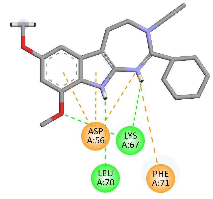
Int. J. Mol. Sci. 2019, 20, 488                                                                     2 of 11

      Sigma-2 σ2 receptor is a poorly understood protein whose identification has been controversial.
For a long time its binding site has been postulated to be located in the progesterone receptor
membrane component 1 (PGRMC1). A recent study has highlighted that σ2 receptor and PGRMC1
are different proteins since the presence or absence of PGRMC1 has no impact on σ2 ligands binding
ability [11]. In 2017, Alon and coworkers [12] identified the σ2 receptor as an endoplasmic reticulum-
resident transmembrane protein (TMEM97) playing a role in the cholesterol homeostasis and the
sterol transporter Niemann–Pick disease type C1. Despite the challenges in identifying its true
identity, σ2 receptor has earned a growing scientific interest due to its involvement in several disease
states. High levels of the σ2 receptor have been found in several cancer cells and proliferating tumors
such as lung, colorectal, ovarian, and breast cancers [13]. Extensive data support the utility of sigma-
receptor ligands as cancer therapeutics and diagnostic tools [14,15]. Due to a 10-fold higher density
in proliferating tumor cells than in quiescent tumor cells, σ2 receptor also represents an important
clinical biomarker for determining the proliferative status of solid tumors [14]. The fluorine (F18) ISO-
1 is a promising positron emission tomography (PET) ligand evaluated in clinical trials for the
imaging of σ2 receptor binding in primary breast cancer [16].
      More recently, σ2 receptor has been implicated in neurological disorders, including Alzheimer’s
disease, schizophrenia, alcohol abuse, and pain [17–20]. The small-molecule CT1812, a σ2 receptor
antagonist whose structure has not been disclosed, is currently under clinical trial in patients with
mild to moderate Alzheimer’s disease [21]. Also, Roluperidone (MIN-101) is in phase III clinical trials
for the treatment of negative symptoms of schizophrenia [21].
      Ibogaine (Figure 1), a psychoactive indole alkaloid, is a typical “dirty drug” with high affinity
for a panel of targets including NMDA, κ- and µ-opioid receptors and σ2 receptor sites [22]. Ibogaine
has also shown to interact with the acetylcholine, serotonin, and dopamine systems and to modify
the expression of some proteins including substance P and brain-derived neurotrophic factor (BDNF)
[23,24]. Initially used for its hallucinogenic properties, ibogaine has been then investigated for its
potential in treating drug abuse [25]. However, little research in humans has been done due to the
severe side effects and death following its ingestion, including tremors, neurotoxicity, and
cardiotoxicity [26].
      As mentioned above, the receptor sites through which ibogaine mediates its effects are not
known with certainty even if clear evidence indicates that ibogaine and other iboga alkaloids interact
with σ2 receptors. Indeed, ibogaine shows a moderate nanomolar affinity for σ2 receptor with a Ki
value of 201 nM and good selectivity over σ1 site (Ki 8554 nM) [22]. Lower affinity values for other
neurotransmitter receptors have been showed [27–29]. Similar Ki values have been reported for other
iboga alkaloid analogs in which the presence or the position of the methoxy group on the aromatic
ring of the indole moiety as the presence of another substituent appears to be not critical for σ2 affinity
[30–32].
      In light of the neurotoxic and tremorigenic effects, which associated with a complex structure
prevent ibogaine from potential clinical applications, synthetic σ2 receptor analogs with low toxicity
may be particularly beneficial. However, the development of ligands endowed with high affinity and
selectivity can often run into several limitations and challenges. With the aim to overcome this issue,
we      recently    reported     the    σ2     receptor   selective    ligand     database      (S2RSLDB,
http://www.researchdsf.unict.it/S2RSLDB, accessed on 23 January 2019), a manually curated
collection of the whole set of selective σ2 receptor ligands published in the literature [33]. At the same
time, we also developed a 2D-QSAR affinity filter [34], built-up with 548 compounds, and a 3D-QSAR
model for the identification of potentially selective σ2 receptor ligands [35,36].
      With the interest to find new and easily synthesizable skeletons able to interact with the σ2
receptor, we have performed a deconstruction study of the ibogaine structure. In the study we report
here, we have systematically modified the tricyclic moiety of ibogaine and its indole counterpart
using a scaffold-hopping approach, and investigated the ability of the new obtained fragments to
bind to the σ2 receptor. The σ2 pKi values of these new compounds were predicted applying the
abovementioned 3D-QSAR model and the potency of the best-scored molecules were further
Int. J. Mol. Sci. 2019, 20, 488 3 of 11
validated by a molecular docking analysis using the σ2 receptor homology model already reported
by us [37].
2. Results and Discussion
2.1. 3D-Ligand Evaluation and Scaffold-Hopping Analysis
With the aim to produce a library of virtual compounds to further guide us in development of
new hit ibogaine derived σ2 receptor ligands, we proceed to deconstruct the tricyclic ibogaine system
containing the azepane moiety by a first scaffold-hopping [38–40] approach set out to maintain an
indolo fused six- or seven-membered ring (Figure 1, Series 1). Successively, on the best-scored
compound was performed a second scaffold-hopping cycle to alter the external aromatic (Figure 1,
Series 2). As expected, this second series of compounds results as potentially more effective; in fact,
in this series the ibogaine scaffold was optimized in both the selected components in Figure 1,
differently in the series only one component was optimized.
Figure 1. Series 1 and 2 derived from ibogaine.
Then the resulted molecules (1055 from Series 1 and 500 from Series 2) were filtered through
statistical/2D descriptors filters using DataWarrior software [41]. To perform this, we analyzed the
most potent and selective compounds present in the S2RSLDB [33] retrieving only the ligands
presenting a σ2 Ki value ≤ 10 nM and a σ1/σ2 selectivity ≥ 1, for a total of 115 entities. The ranges of
molecular weight (up to 651), cLogP (1.76/8.43), cLogS (−9.51/−2.26), H-acceptors (1/9), H-donor (0/2),
Druglikeness (−15.1/8.2), DrugScore (0.04/0.86), topological polar surface area (3/96) belonging to the
115 potent and selective compounds were all chosen as 2D descriptors and the dataset of 1555
molecules was further filtered using these interval values to give 179 molecules from the Series 1 and
319 molecules from the Series 2.
The resulting 498 filtered molecules and the ibogaine were aligned in the 3D-QSAR model using
Forge (v10.4.2, Cresset, New Cambridge House, United Kingdom) as a software [42], by adopting
parameters reported in Figures S1 and S2. Once aligned, these compounds were scored assuming
that if the fields (defined as the local extrema of the electrostatic, van der Waals, and hydrophobic
potentials of each molecule) of the newly designed molecules are very similar to that of the original
compounds, the resulting compounds will have similar biological properties [37,43–45]. The
evaluation of the ibogaine in the 3D-QSAR model resulted in a predicted pKi value of 6.8, which is in
excellent agreement with the experimental one (6.69) [22]. Some selected compounds resulted from
the 3D-ligand based filter are reported in Table 1; Table 2 while the full set of compounds is present
in the Supplementary Materials (Tables S1 and S2). Overall, the results indicate that the double
scaffold-hopping approach and the following 3D-QSAR model evaluation generate compounds with
a suitable chemical structure for the σ2 receptor binding. Most importantly, several of the new
generated compounds are predicted to be more effective than the parent hit compound ibogaine.
Interestingly, among the 179 molecules of Series 1 we found a simplified analogue (Table 1) of
incazane (metralindole, Figure 2), a reversible inhibitor of the monoamine oxidase A possessing an
antidepressant activity [46], and the natural product pinoline (Table 1), another inhibitor of the
monoamine oxidase A [47].Int. J. Mol. Sci. 2019, 20, 488 4 of 11
Figure 2. Structure of incazane.
Table 1. Structure and predicted pKi values of the selected ibogaine derivatives resulted from the
scaffold-hopping study of Series 1.
Entry ID Structure Predicted pKi
1 7.4
6 7.0
45 6.9
125 (Incazane derivative) 6.5
179 (Pinoline) 4.7
Table 2. Structure and predicted pKi values of the selected ibogaine derivatives resulted from the
scaffold-hopping study of Series 2.
Entry Structure Predicted pKi
1 8.3
4 8.1Int. J. Mol. Sci. 2019, 20, 488 5 of 11
35 7.8
2.2. Molecular Docking Analysis
To further validate the predicted pKi values of the 3D-QSAR model and to investigate the
interactions of the new ligands within the σ2 receptor active site, we conducted a docking study on
the selected compounds reported in Table 3. Each ligand was docked in the binding pocket of the σ2
receptor structure already built, in our group, by homology modeling [37]; successively, the best
pose/receptor complex structure was minimized to allow the ligand to better adapt to the pocket of
the active site and then a re-docking was performed using the same procedure already reported by
us [48]. The values of the calculated pKi, reported in Table 3, are well in accord to the predicted ones
by the 3D-QSAR model with the exception for the incazane derivative and compound 2_1.
Interestingly, the pKi value calculated by docking for compound 2_1 is the same as DTG (1,3-di(2-
tolyl)guanidine) [51], a selective sigma receptor ligand used for the binding assays, of which it shares
the portion similar to guanidine.
Moreover, to investigate the σ1/σ2 selectivity (SI) of this set of compounds, we conducted a
molecular docking study using the crystal structure of the human σ1 receptor model bound to the
high-affinity and selective σ1 antagonist PD144418 (PDB ID: 5HK1), employing the same
methodology already validated by us [48]. The SI values reported in Table 3 for reference compounds
(ibogaine and DTG) indicate that the computational models are efficient in the prediction and the
new compounds should possess an effective σ2 selectivity.
Table 3. Docking calculated σ2 pKi values compared to the 3D-QSAR predicted ones and docking
calculated σ1 pKi values with σ1/σ2 selectivity index for selected compounds.
3D-QSAR Docking Calculated Docking Calculated SI a
Series ID_Entry ID
Predicted σ2 pKi σ2 pKi σ1 pKi
1_1 7.4 7.24 6.50 5.5
1_6 7.0 6.98 6.81 1.5
1_45 6.9 7.19 6.77 2.6
1_125 (Incazane
6.5 7.40 5.13 186.2
derivative)
1_179 (Pinoline) 4.7 4.53 3.81 5.2
2_1 8.3 7.56 6.15 25.7
2_4 8.1 8.39 6.65 55.0
2_35 7.8 8.14 6.58 36.3
Incazane 6.4 6.63 5.35 19.1
Ibogaine 6.8 6.89 5.06 67.6 b
DTG 6.8 7.27 7.32 0.9 c
aSI: Selectivity index calculated as σ1 Ki/σ2 Ki. b SI = 42.5 from Reference [22]. c SI = 1.1 from Reference
[49].
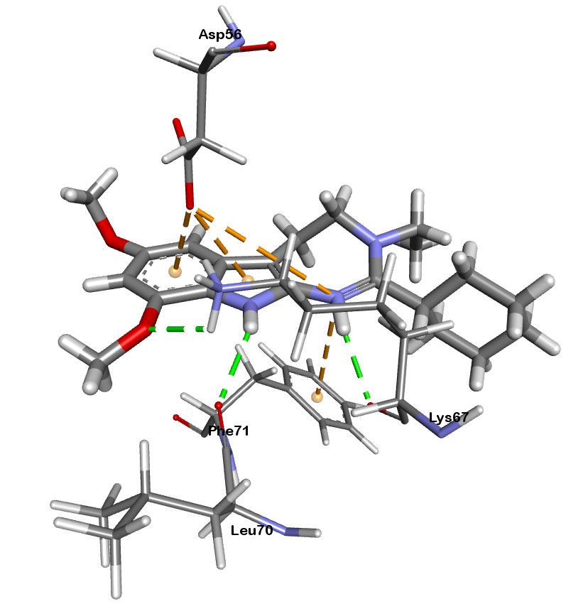
A representation of the best docked pose for compound 2_4 is depicted in Figure 3. There are
clearly visible two hydrogen bonds between the LYS67 and LEU70 with the two hydrogen atoms at
nitrogens and another one between LYS67 and a methoxyl oxygen atom. Moreover, two π-ion
interactions were established between the ASP56 and the two aromatic rings of the indole and
another two between the ASP56 and PHE71 with the π-orbital of the nitrogen atom of the 1,3-
diazepine ring. A comparison of the best docked poses for ibogaine, pinoline and compound 2_4 are
reported in Figure S3.Int. J. Mol. Sci. 2019, 20, 488 6 of 11
Figure 3. 3D (left) and 2D (right) representations of the docked pose for compound 2_4. Green dotted
lines represent hydrogen bonds and orange dotted lines π-ion interactions.
2.3. Pinoline Biological Assay
Among the compounds with the best 3D-QSAR predicted and docking calculated pKi values,
we decided to evaluate pinoline (compound 1_179) for affinity at both σ1 and σ2 receptors, with
haloperidol as reference standard. Our choice was grounded on a structural simplicity, ease of
commercial availability, and based on the fact that literature data for sigma binding affinity for
pinoline have not been provided yet. However, the lack of a substituent on the N-atom of the
piperidine appears to be critical for σ2 affinity since a Ki of 35.4 ± 2.6 µM (pKi = 4.45) has been shown
(Figure S4), thus confirming the range of magnitude for this displacement assay predicted by the in
silico models. Moreover, the measure of the σ1 affinity for pinoline give a Ki value > 100 (pKi < 4.00)
accordingly with the calculated selectivity.
3. Materials and Methods
3.1. 2D to 3D Building and Minimization of Structures
The structures of ibogaine and related compounds were built using Marvin Sketch (ChemAxon,
Budapest, Hungary). The 2D structures were subjected to molecular mechanics energy minimization
by Merck molecular force field (MMFF94) using the Marvin Sketch geometrical descriptors plugin.
The protonation states of the molecules were calculated considering a neutral pH. Before the
alignment for the 3D-QSAR filter, the geometry of the obtained 3D structures was further optimized
at semi-empirical level using the parameterized model number 3 (PM3) Hamiltonian [50,51] as
implemented in MOPAC package (vMOPAC2016, Stewart Computational Chemistry, Colorado
Springs, CO, USA) [52].
3.2. Compound Alignment and Scaffold-Hopping Analysis
All the optimized three-dimensional structures were imported into the software Forge (v10.4.2,
Cresset, New Cambridge House, UK). The computational evaluation of all the molecules in the
imported dataset was made by the field-based 3D-QSAR model previously published [35], after a
careful alignment with the training set of the model (see Supplementary Material for more
information). The molecules were described by means of field points (negative, positive, shape and
hydrophobic), and all of them were generated using the extended electron distribution (XED) force
field in Forge. In Figures S1 and S2 (Supplementary Material) are shown the software’s parameters
used for the conformation hunt and the alignment. 500 was set as maximum number of conformations
generated for each molecule. The root-mean-square deviation of atomic positions cutoff for duplicateInt. J. Mol. Sci. 2019, 20, 488 7 of 11
conformers was set to 0.5 Å (the similarity threshold below which two conformers are assumed
identical). The gradient cutoff for conformer minimization was set to 0.1 kcal/mol. The energy
window was set to 2.5 kcal/mol. Conformers with a minimized energy outside the energy window
were discarded. The scaffold-hopping analysis was performed using Spark as a software (v10.4.0)
using the same 511717 fragments [38–40,45].
3.3. Molecular Docking
Docking experiments were performed employing AutoDock 4.2.5.2 software implemented in
YASARA (v. 18.12.7, YASARA Biosciences GmbH, Vienna, Austria) [53,54] using the homology
model of the σ2 receptor previously built by the same authors. The maps were generated by the
program AutoGrid (4.2.5.2) with a spacing of 0.375 Å and dimensions that encompass all the surface
of the active site. All the parameters were inserted at their default settings as previously reported
[37].
To allow each ligand to adapt to the binding pocket, we carried out this study utilizing a three-
step sequence already validated by us [48]: (i) ligand was docked upon σ1 or σ2 receptor, (ii) 5 ns of
molecular dynamic (MD) simulation of the best pose obtained for the ligand/σ receptor complex, in
order to accommodate the ligand, and (iii) redocking of the complex obtained from the last 3 ns of
MD simulation averaged frames. The MD simulation was performed as described in Reference [48].
3.4. Radioligand Binding Assay
Sigma-2 binding experiments were performed as previously reported by Matsumoto et al. [55]
and Mach et al. [56]. Briefly, each tube containing 360 µg of membrane protein was incubated with
3.26 nM [3H]DTG (1,3-di-2-tolylguanidine, Perkin Elmer, Waltham, MA, USA) (31 Ci/mM) in the
presence of 400 nM (+)-SKF10,047 (Sigma-Aldrich, Saint Louis, MO, USA) to mask the σ1 sites. Test
compounds were dissolved in dimethyl sulfoxide and then diluted in buffer to a final volume of 1
mL. Pinoline (Sigma-Aldrich, Saint Louis, MO, USA) was added to give a concentration in the range
of 10−3–10−10 M, while haloperidol (Sigma-Aldrich, Saint Louis, MO, USA) was added to give a
concentration in the range of 10−5–10−10 M. Incubation was carried out in 50 mM Tris-HCl (pH 8.0) for
120 min at room temperature. Each assay was terminated by the addition of ice-cold 10 mM Tris-HCl,
pH 8.0, followed by filtration through a Whatman GF/B glass fiber filter that had been presoaked for
1 h in a 0.5% polyethylenimine (PEI) (Sigma-Aldrich, Saint Louis, MO, USA)solution. Filters were
washed twice with 4 mL of ice-cold buffer. Non-specific binding was assessed in the presence of 5
µM DTG (Tocris, Minneapolis, MN, USA).
Sigma-1 binding assays were carried out according to DeHaven et al. [57]. Each tube containing
500 µg of membrane protein was incubated with 3.26 nM [3H]-(+)-pentazocine (Perkin Elmer,
Waltham, MA, USA) (45 Ci/mmol) in 50 mM Tris-HCl (pH 7.4). Non-specific binding was evaluated
in the presence of 10 µM haloperidol. Test compounds were dissolved in dimethyl sulfoxide and then
diluted in buffer to a final volume of 1 mL. Pinoline was added to give a concentration of 10−4 M,
while haloperidol was added to give a concentration in the range of 10−5–10−10 M. After incubation
(150 min at 37 °C), the samples were filtered through Whatman GF/B glass fiber filters that were
presoaked in a 0.5% PEI solution using a millipore filter apparatus. The filters were washed twice
with 4 mL of ice-cold buffer and the amount of bound radioactivity on the filters air-dried and then
soaked in Scintillation cocktail (Ultima Gold MV, Perkin Elmer, Waltham, MA, USA) was measured
using a liquid scintillation counter (Beckman LS6500). Results are expressed as inhibition constants
(Ki values) and calculated using GraphPad Prism (GraphPad Software, San Diego, CA, USA).
4. Conclusions
Ibogaine simplified analogs with high affinity for σ2 receptor represent an attractive and useful
field to investigate. However, the development of ligands endowed with high affinity and selectivity
has often several challenges. In this view, in silico methods have become essential tools in the drug
design process. With the aim to find new, easily synthesizable skeletons able to interact with σ2Int. J. Mol. Sci. 2019, 20, 488 8 of 11
receptor, we here reported a deconstruction study on the ibogaine tricyclic moiety and a successive
scaffold-hopping of the indole counterpart that indicated two new scaffolds that further decorated
could constitute an excellent alternative for the synthesis of powerful σ2 receptor ligands. In
particular, compound 2_4 emerged for the predicted/calculated pKi values of 8.1 and 8.39,
respectively, which are about 1.6 units higher than that of ibogaine. We eventually evaluated
pinoline, a carboline derivative, for σ2 receptor affinity through radioligand binding assay and the
result confirmed the predicted high µM range of affinity and even a good selectivity. The obtained
results will be used by our research group for the next step in the development of new ibogaine
simplified analogs with improved σ2 receptor binding capabilities.
Supplementary Materials: Supplementary materials can be found at www.mdpi.com/xxx/s1.
Author Contributions: Conceptualization, G.F., A.R. and E.A.; Data curation, E.A., M.D. and D.G.; Formal
analysis, G.F., D.G., A.R. and E.A.; Investigation, G.F., A.R. and E.A.; Methodology, G.F., A.R. and E.A.; Project
administration, A.R. and E.A.; Resources, G.F., D.G., M.D. and A.R.; Supervision, A.R. and E.A.; Validation, G.F.
M.D. A.M. and E.A.; Biological assays, O.P.; Writing—original draft, G.F., M.D., O.P., A.R. and E.A.; Writing—
review & editing, G.F., M.D., D.G., O.P., A.M, A.R. and E.A.
Funding: This research received no external funding.
Acknowledgments: This work was supported by the University of Catania (Piano per la Ricerca 2016–2018 —
Linea di Intervento 2 “Dotazione Ordinaria”). Free academic licenses from ChemAxon and Cresset for their
suites of programs are gratefully acknowledged.
Conflicts of Interest: The authors declare no conflict of interest.
References
1. Hayashi, T.; Su, T.P. Sigma-1 receptor chaperones at the ER-mitochondrion interface regulate Ca(2+)
signaling and cell survival. Cell 2007, 131, 596–610.
2. Schmidt, H.R.; Zheng, S.; Gurpinar, E.; Koehl, A.; Manglik, A.; Kruse, A.C. Crystal structure of the human
sigma1 receptor. Nature 2016, 532, 527–530.
3. Fujimoto, M.; Hayashi, T.; Urfer, R.; Mita, S.; Su, T.P. Sigma-1 receptor chaperones regulate the secretion of
brain-derived neurotrophic factor. Synapse 2012, 66, 630–639.
4. Weng, T.Y.; Tsai, S.A.; Su, T.P. Roles of sigma-1 receptors on mitochondrial functions relevant to
neurodegenerative diseases. J. Biomed. Sci. 2017, 24, 74.
5. Maurice, T. Improving Alzheimer’s Disease-Related Cognitive Deficits with sigma1 Receptor Agonists.
Drug News Perspect. 2002, 15, 617–625.
6. Albayrak, Y.; Hashimoto, K. Sigma-1 Receptor Agonists and Their Clinical Implications in
Neuropsychiatric Disorders. Adv. Exp. Med. Biol. 2017, 964, 153–161.
7. Olivieri, M.; Amata, E.; Vinciguerra, S.; Fiorito, J.; Giurdanella, G.; Drago, F.; Caporarello, N.; Prezzavento,
O.; Arena, E.; Salerno, L.; et al. Antiangiogenic Effect of (+/-)-Haloperidol Metabolite II Valproate Ester [(+/-
)-MRJF22] in Human Microvascular Retinal Endothelial Cells. J. Med. Chem. 2016, 59, 9960–9966.
8. Amata, E.; Dichiara, M.; Arena, E.; Pittala, V.; Pistara, V.; Cardile, V.; Graziano, A.C.E.; Fraix, A.; Marrazzo,
A.; Sortino, S.; et al. Novel Sigma Receptor Ligand-Nitric Oxide Photodonors: Molecular Hybrids for
Double-Targeted Antiproliferative Effect. J. Med. Chem. 2017, 60, 9531–9544.
9. Arena, E.; Dichiara, M.; Floresta, G.; Parenti, C.; Marrazzo, A.; Pittalà, V.; Amata, E.; Prezzavento, O. Novel
Sigma-1 receptor antagonists: From opioids to small molecules: What is new? Future Med. Chem. 2018, 10,
231–256.
10. Schinina, B.; Martoran, A.; Colabufo, N.A.; Contino, M.; Niso, M.; Perrone, M.G.; De Guidi, G.; Catalfo, A.;
Rappazzo, G.; Zuccarello, E.; et al. 4-Nitro-2,1,3-benzoxadiazole derivatives as potential fluorescent sigma
receptor probes. RSC Adv. 2015, 5, 47108–47116.
11. Pati, M.L.; Groza, D.; Riganti, C.; Kopecka, J.; Niso, M.; Berardi, F.; Hager, S.; Heffeter, P.; Hirai, M.;
Tsugawa, H.; et al. Sigma-2 receptor and progesterone receptor membrane component 1 (PGRMC1) are
two different proteins: Proofs by fluorescent labeling and binding of sigma-2 receptor ligands to PGRMC1.
Pharmacol. Res. 2017, 117, 67–74.
12. Alon, A.; Schmidt, H.R.; Wood, M.D.; Sahn, J.J.; Martin, S.F.; Kruse, A.C. Identification of the gene that
codes for the sigma2 receptor. Proc. Natl. Acad. Sci. USA 2017, 114, 7160–7165.Int. J. Mol. Sci. 2019, 20, 488 9 of 11
13. Crawford, K.W.; Bowen, W.D. Sigma-2 receptor agonists activate a novel apoptotic pathway and potentiate
antineoplastic drugs in breast tumor cell lines. Cancer Res. 2002, 62, 313–322.
14. Zeng, C.; McDonald, E.S.; Mach, R.H. Molecular Probes for Imaging the Sigma-2 Receptor: In Vitro and In
Vivo Imaging Studies. Handb. Exp. Pharmacol. 2017, 244, 309–330.
15. van Waarde, A.; Rybczynska, A.A.; Ramakrishnan, N.K.; Ishiwata, K.; Elsinga, P.H.; Dierckx, R.A. Potential
applications for sigma receptor ligands in cancer diagnosis and therapy. Biochim. Biophys. Acta 2015, 1848,
2703–2714.
16. Washington University School of Medicine. [18F]ISO-1 PET/CT in Breast Cancer. Available online:
https://clinicaltrials.gov/ct2/show/NCT02762110 (accessed on 29 December 2018).
17. Sahn, J.J.; Mejia, G.L.; Ray, P.R.; Martin, S.F.; Price, T.J. Sigma 2 Receptor/Tmem97 Agonists Produce Long
Lasting Antineuropathic Pain Effects in Mice. ACS Chem. Neurosci. 2017, 8, 1801–1811.
18. Vazquez-Rosa, E.; Watson, M.R.; Sahn, J.J.; Hodges, T.R.; Schroeder, R.E.; Cintron-Perez, C.J.; Shin, M.K.;
Yin, T.C.; Emery, J.L.; Martin, S.F.; et al. Neuroprotective Efficacy of a Novel Sigma 2 Receptor/TMEM97
Modulator (DKR-1677) after Traumatic Brain Injury. ACS Chem. Neurosci. 2018,
doi:10.1021/acschemneuro.8b00543.
19. Yi, B.; Sahn, J.J.; Ardestani, P.M.; Evans, A.K.; Scott, L.L.; Chan, J.Z.; Iyer, S.; Crisp, A.; Zuniga, G.; Pierce,
J.T.; et al. Small molecule modulator of sigma 2 receptor is neuroprotective and reduces cognitive deficits
and neuroinflammation in experimental models of Alzheimer’s disease. J. Neurochem. 2017, 140, 561–575.
20. Scott, L.L.; Sahn, J.J.; Ferragud, A.; Yen, R.C.; Satarasinghe, P.N.; Wood, M.D.; Hodges, T.R.; Shi, T.;
Prakash, B.A.; Friese, K.M.; et al. Small molecule modulators of sigma2R/Tmem97 reduce alcohol
withdrawal-induced behaviors. Neuropsychopharmacology 2018, 43, 1867–1875.
21. Washington University School of Medicine. Study to Evaluate Efficacy and Safety of Roluperidone (MIN-
101) in Adult Patients with Negative Symptoms of Schizophrenia. Available online:
https://clinicaltrials.gov/ct2/show/NCT03397134 (accessed on 29 December 2018).
22. Bowen, W.D.; Vilner, B.J.; Williams, W.; Bertha, C.M.; Kuehne, M.E.; Jacobson, A.E. Ibogaine and its
congeners are sigma 2 receptor-selective ligands with moderate affinity. Eur. J. Pharmacol. 1995, 279, R1–
R3.
23. Popik, P.; Layer, R.T.; Skolnick, P. 100 years of ibogaine: Neurochemical and pharmacological actions of a
putative anti-addictive drug. Pharmacol. Rev. 1995, 47, 235–253.
24. He, D.Y.; Ron, D. Autoregulation of glial cell line-derived neurotrophic factor expression: Implications for
the long-lasting actions of the anti-addiction drug, Ibogaine. FASEB J. 2006, 20, 2420–2422.
25. Maciulaitis, R.; Kontrimaviciute, V.; Bressolle, F.M.; Briedis, V. Ibogaine, an anti-addictive drug:
Pharmacology and time to go further in development. A narrative review. Hum. Exp. Toxicol. 2008, 27, 181–
194.
26. Litjens, R.P.; Brunt, T.M. How toxic is ibogaine? Clin. Toxicol. 2016, 54, 297–302.
27. Deecher, D.C.; Teitler, M.; Soderlund, D.M.; Bornmann, W.G.; Kuehne, M.E.; Glick, S.D. Mechanisms of
action of ibogaine and harmaline congeners based on radioligand binding studies. Brain Res. 1992, 571, 242–
247.
28. Popik, P.; Layer, R.T.; Skolnick, P. The putative anti-addictive drug ibogaine is a competitive inhibitor of
[3H]MK-801 binding to the NMDA receptor complex. Psychopharmacology 1994, 114, 672–674.
29. Sweetnam, P.M.; Lancaster, J.; Snowman, A.; Collins, J.L.; Perschke, S.; Bauer, C.; Ferkany, J. Receptor
binding profile suggests multiple mechanisms of action are responsible for ibogaine’s putative anti-
addictive activity. Psychopharmacology 1995, 118, 369–376.
30. Bowen, W.D. Sigma receptors and iboga alkaloids. Alkaloids Chem. Biol. 2001, 56, 173–191.
31. Mésangeau, C.; Amata, E.; Alsharif, W.; Seminerio, M.J.; Robson, M.J.; Matsumoto, R.R.; Poupaert, J.H.;
McCurdy, C.R. Synthesis and pharmacological evaluation of indole-based sigma receptor ligands. Eur. J.
Med. Chem. 2011, 46, 5154–5161.
32. Prezzavento, O.; Arena, E.; Sánchez-Fernández, C.; Turnaturi, R.; Parenti, C.; Marrazzo, A.; Catalano, R.;
Amata, E.; Pasquinucci, L.; Cobos, E.J. (+)-and (−)-Phenazocine enantiomers: Evaluation of their dual opioid
agonist/σ1 antagonist properties and antinociceptive effects. Eur. J. Med. Chem. 2017, 125, 603–610.
33. Nastasi, G.; Miceli, C.; Pittala, V.; Modica, M.N.; Prezzavento, O.; Romeo, G.; Rescifina, A.; Marrazzo, A.;
Amata, E. S2RSLDB: A comprehensive manually curated, internet-accessible database of the sigma-2
receptor selective ligands. J. Cheminform. 2017, 9, 3.Int. J. Mol. Sci. 2019, 20, 488 10 of 11
34. Rescifina, A.; Floresta, G.; Marrazzo, A.; Parenti, C.; Prezzavento, O.; Nastasi, G.; Dichiara, M.; Amata, E.
Development of a Sigma-2 Receptor affinity filter through a Monte Carlo based QSAR analysis. Eur. J.
Pharm. Sci. 2017, 106, 94–101.
35. Floresta, G.; Rescifina, A.; Marrazzo, A.; Dichiara, M.; Pistara, V.; Pittala, V.; Prezzavento, O.; Amata, E.
Hyphenated 3D-QSAR statistical model-scaffold hopping analysis for the identification of potentially
potent and selective sigma-2 receptor ligands. Eur. J. Med. Chem. 2017, 139, 884–891.
36. Rescifina, A.; Floresta, G.; Marrazzo, A.; Parenti, C.; Prezzavento, O.; Nastasi, G.; Dichiara, M.; Amata, E.
Sigma-2 receptor ligands QSAR model dataset. Data Brief 2017, 13, 514–535.
37. Floresta, G.; Amata, E.; Barbaraci, C.; Gentile, D.; Turnaturi, R.; Marrazzo, A.; Rescifina, A. A Structure-
and Ligand-Based Virtual Screening of a Database of “Small” Marine Natural Products for the
Identification of “Blue” Sigma-2 Receptor Ligands. Mar. Drugs 2018, 16, 384.
38. Floresta, G.; Apirakkan, O.; Rescifina, A.; Abbate, V. Discovery of High-Affinity Cannabinoid Receptors
Ligands through a 3D-QSAR Ushered by Scaffold-Hopping Analysis. Molecules 2018, 23, 2183.
39. Floresta, G.; Pittala, V.; Sorrenti, V.; Romeo, G.; Salerno, L.; Rescifina, A. Development of new HO-1
inhibitors by a thorough scaffold-hopping analysis. Bioorg. Chem. 2018, 81, 334–339.
40. Floresta, G.; Amata, E.; Dichiara, M.; Marrazzo, A.; Salerno, L.; Romeo, G.; Prezzavento, O.; Pittala, V.;
Rescifina, A. Identification of Potentially Potent Heme Oxygenase 1 Inhibitors through 3D-QSAR Coupled
to Scaffold-Hopping Analysis. ChemMedChem 2018, 13, 1336–1342.
41. Sander, T.; Freyss, J.; von Korff, M.; Rufener, C. DataWarrior: An open-source program for chemistry aware
data visualization and analysis. J. Chem. Inf. Model. 2015, 55, 460–473.
42. Cheeseright, T.; Mackey, M.; Rose, S.; Vinter, A. Molecular field extrema as descriptors of biological
activity: Definition and validation. J. Chem. Inf. Model. 2006, 46, 665–676.
43. Greish, K.F.; Salerno, L.; Al Zahrani, R.; Amata, E.; Modica, M.N.; Romeo, G.; Marrazzo, A.; Prezzavento,
O.; Sorrenti, V.; Rescifina, A.; et al. Novel Structural Insight into Inhibitors of Heme Oxygenase-1 (HO-1)
by New Imidazole-Based Compounds: Biochemical and In Vitro Anticancer Activity Evaluation. Molecules
2018, 23, 1209.
44. Floresta, G.; Cilibrizzi, A.; Abbate, V.; Spampinato, A.; Zagni, C.; Rescifina, A. FABP4 inhibitors 3D-QSAR
model and isosteric replacement of BMS309403 datasets. Data Brief 2018, 22, 471–483.
45. Floresta, G.; Cilibrizzi, A.; Abbate, V.; Spampinato, A.; Zagni, C.; Rescifina, A. 3D-QSAR assisted
identification of FABP4 inhibitors: An effective scaffold hopping analysis/QSAR evaluation. Bioorg. Chem.
2019, 84, 276–284.
46. Andreeva, N.I.; Asnina, V.V.; Liberman, S.S. Domestic Antidepressants. 3. Incazane (Metralindole). Pharm.
Chem. J. 2001, 35, 59–62.
47. Airaksinen, M.M.; Huang, J.T.; Ho, B.T.; Taylor, D.; Walker, K. The uptake of 6-methoxy-1,2,3,4-tetrahydro-
beta-carboline and its effect on 5-hydroxytryptamine uptake and release in blood platelets. Acta Pharmacol.
Toxicol. 1978, 43, 375–380.
48. Amata, E.; Rescifina, A.; Prezzavento, O.; Arena, E.; Dichiara, M.; Pittalà, V.; Montilla-Garcia, A.; Punzo, F.;
Merino, P.; Cobos, E.J.; Marrazzo, A. (+)-Methyl (1R,2S)-2-{[4-(4-Chlorophenyl)-4-hydroxypiperidin-1-
yl]methyl}-1-phenylcyclopropa necarboxylate [(+)-MR200] Derivatives as Potent and Selective Sigma
Receptor Ligands: Stereochemistry and Pharmacological Properties. J. Med. Chem. 2018, 61, 372–384.
49. Lever, J.R.; Gustafson, J.L.; Xu, R.; Allmon, R.L.; Lever, S.Z. σ1 and σ2 receptor binding affinity and
selectivity of SA4503 and fluoroethyl SA4503. Synapse 2006, 59, 350–358.
50. Stewart, J.J.P. Optimization of Parameters for Semiempirical Methods 1. Method. J. Comput. Chem. 1989, 10,
209–220.
51. Stewart, J.J.P. Optimization of parameters for semiempirical methods IV: Extension of MNDO, AM1, and
PM3 to more main group elements. J. Mol. Model. 2004, 10, 155–164.
52. Stewart, J.J.P. MOPAC2016. Available online: http://OpenMOPAC.net (accessed on 29 December 2018).
53. Krieger, E.; Vriend, G. YASARA View—Molecular graphics for all devices—From smartphones to
workstations. Bioinformatics 2014, 30, 2981–2982.
54. Krieger, E.; Koraimann, G.; Vriend, G. Increasing the precision of comparative models with YASARA
NOVA—A self-parameterizing force field. Proteins 2002, 47, 393–402.
55. Matsumoto, R.R.; Bowen, W.D.; Tom, M.A.; Vo, V.N.; Truong, D.D.; De Costa, B.R. Characterization of two
novel sigma receptor ligands: Antidystonic effects in rats suggest sigma receptor antagonism. Eur. J.
Pharmacol. 1995, 280, 301–310.Int. J. Mol. Sci. 2019, 20, 488 11 of 11
56. Mach, R.H.; Smith, C.R.; Childers, S.R. Ibogaine possesses a selective affinity for sigma 2 receptors. Life Sci.
1995, 57, 57–62.
57. Dehavenhudkins, D.L.; Fleissner, L.C.; Fordrice, F.Y. Characterization of the Binding of [H-3] (+)-
Pentazocine to Sigma-Recognition Sites in Guinea-Pig Brain. Eur. J. Pharmacol. 1992, 227, 371–378.
© 2019 by the authors. Licensee MDPI, Basel, Switzerland. This article is an open access
article distributed under the terms and conditions of the Creative Commons Attribution
(CC BY) license (http://creativecommons.org/licenses/by/4.0/).You can also read