Morphometric Analysis of Infraorbital Foramen Using Cone Beam Computed Tomography in a Cohort of Sri Lankan Adults
←
→
Page content transcription
If your browser does not render page correctly, please read the page content below
Int. J. Morphol.,
39(2):489-496, 2021.

Morphometric Analysis of Infraorbital Foramen Using Cone Beam
 Computed Tomography in a Cohort of Sri Lankan Adults

                   Análisis Morfométrico del Foramen Infraorbitario Mediante Tomografía
                 Computarizada de Haz Cónico en una Cohorte de Adultos de Sri Lanka

                                   I. P. Thilakumara1; P. V. K. S. Hettiarachchi2; R. M. Jayasinghe1;
                                      M. C. N. Fonseka3; R. D. Jayasinghe2 & C. D. Nanayakkara4

THILAKUMARA, I. P.; HETTIARACHCHI, P. V. K. S.; JAYASINGHE, R. M.; FONSEKA, M. C. N.; JAYASINGHE, R. D. &
NANAYAKKARA, C. D. Morphometric analysis of infraorbital foramen using cone beam computed tomography in a cohort of Sri
Lankan adults. Int. J. Morphol., 39(2):489-496, 2021.

         SUMMARY: Infraorbital foramen (IOF) located bilaterally within the maxillary bone about 1 cm inferior to the infraorbital
margin is a vital landmark when delivering local anesthesia and during surgical interventions in the midface region. A total of 122
infraorbital foramina in 61 cone beam computed tomographic (CBCT) images of 32 females and 29 males in the age range of 17 to 32
were analyzed to determine the shape, direction, presence of accessory foramina, size and the precise position of IOF in relation to the
inferior orbital margin (IOM), maxillary midline (MM), lateral nasal wall (LNW), alveolus (ALV) and maxillary teeth in a group of Sri
Lankan people. The IOF was oval in shape (80.3 % and 88.5 % on the right and left side, respectively) in a majority of individuals. The
infraorbital foramina were located at a mean distance of 5.56 ± 3.95 and 4.91 ± 2.08 mm, below the IOM on the right and left side, 27.13
± 2.6 and 26.99 ± 2.73 on the right and left side from the mid maxillary line, 11.96 ± 3.45 mm and 12.18 ± 3.35 from the LNW on the
right and left side and 29.59 ± 3.59 and 29.65 ± 3.28 above the alveolar crest on the right and left side. There were no statistically
significant differences between the left and right sides or between sexes. Majority of IOF (37.5 % and 55.9 % on the right and left side,
respectively) were located in the vertical plane passing though the maxillary second premolar tooth.

        KEY WORDS: Infraorbital foramen; Infraorbital nerve; Morphometry.

INTRODUCTION

        The infraorbital foramen (IOF) is located bilaterally              region. The infraorbital nerve block is often used to
within the maxillary bone about 1 cm inferior to the                       accomplish regional anesthesia of the face. Nerve blocks are
infraorbital margin (Standring, 2008). The infraorbital nerve              useful when repair of a large area is needed in an area
and vessels traverse through this foramen. It is relatively                innervated by one nerve. Further, this procedure offers several
larger than the supraorbital foramen and varies in form and                advantages over local tissue infiltration. Blockage of a nerve
position (Berge & Bergman, 2001). The infraorbital nerve,                  is also useful when local infiltration may not be possible,
the continuation of the maxillary, the second division of the              ineffective or could result in tissue damage or distortion.
trigeminal nerve, is exclusively a sensory nerve. It traverses
the inferior orbital fissure into the inferior orbital canal and                   An infraorbital nerve block is very useful for
emerges on the face at the IOF. It divides into several                    procedures which involve the skin between the lower eyelid,
branches that innervate the skin and the mucous membrane                   upper lip and for dental procedures on the ipsilateral
of the midface, such as the lower eyelid, cheek, lateral aspect            maxillary teeth (Kothari et al., 2019). The direction and
of the nose, upper lip and the labial gum which are important              position of IOF also determine the acupuncture point used
in oral and maxillofacial surgical practice (Standring).                   in the treatment of trigeminal neuralgia (Wilkinson, 1999).
                                                                           Infraorbital nerve injuries might occur in the anterior and
       The IOF is a vital landmark when delivering local                   superior walls during the surgical treatment such as
anesthesia in carrying out surgical interventions in the midface           rhinoplasty, Caldwell-Luc surgical procedures, tumor
1
  Department of Prosthetic Dentistry, Faculty of Dental Sciences, University of Peradeniya, Sri Lanka.
2
  Department of Oral Medicine and Periodontology, Faculty of Dental Sciences, University of Peradeniya, Sri Lanka.
3
  Department of Restorative Dentistry, Faculty of Dental Sciences, University of Peradeniya, Sri Lanka.
4
  Department of Basic Sciences, Faculty of Dental Sciences, University of Peradeniya,Sri Lanka.

                                                                                                                                    489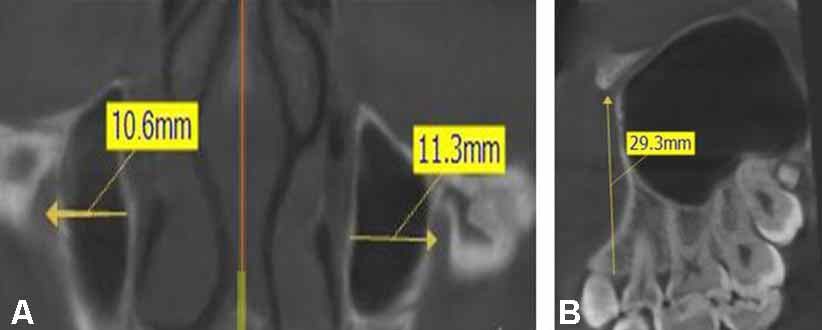
THILAKUMARA, I. P.; HETTIARACHCHI, P. V. K. S.; JAYASINGHE, R. M.; FONSEKA, M. C. N.; JAYASINGHE, R. D. & NANAYAKKARA, C. D. Morphometric analysis of infraorbital
foramen using cone beam computed tomography in a cohort of Sri Lankan adults. Int. J. Morphol., 39(2):489-496, 2021.
surgery, orbital basis reduction (blow-out), zygomatic region of Oral Medicine and Radiology, Faculty of Dental Sciences
fractures and Le Fort type-I osteotomy (Mozsary & University of Peradeniya for routine CBCT imaging. The
Middleton, 1983). Therefore, knowing the exact anatomical de-identified digital imaging and communications of medi-
location is important in order to guarantee safe and successful cine (DICOM) files were used for the assessment. Images
delivery of regional anesthesia and to evade the risk of demonstrating a clear image of the maxilla, devoid of gross
iatrogenic injuries. Multiple studies carried out using dry malocclusions, craniofacial anomalies and bony pathology
skulls have demonstrated that the dimensions and relative were selected for the analysis. In addition, images belonging
positions of the IOF vary between different populations and to patients less than 17 years, images with incomplete clinical
sex (Apinhasmit et al., 2006; Boopathi et al., 2010; Elsheikh records, maxillofacial trauma, craniofacial anomalies such
et al., 2013; Potu et al., 2019). Cone-beam computed as cleft lip and palate and those who had undergone
tomography (CBCT) has also been considered in previous orthodontic correction of malocclusion were excluded.
research to ascertain the same (Dagistan et al., 2017; Bahsi Ethical clearance for this study was obtained from the Ethics
et al., 2019; Sokhn et al., 2019). Review Committee of the Faculty of Dental Sciences,
University of Peradeniya, prior to commencement of the
Little information is available on the dimensions and study. (ERC/FDS/UOP/1/2017/06) Written informed consent
relative position of the IOF in the Sri Lankan population, had been obtained from the patients to use the images for
which have been based on studies carried out by using dry study purposes prior to carrying out the CBCT scans. The
human skulls (Ilayperuma et al., 2010; Nanayakkara et al., study was carried out conforming the STROBE Guidelines
2016). However, to the best of our knowledge, no published and ethical standards of the 1964 Helsinki Declaration.
data was retrieved regarding the dimensions of IOF using
CBCT analysis in the Sri Lankan population. Hence the The methods described in our previous study
present study was undertaken to ascertain the shape, (Jayasinghe et al., 2020) were adopted for CBCT image
dimensions and the position of IOF in relation to clinically acquisition and interpretation. These images were
relevant landmarks, the maxillary midline (MM), infraorbital retrospectively analyzed in coronal, sagittal and axial
margin (IOM) lateral nasal wall (LNW), alveolus (ALV) and sections. In the coronal sections, the shape of IOF and its
maxillary teeth and to determine any variation in the position direction in relation to the midline were assessed. The shape
of IOF between the left and right sides and the sex in a sample of the foramen was categorized as either oval or circular in
of Sri Lankan adults using CBCT imaging. outline and foramina with oval shape was sub classified as
oblique, vertical, and horizontal, considering its direction in
relation to midline. Maximum horizontal (medio-lateral)
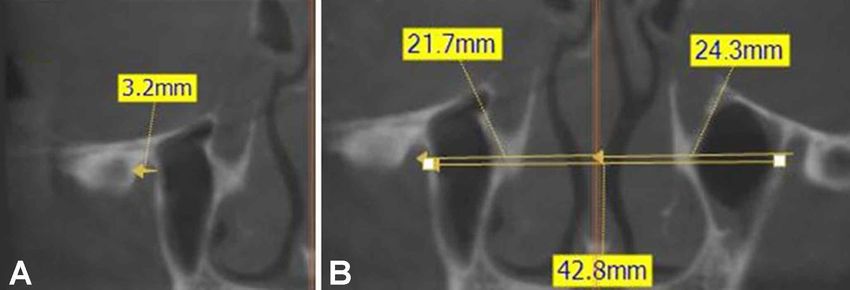
MATERIAL AND METHOD diameter of the IOF (Fig. 1a) and the horizontal distance
between right and left IOFs were also assessed (Fig. 1b).
Sagittal sections were assessed for the maximum vertical
A total of 61 CBCT images of patients (32 females diameter of the IOF and the soft tissue thickness overlying
and 29 males) in the age range of 17 to 32 years were selected IOF. Further, the presence and number of accessory foramina
randomly from the records of patients referred to the Division were assessed in axial sections.
Fig. 1. The coronal section demonstrating (a) Maximum horizontal (Medio-lateral) diameter of the IOF (b) the measurement of distance
from right and left infra orbital foramen (IOF) to the mid maxillary line (MML) and the horizontal distance between the right and left
infra orbital foramina.
490THILAKUMARA, I. P.; HETTIARACHCHI, P. V. K. S.; JAYASINGHE, R. M.; FONSEKA, M. C. N.; JAYASINGHE, R. D. & NANAYAKKARA, C. D. Morphometric analysis of infraorbital
foramen using cone beam computed tomography in a cohort of Sri Lankan adults. Int. J. Morphol., 39(2):489-496, 2021.
The position of IOF in relation to important parameter, the relative position of IOF in relation to
anatomical landmarks were evaluated as: maxillary teeth was recorded, according to the guide
shown in Figure 3 (either as in line with the same verti-
I. The horizontal distance between the mid maxillary line cal axis passing through the long axis of the tooth or in
(MML) and the center of the IOF on each side (Fig. 1b). relation to the cusp tips).
II. Horizontal distance from the lateral nasal wall (LNW)
to the center of IOF bilaterally (Fig. 2a). Descriptive statistics including the means, standard
III. Vertical distance from the inferior orbital margin (IOM) deviations, minimum and maximum values were computed
to the mid-point of the IOF, for each variable, the differences between the left and right
IV. Vertical distance to the crest of the alveolar bone (ALV) sides and males and females were analyzed using the
from the mid-point of the IOF (Fig. 2b). Statistical Package for Social Sciences (SPSS), 20th version.
V. The tooth that is associated when a vertical line is dropped Students’ t-test was used for the analysis and P < 0.05 was
from the mid-point of the IOF. When assessing this considered as statistically significant.
Fig. 2. (a) The coronal section demonstrating horizontal distance from the lateral nasal wall (LNW) to the center of left infra orbital
foramen (IOF) bilaterally; (b) the sagittal section demonstrating the vertical distance to the crest of the alveolar bone (ALV) from the mid
– point of the IOF.
RESULTS
The sample comprised of 122 infraorbital foramina
of 32 females and 29 males in the age range of 17 to 32
years. In females on both right and left sides, the
predominant shape of the IOF was oval (87.5 %) followed
by the circular shape (12.5 %). In males, the pattern was
similar with 82.8 % having an oval shaped IOF on the right
side and 89.7 % on the left side. The difference of having an
oval or a circular shaped IOF between females and males
was not statistically significant. The difference between the
sides was also not statistically significant (Table I).
Fig. 3. The midpoint of the infraorbital foramen is in line; along
the long axis of the canine (A), between the long axis of the canine
A majority of females and males had an obliquely
and 1st pre molar (B), along the long axis of the first premolar tooth
(C), between the long axis of the 1st and the 2nd pre molar (D), directed infraorbital foramen on both left and right sides and
along the long axis of the second premolar tooth (E), between the the difference between sexes was not statistically significant
long axis of the 2nd premolar and the molar tooth (F), along the (P≥0.001). Difference between the sides was also not
long axis of the first molar tooth (G) statistically significant (Table II).
491THILAKUMARA, I. P.; HETTIARACHCHI, P. V. K. S.; JAYASINGHE, R. M.; FONSEKA, M. C. N.; JAYASINGHE, R. D. & NANAYAKKARA, C. D. Morphometric analysis of infraorbital
foramen using cone beam computed tomography in a cohort of Sri Lankan adults. Int. J. Morphol., 39(2):489-496, 2021.
Table I. Shape of IOF in females and males.
Shape Femal es (N= 32) Males (N= 29.) Total sample ( N=61) P value Sex P value Side
Left % Right % Left % Right % Left % Right % difference difference
Circle 12.5 % (4) 12.5 % (4) 10.3 % (3) 17.2 % (5) 11.5 % (7) 29.7 % (9) 0.678 0.385
Oval 87.5 % (28) 87.5 % (28) 89.7 % (26) 82.8 % (24) 88.5 % (54) 80.3 % (52) 0.563 0.462
Table II. Direction of IOF in females and males.
Direction Female s (N= 28) Males (N= 26) Total sample (N=54) P value Sex P value
Left % Right % Left % Right % Left % Right % difference Side
difference
Horizontal 10.7 % (3) 0.7 % (2) 0 % (0) 3.8 % (1) 9.3 %(5) 1.9 %( 1) - 0.232
Oblique 46.4 %(13) 53.6 %(15) 69.2 %(18) 69.2 %(18) 57.4 %(31) 61.1 %(33) 0.212 0.456
Vertical 42.8 %(12) 39.3 %(11) 30.8 %(08) 19.2 %(5) 37.04 %(20) 29.6 %(16) 0.086 0.322
All CBCT images demonstrated the presence of a sin- between left and right sides also were not statistically
gle IOF on both right and left sides. In females 3.1 % showed significant (Table IV).
an accessory foramen on the left side while none were present
on the right side. In males 6.9 % showed accessory foramina Majority of IOF on the left side were positioned in
on the left side while 3.1 % had accessory foramina on the relation to a vertical line passing through the second premolar
right side. The differences in relation to sex and side were tooth (31.7 %) followed by a position in relation to a verti-
found to be statistically not significant (p>0.05) (Table III). cal line passing between the first and second premolars. (30
%). The pattern was similar on the right side with 35 % of
The horizontal and vertical diameters of the IOF and IOF were located in relation to the vertical line passing
the linear distances from the IOF to the inferior orbital margin, through the second premolar, and 23.3 % located in relation
alveolus, maxillary midline and to the lateral wall of nose of to the vertical line passing through the first and second
both right and left sides are shown in Table IV. According to premolars. Statistical significance was not considered as the
the analysis, the differences between sexes for each variable number for each category was smaller than needed for
were not statistically significant. Further, the differences comparison (Table V).
Table III. Presence of accessory foramina on right and left sides of females and males.
Presence of Females (N= 32) Males (N= 29) Total sample (N=61) P value Sex P value Side
accessory difference d ifference
foramen
Left % Right % Left % Right % Left % Right %
No 96.9 % (31) 100 % (32) 93.1 % (27) 96.6 % (28) 95.1 % (58) 98.4 % (60) 0.466 0.532
Yes 3.1 %(1) 0 %(0) 6.9 %(02) 3.4 %(1) 4.9 % (3) 1.6 % (1) - -
Table IV. Measurements of the left and right infraorbital foramina in males and females and total sample (in mm).
Mea surement Female Male Total sample P value P value
Minimum Maximum Mean ± SD Minimum Maximum Mean ± SD Sex difference Side difference
L eft infraorbital foramen
Maximum vertical diameter 1.60 5.50 3.58±0.85 1.70 6.00 3.50±0. 95 3.54±0.99 0.205 0.758
Maximum horizontal diameter 1.90 5.40 3.17±0.75 2.21 5.1 3.28±0. 76 3.22±0.83 0.247 0.786
Dis tance from IOF to MM 22.4 31.5 26.77±2.4 22.1 33 27.24±2.7 26.99±2.73 0.537 0.673
Dis tance from IOF to IOM 1.05 9.5 4.63±2 .1 1 .1 8.5 5.2±1.61 4.91±2.08 0.616 0.512
Dis tance from IOF to LNW 1.21 15.7 11.92±2.89 1.59 16.2 1 2.44±2 .74 12.18±3.35 0.520 0.894
Dis tance from IOF to ALV 24.1 34 29.46±2.48 21.2 34.8 2 9.85±3 .48 29.65±3.28 0.528 0.839
Soft tissue thickness 1.54 15.9 10.98±3.18 7 16.7 1 0.98±1 .73 10.94±2.89 0.238 0.896
Right infraorbital foramen
Maximum vertical diameter 1.27 4.70 3.54±0.83 1.60 5.00 3.49±0. 81 3.52±0.90 0.277 -
Maximum horizontal diameter 1.88 4.6 2.98±0.73 1.88 5.1 3.24±0. 79 3.11±0.83 0.271 -
Dis tance from IOF to MM 22.4 30.7 26.54±2.1 22.4 34 2 7.73±2 .43 27.13±2.6 0.477 -
Dis tance from IOF to IOM 1.37 25.3 5.45±4 .1 2.31 10.4 5.67±1. 88 5.56±3.95 0.495 -
Dis tance from IOF to LNW 1.23 15.9 11.52±2.85 1.65 20 1 2.42±2 .74 11.96±3.45 0.184 -
Dis tance from IOF to ALV 24.4 34.8 29.58±2.94 20 34.4 2 9.60±3 .66 29.59±3.59 0.528 -
Soft tissue thickness 1.65 16.7 11.23±3.16 5 .7 16.4 1 1.04±2 .26 11.14±3.08 0.371 -
Dis tance from IOF to IOF 41.9 57.6 49.98±4.03 43.4 57.6 4 9.80±3 .07 49.89±3.89 0.603 -
492THILAKUMARA, I. P.; HETTIARACHCHI, P. V. K. S.; JAYASINGHE, R. M.; FONSEKA, M. C. N.; JAYASINGHE, R. D. & NANAYAKKARA, C. D. Morphometric analysis of infraorbital
foramen using cone beam computed tomography in a cohort of Sri Lankan adults. Int. J. Morphol., 39(2):489-496, 2021.
Table V. Position of IOF in relation to maxillary teeth in females and males.
Position of IOF in Females (N= 32) Males (N= 28) Total sample (N=60)
relation to maxillary teeth
Left % Right % Left % Right % Left % Right %
A 0 % (0) 0 % (0) 3.6 % (1) 3.6 % (1) 1.7 % (1) 1.7 % (1)
B 12.5 %(4) 12.5 %(4) 14.3 %(4) 10.8 %(3) 13.3 %(8) 11.7 %(7)
C 18.75 %( 6) 21.9 %(7) 10.8 %(3) 14.3 %(4) 15 %(9) 18.3 %(11)
D 28.1 %(9) 18.75 %(6) 32.1 %(9) 28.6 %(8) 30 %(18) 23.3 %(14)
E 28.1 %(9) 31.2 %(10) 35.8 %(10) 39.3 %(11) 31.7 %(19) 35 %(21)
F 9.4 %(3) 9.4 %(3) 0 %(0) 0 %(0) 5 %(3) 5 %(3)
G 3.1 %(1) 3.1 %(1) 3.6 %(1) 7.1 %(2) 3.3 %(2) 5 %(3)
DISCUSSION
The infraorbital nerve, which emerges through the Studies carried out to identify the precise location
IOF to appear on the face, is responsible for the sensory of the IOF have made use of various reference points. Bony
innervation to the skin of the malar area between the lower landmarks such as the IOM, nasion and anterior nasal spine
eyelid and the upper lip (Standring). The infraorbital nerve which can be easily palpated are useful to locate the IOF
being responsible for the sensory innervation of an apparently while landmarks such as the lateral margin of the piriform
larger area, serves as an ideal candidate for a regional nerve aperture which is neither seen nor clinically palpable may
block. An injury to the infraorbital nerve might lead to be of limited help to be used for the same. In the present
numbness of the upper lip, lateral wall of the nose, lower lid study, clinically relevant landmarks and the positional
and the infraorbital region of the affected side. Identifying relationship with the maxillary teeth have been used to
the precise location of the IOF is therefore essential both determine the location of IOF.
during surgery involving the midface region and when
administering the infraorbital nerve block. Numerous studies have been carried out with the aim
of identifying satisfactory reference points for locating the
It is widely agreed that the shape of the IOF shows IOF. The IOM is the widely used reference point to predict
variability (Apinhasmit et al.; Boopathi et al.; Elsheikh et the precise location of the IOF (Apinhasmit et al.; Boopathi
al.; Nanayakkara et al.; Dagistan et al.; Potu et al.). The et al.; Elsheikh et al.; Nanayakkara et al.; Dagistan et al.;
shape of the IOF was predominantly oval in our sample (88.5 Potu et al.). A wide variation has been documented regarding
% on the left side and 80.3 % on the right side) followed by the location of the IOF in relation to the IOM (Table VI).
the circular type (11.5 % on the left side and 29.7 %). This The distance of IOF–IOM has been shown to vary
is in agreement with the findings of previous studies where approximately between 4 mm and over 10 mm in different
the most frequent shape reported was oval (Apinhasmit et population groups (Aziz et al., 2000). The mean distances
al.; Boopathi et al.; Elsheikh et al.; Nanayakkara et al.; of the IOF-IOM in the present study, 5.56 ± 3.95 and 4.91 ±
Dagistan et al.; Potu et al.). However, in a study by Sokhn et 2.08 mm on the right and left sides, respectively, are
al. in the Lebanese population, the circular shape was the somewhat lower to those reported in certain other populations
predominant type in females and with regard to sides, the cir- (Canan et al., 1999; Apinhasmit et al.; Bahsi et al.). Although
cular shape was more common on the right side. The semilunar the distance from the IOF to the IOM in our study was greater
and triangular shaped IOFs reported in some previous studies on the right side in both males and females, none of the
(Apinhasmit et al.; Aggarwal et al., 2015; Nanayakkara et al.; differences between the sides were statistically significant.
Potu et al.) were not encountered in this study. Further, the results demonstrate that the distance between
IOF-IOM is greater in the males than in females. However,
The mean vertical and horizontal diameter of the the difference between the sexes was not statistically
IOF on the right side were 3.52 ± 0.90 and 3.11±0.83, while significant. This is in agreement with the findings of most
those on the left side were 3.50±0.95 and 3.28±0.76. The previous studies but it contradicts with the findings of
average size found in our study was comparable with that Apinhasmit et al. where the difference between the IOF-
observed by Apinhamit et al., (2006) in a Thai population IOM of males and females was found to be statistically
and Varshney & Sharma (2013) in an Indian population, significant. An IOF situated closer to the IOM as in our study
but smaller than that reported by Aziz et al., (2000) in a indicates the close proximity of the infraorbital canal to the
study on White, Black and Hispanic skulls. floor of the orbit. In such a situation, a penetrating globe
493THILAKUMARA, I. P.; HETTIARACHCHI, P. V. K. S.; JAYASINGHE, R. M.; FONSEKA, M. C. N.; JAYASINGHE, R. D. & NANAYAKKARA, C. D. Morphometric analysis of infraorbital
foramen using cone beam computed tomography in a cohort of Sri Lankan adults. Int. J. Morphol., 39(2):489-496, 2021.
injury is a possible complication during an extra oral infraorbital block due to acci-
25.14 ± 2.21 right. 25.06 ± 2.13 left
23.52 ± 2.26 right, 23.43 ± 2.39 left
27.13 ± 2.6 right, 26.99 ± 2.73 left
dental advancement of the needle through the infraorbital canal (Saeedi et al., 2011).
29.10 ± 2.13 males, 27.29 ± 2.12
25.15 ± 2.08 males, 24.43 ± 2.06
This knowledge is vital to clinicians and indicates that extra care should be taken
Distance from IOF-MM (mm)
when administering regional nerve block in the infraorbital region to avoid
complications.
With regard to the distance from the IOF to LNW and IOF to mid maxillary
line, our results showed that the mean distance of the IOF-LNW in the total sample
was 11.96 ± 3.45 mm on the right side and 12.18 ± 3.35 mm on the left side, while the
mean distance of IOF-MM was 27.13 ± 2.6 and 26.99 ± 2.73 on the right and left
side, respectively. Although both the measurements of IOF-LNW and IOF-MM were
greater in males when compared with those of females and greater on the left side
than on the right side, no significant sex or side difference could be observed.
11.96 ± 3.45 right, 12.18 ± 3.35 left
8.95 ± 2.54 right, 9.01 ± 2.46 left
9.57 ± 2.39 right, 9.34 ± 2.36 left
On the other hand, Dagistan et al. conducting a study in a Turkish population
11.63 ± 2.40 males, 10.02 ± 2.17
Distance from IOF-LNW (mm)
Table VI. Mean distances from the IOF-IOM, IOF-LNW and IOF-MM in different populations as reported in previous studies.
using CBCT images reported that although the differences between males and females
with regard IOF-LNW measurement were statistically not significant, the differences
between the right and left sides were shown to be statistically significant. Contrary to
these findings of Dagistan et al., in a study using CBCT images in the Lebanese
population Sokhn et al. reported that the values observed for the distance from the
IOF to LNW varied significantly between the sexes while the side differences were
not significant.
Our results showed that the mean distances from the IOF to mid maxillary
line of males (27.24 ±2.7 and 27.73 ± 2.43 on the left and right side respectively)
appeared to be greater relative to those of females (26.77 ± 2.4 and 26.54 ± 2.1left
and right side, respectively). The similar finding that this distance in males was
5.64 ± 1.78 right, 5.63 ± 1.76 left
7.47 ± 1.40 right, 7.39 ± 1.41 left
5.56 ± 3.95 right, 4.91 ± 2.08 left
6.52 ± 2.03 right, 7.30 ± 1.57 left
5.49 ± 1.10 right, 5.85 ±1.06 left
greater has been previously reported (Apinhasmit et al.; Sokhn et al.). Comparison of Distance from IOF-IOM (mm)
9.53 ± 2.23 males, 8.71 ± 1.51
7.94 ± 1.31 males, 8.01 ± 1.47
6.37 ± 1.4 right, 6.7 ± 1.6 left
the mean distances of the IOF from the mid maxillary line in studies across different
population groups showed that this measurement varied approximately between 23 -
10.9 males, 8.3 females
28 mm. The mean distance recorded in our study was consistent with these values.
Likewise, the mean distance from the IOF to the alveolar crest in the present
study was 29.59 ± 3.59 and 29.65 ± 3.28 on the right and left side respectively.
6.57 ± 1.28
Analyzing dry skulls in an Indian population Aggarwal et al. reported the mean distance
of IOF-ALV as 28.41 ± 62.82 mm. This distance is useful during an intraoral approach
to an infraorbital nerve block.
Dry skulls
Dry skulls
Dry skulls
Dry skulls
Dry skulls
Specimen
Cadavers
The position of IOF in relation to various anatomical landmarks are variable
CBCT
CBCT
CBCT
CBCT
within the population and among different population groups. Knowing the most
accurate position is therefore mandatory to ensure successful regional anesthesia,
and to avoid iatrogenic nerve injuries during surgery of the midface region.
Apinhasmit et al., 2006 - Thailand
The IOF may have an accessory foramen by its side, which may be single or
Dagistan et al., 2017 - Turkey
Nanayakkara et al., 2016 - Sri
Potu et al., 2019 - South India
Sokhn et al., 2019 - Lebanon
Elsheikh et al., 2013 - Egypt
Boopathi et al., 2010 - India
multiple. The occurrence of multiple/ accessory infraorbital foramina (AIOF) is
Canan et al., 1999 - Turkey
Bah_i et al., 2018 - Turkey
extensively reported in the literature (Apinhasmit et al., 2006; Boopathi et al.;
Ilayperuma et al.; Elsheikh et al.; Nanayakkara et al.; Dagistan et al.; Potu et al.;
Study and population
Sokhn et al.). As early as in 1875 Gruber reported that the number of accessory IOF
may vary from 1 to 5 (Leo et al., 1995). In a recent study Hwang et al. (2015)
Present study
analyzing the results published in the literature reported that the occurrence of AIOF
varied between 0.8 % and 27.3 %. Nanayakkara et al. and Ilayperuma et al. reported
that the incidence of AIOF in Sri Lankan dry skulls to be 7.4 % and 3.7 %,
respectively.
494THILAKUMARA, I. P.; HETTIARACHCHI, P. V. K. S.; JAYASINGHE, R. M.; FONSEKA, M. C. N.; JAYASINGHE, R. D. & NANAYAKKARA, C. D. Morphometric analysis of infraorbital
foramen using cone beam computed tomography in a cohort of Sri Lankan adults. Int. J. Morphol., 39(2):489-496, 2021.
The incidence of accessory foramina in the present nerve during surgery of the midface region. As per the
study was 4.9 % and 1.6 % on the left and right side, measurements obtained in our study, the IOF is most likely
respectively. Although the existence of AIOF was greater to be located approximately in a position which is about 5
on the left side and greater in males, the differences between mm inferior to the infraorbital margin, about 27 mm lateral
sides and sexes were found to be statistically not significant. to mid maxillary line in a line perpendicular to the mid
Knowledge regarding the presence of AIOF is an important maxillary line, about 12 mm lateral to the lateral nasal wall,
consideration for the surgeon as it has been reported 29 mm from the maxillary alveolar crest and in the same
previously the possibility of having an accessory branch of vertical plane as the maxillary second premolar tooth. The
the infraorbital nerve passing though the AIOF. Previous existence of accessory foramina in a minority of individuals
studies have reported that both the IOF and AIOF were supports the possibility of either a partial or failed infraorbital
observed to be transmitting their own individual nerve block or injury to the accessory neurovascular bundle
neurovascular bundles (Tubbs et al., 2010; Bahrami et al., resulting in sensory deficits.
2016). The presence of an AIOF and the accessory branch
of the infraorbital nerve raises a mark of caution to both the
surgeons and anesthetists as an injury to this branch during THILAKUMARA, I. P.; HETTIARACHCHI, P. V. K. S.;
surgical maneuvering in the maxillofacial region can result JAYASINGHE, R. M.; FONSEKA, M. C. N.; JAYASINGHE,
in a sensory deficit and when administering the infraorbital R. D. & NANAYAKKARA, C. D. Análisis morfométrico del fo-
nerve block it may result in insufficient anaesthesia in the ramen infraorbitario mediante tomografía computarizada de haz
cónico en una cohorte de adultos de Sri Lanka. Int. J. Morphol.,
region. Double and triple IOF, or accessory foramina were
39(2):489-496, 2021.
not encountered in the present study, although an
occurrence as 2.2–18.2 and 0.5–1.28 %, respectively, has RESUMEN: El foramen infraorbitario (FIO) ubicado
been reported in several previous studies (Leo et al.; Aziz et bilateralmente dentro de la maxila, aproximadamente 1 cm inferior
al., 2000; Apinhasmit et al.; Boopathi et al.; Ilayperuma et al margen infraorbitario, es un punto de referencia vital cuando se
al.; Elsheikh et al.; Nanayakkara et al.; Dagistan et al.; Potu administra anestesia local y durante intervenciones quirúrgicas en la
et al.; Sokhn et al.). región media de la cara. Se analizaron un total de 122 forámenes
infraorbitarios en 61 imágenes de tomografía computarizada de haz
The results of the present study show that in relation cónico (CBCT) de 32 mujeres y 29 hombres en un rango etario de 17
a 32 años para determinar la forma, dirección, presencia de forámenes
to maxillary teeth, the IOF is frequently located in a vertical
accesorios, tamaño y posición precisa de FIO en relación con el mar-
plane passing through the maxillary second premolar tooth gen orbitario inferior (MOI), la línea mediana maxilar (MM), la pa-
(31.7 % and 35 % on the left and right side respectively) red nasal lateral (PNL), el alvéolo (ALV) y los dientes maxilares en
followed by a position in a plane passing between the first un grupo de personas de Sri Lanka. En la mayoría de los adultos se
and second maxillary premolar teeth (30 % and 23.3 % on observó que el FIO tenía forma ovalada (80,3 % y 88,5 % en el lado
the left and right side respectively). This observation is in derecho e izquierdo, respectivamente) Los forámenes infraorbitarios
agreement with the findings of previous studies done using se ubicaron a una distancia media de 5,56 ± 3,95 y 4,91 ± 2,08 mm,
dry skulls (Nanayakkara et al.; Aggarwal et al.). However, por debajo del MOI en los lados derecho e izquierdo; 27,13 ± 2,6 y
according to Aziz et al. (2000) the IOF was frequently located 26,99 ± 2,73 en el lado derecho e izquierdo desde la línea maxilar
mediana, 11,96 ± 3,45 mm y 12,18 ± 3,35 de la PNL en el lado dere-
in a vertical plane passing through the maxillary 1st premolar
cho e izquierdo y 29,59 ± 3,59 y 29,65 ± 3,28 por encima de la cresta
tooth. In the present study, the IOF was observed at the alveolar en los lados derecho e izquierdo. No hubo diferencias
maxillary first molar tooth in a small percentage of the estadísticamente significativas entre los lados izquierdo y derecho o
sample (3.3 % and 5 % on the left and right side respectively); entre sexos. La mayoría de IOF (37,5 % y 55,9 % en el lado derecho
a situation that is likely to cause an unsuccessful infraorbital e izquierdo, respectivamente) se ubicaron en el plano vertical que
nerve block. pasa por el segundo premolar maxilar.
PALABRAS CLAVE: Foramen infraorbitario; Nervio
CONCLUSION infraorbitario; Morfometría.
The aforesaid description regarding the IOF REFERENCES
demonstrates wide variation in the form, position and the
occurrence of accessory foramina among different
Aggarwal, A.; Kaur, H.; Gupta, T.; Tubbs, R. S.; Sahni, D.; Batra, Y. K. &
population groups. Locating accurately the position of the Sondekoppam, R. V. Anatomical study of the infraorbital foramen: A
IOF is important to ensure safe and successful regional basis for successful infraorbital nerve block. Clin. Anat., 28(6):753-
anesthesia and to avoid iatrogenic injury to the infraorbital 60, 2015.
495THILAKUMARA, I. P.; HETTIARACHCHI, P. V. K. S.; JAYASINGHE, R. M.; FONSEKA, M. C. N.; JAYASINGHE, R. D. & NANAYAKKARA, C. D. Morphometric analysis of infraorbital
foramen using cone beam computed tomography in a cohort of Sri Lankan adults. Int. J. Morphol., 39(2):489-496, 2021.
Apinhasmit, W.; Chompoopong, S.; Methathrathip, D.; Sansuk, R. & Varshney, R. & Sharma, N. Infraorbital foramen - Morphometric study
Phetphunphiphat, W. Supraorbital notch/foramen, infraorbital fora- and clinical application in adult Indian skulls. Saudi J. Health Sci.,
men and mental foramen in Thais: anthropometric measurements and 2(3):151-5, 2013.
surgical relevance. J. Med. Assoc. Thai., 89(5):675-82, 2006. Wilkinson, H. A. Trigeminal nerve peripheral branch phenol/glycerol
Aziz, S. R.; Marchena, J. M. & Puran, A. Anatomic characteristics of the injections for tic douloureux. J. Neurosurg., 90(5):828-32, 1999.
infraorbital foramen: a cadaver study. J. Oral Maxillofac. Surg.,
58(9):992-6, 2000.
Bahrami, A.; Saman, M. & Ducic, Y. Duplicate infraorbital nerve an
uncommon anatomical variation. JSM Oro Facial Surg., 1(1):1001, Corresponding author:
2016. Dr. IP Thilakumara
Bahsi, I.; Orhan, M.; Kervancıoglu, P. & Yalçın, E. D. Morphometric Senior Lecturer
evaluation and surgical implications of the infraorbital groove, canal Department of Prosthetic Dentistry,
and foramen on cone-beam computed tomography and a review of Faculty of Dental Sciences,
literature. Folia Morphol. (Warsz.), 78(2):331-43, 2019.
University of Peradeniya
Berge, J. K. & Bergman, R. A. Variations in size and in symmetry of
foramina of the human skull. Clin. Anat., 14(6):406-13, 2001.
SRI LANKA
Boopathi, S.; Chakravarthy Marx, S.; Dhalapathy, S. L. & Anupa, S.
Anthropometric analysis of the infraorbital foramen in a South Indian
population. Singapore Med. J., 51(9):730-5, 2010. Email: ithilakumara@yahoo.com
Canan, S.; Asim, O. M.; Okan, B.; Ozek, C. & Alper, M. Anatomic
variations of the infraorbital foramen. Ann. Plast. Surg., 43(6):613-
7, 1999. Received: 02-12-2020
Dagistan, S.; Milolu, O.; Altun, O. & Umar, E. K. Retrospective
Accepted: 10-01-2021
morphometric analysis of the infraorbital foramen with cone beam
computed tomography. Niger. J. Clin. Pract., 20(9):1053-64, 2017.
Elsheikh, E. M.; Nasr, W. F. & Ibrahim, A. A. S. Anatomical variations
of infraorbital foramen in dry human adult Egyptian skulls,
anthropometric measurements and surgical relevance.
Otorhinolaryngol. Clin., 5(3):125-9, 2013.
Hwang, K.; Lee, S. J.; Kim, S. Y. & Hwang, S. W. Frequency of existence,
numbers, and location of the accessory infraorbital foramen. J.
Craniofac. Surg., 26(1):274-6, 2015.
Ilayperuma, I.; Nanayakkara, G. & Palahepitiya, N. Morphometric
analysis of the infraorbital foramen in adult Sri Lankan skulls. Int. J.
Morphol., 28(3):777-82, 2010.
Jayasinghe, R. M.; Hettiarachchi, P. V. K. S.; Fonseka, M. C. N.;
Nanayakkara, D. & Jayasighe, R. D. Morphometric analysis of
nasopalatine foramen in Sri Lankan population using CBCT. J. Oral
Biol. Craniofac. Res., 10(2):238-40, 2020.
Kothari, S. F.; Shimosaka, M.; Iida, T.; Komiyama, O.; Shibutani, K.;
Svensson, P. & Baad-Hansen, L. Quantitative and qualitative
assessment of sensory changes induced by local anesthetics block of
two different trigeminal nerve branches. Clin. Oral Investig.,
23(6):2637-49, 2019.
Leo, J. T.; Cassell, M. D. & Bergman, R. A. Variation in human infraorbital
nerve, canal and foramen. Ann. Anat., 177(1):93-5, 1995.
Mozsary, P. G. & Middleton, R. A. Microsurgical reconstruction of the
infraorbital nerves. J. Oral Maxillofac. Surg., 41(11):697-700, 1983.
Nanayakkara, D.; Peiris, R.; Mannapperuma, N. & Vadysinghe, A.
Morphometric analysis of the infraorbital foramen: the clinical
relevance. Anat. Res. Int., 2016:7917343, 2016.
Potu, B. K.; Srungavarapu, G. C. & Pulakunta, T. Morphometric
evaluation of the infraorbital foramen in human dry skulls of South
Indian population. Ital. J. Anat. Embryol., 124(3):382-91, 2019.
Saeedi, O. J.; Wang, H. & Blomquist, P. H. Penetrating globe injury during
infraorbital nerve block. Arch. Otolaryngol. Head Neck Surg.,
137(4):396-7, 2011.
Sokhn, S.; Challita, R.; Challita A. & Challita, R. The infraorbital fora-
men in a sample of the Lebanese population: a radiographic study.
Cureus, 11(12):e6381, 2019.
Standring, S. Gray’s Anatomy: Anatomical Basis of Clinical Practice.
40th ed. London, Churchill Livingstone Elsevier, 2008.
Tubbs, R. S.; Loukas, M.; May, W. R. & Cohen-Gadol, A. A. A variation
of the infraorbital nerve: its potential clinical consequence especially
in the treatment of trigeminal neuralgia: case report. Neurosurgery,
67(3 Suppl. Operative):onsE315, 2010.
496You can also read