Multiomics integrative analysis identifies APOE allele-specific blood biomarkers associated to Alzheimer's disease etiopathogenesis
←
→
Page content transcription
If your browser does not render page correctly, please read the page content below
www.aging-us.com                                                           AGING 2021, Vol. 13, No. 7
                                                                                                Research Paper
Multiomics integrative analysis identifies APOE allele-specific blood
biomarkers associated to Alzheimer’s disease etiopathogenesis
Laura Madrid1, Sonia Moreno-Grau2,3, Shahzad Ahmad4, Antonio González-Pérez1, Itziar de
Rojas2,3, Rui Xia5, Pamela V. Martino Adami6, Pablo García-González2, Luca Kleineidam6,7,8, Qiong
Yang9, Vincent Damotte10, Joshua C. Bis11, Fuensanta Noguera-Perea12, Céline Bellenguez10,
Xueqiu Jian5, Juan Marín-Muñoz12, Benjamin Grenier-Boley10, Adela Orellana2,3, M. Arfan Ikram4,
Philippe Amouyel10, Claudia L. Satizabal13,14, Alzheimer’s Disease Neuroimaging Initiative
(ADNI)*, EADI consortium, CHARGE consortium, GERAD consortium, GR@ACE/DEGESCO
consortium, Luis Miguel Real15,16, Carmen Antúnez-Almagro12, Anita DeStefano7,14, Alfredo
Cabrera-Socorro17, Rebecca Sims18, Cornelia M. Van Duijn4, Eric Boerwinkle19, Alfredo
Ramírez6,7,8, Myriam Fornage5, Jean-Charles Lambert10, Julie Williams18,20, Sudha Seshadri13,14,
ADAPTED consortium, Janina S. Ried21, Agustín Ruiz2,3, Maria Eugenia Saez1
1
 Andalusion Bioiformatics Research Centre (CAEBi), Sevilla, Spain
2
 Research Center and Memory Clinic Fundació ACE, Institut Català de Neurociències Aplicades, Universitat
Internacional de Catalunya, Barcelona, Spain
3
 CIBERNED, Network Center for Biomedical Research in Neurodegenerative Diseases, National Institute of Health
Carlos III, Madrid, Spain
4
 Department of Epidemiology, Erasmus Medical Centre, Rotterdam, The Netherlands
5
 Institute of Molecular Medicine and Human Genetics Center, University of Texas Health Science Center at
Houston, Houston, TX 77030, USA
6
 Division of Neurogenetics and Molecular Psychiatry, Department of Psychiatry and Psychotherapy, Medical
Faculty, University of Cologne, Cologne, Germany
7
 Department of Neurodegenerative Diseases and Geriatric Psychiatry, University Hospital Bonn, Bonn, Germany
8
 German Center for Neurodegenerative Diseases (DZNE), Bonn, Germany
9
 Department of Biostatistics, Boston University School of Public Health, Boston, MA 02118, USA
10
  University Lille, Inserm, CHU Lille, Institut Pasteur de Lille, U1167-RID-AGE-Facteurs de Risque Et Déterminants
Moléculaires des Maladies Liées au Vieillissement, Lille, France
11
  Cardiovascular Health Research Unit, Department of Medicine, University of Washington, Seattle, WA 98195,
USA
12
  Unidad de Demencias, Hospital Clínico Universitario Virgen de la Arrixaca, Carretera de Madrid-Cartagena s/n,
30120 El Palmar, Murcia, España
13
  Glenn Biggs Institute for Alzheimer's and Neurodegenerative Diseases, UT Health San Antonio, San Antonio, TX
78229, USA
14
  Department of Neurology, Boston University School of Medicine, Boston, MA 02118, USA
15
  Unit of Infectious Diseases and Microbiology, Hospital Universitario de Valme, Sevilla, Spain
16
  Department of Surgery, Biochemistry and Immunology, University of Malaga, Spain
17
  Janssen Research and Development, a Division of Janssen Pharmaceutica N.V., Beerse, Belgium
18
  Division of Psychological Medicine and Clinical Neuroscience, School of Medicine, Cardiff University, Cardiff, UK
19
  Department of Epidemiology, Human Genetics, and Environmental Sciences, School of Public Health, The
University of Texas Health Science Center at Houston, Houston, TX 77030, USA
20
  UKDRI@Cardiff, School of Medicine, Cardiff University, Cardiff, UK
21
  AbbVie Deutschland GmbH & Co. KG, Genomics Research Center, Knollstrasse, Ludwigshafen, Germany

www.aging-us.com                                      9277                                                  AGING
*Data used in preparation of this article were obtained from the Alzheimer’s Disease Neuroimaging Initiative
(ADNI) database (adni.loni.usc.edu). As such, the investigators within the ADNI contributed to the design and
implementation of ADNI and/or provided data but did not participate in analysis or writing of this report. A
complete listing of ADNI investigators can be found at: http://adni.loni.usc.edu/wp-
content/uploads/how_to_apply/ADNI_Acknowledgement_List.pdf

Correspondence to: Maria Eugenia Saez; email: mesaez@caebi.es
Keywords: Alzheimer’s disease, APOE, integrative analysis, biomarkers
Received: February 3, 2021       Accepted: March 26, 2021             Published: April 12, 2021

Copyright: © 2021 Madrid et al. This is an open access article distributed under the terms of the Creative Commons
Attribution License (CC BY 3.0), which permits unrestricted use, distribution, and reproduction in any medium, provided the
original author and source are credited.

ABSTRACT

Alzheimer’s disease (AD) is the most common form of dementia, currently affecting 35 million people
worldwide. Apolipoprotein E (APOE) ε4 allele is the major risk factor for sporadic, late-onset AD (LOAD), which
comprises over 95% of AD cases, increasing the risk of AD 4-12 fold. Despite this, the role of APOE in AD
pathogenesis is still a mystery. Aiming for a better understanding of APOE-specific effects, the ADAPTED
consortium analyzed and integrated publicly available data of multiple OMICS technologies from both plasma
and brain stratified by APOE haplotype (APOE2, APOE3 and APOE4). Combining genome-wide association
studies (GWAS) with differential mRNA and protein expression analyses and single-nuclei transcriptomics, we
identified genes and pathways contributing to AD in both APOE dependent and independent fashion.
Interestingly, we characterized a set of biomarkers showing plasma and brain consistent protein profiles and
opposite trends in APOE2 and APOE4 AD cases that could constitute screening tools for a disease that lacks
specific blood biomarkers. Beside the identification of APOE-specific signatures, our findings advocate that this
novel approach, based on the concordance across OMIC layers and tissues, is an effective strategy for
overcoming the limitations of often underpowered single-OMICS studies.

INTRODUCTION                                                      positions [6–8]. These aminoacidic substitutions result
                                                                  in a conformational change that brings together the N-
Non-Mendelian Alzheimer’s disease (AD) has become                 terminal and C-terminal domains in APOE4, which are
the paradigm of a complex disease for which a major               normally separated in APOE2 and APOE3 isoforms.
genetic determinant is known, the APOE locus. Three               The consequences in downstream signaling of this
linkage studies published in 1993 pointed to the APOE             conformational shift in the APOE4 isoform are still
region at 19q13 as a risk locus for late onset familial           unknown. In fact, it is not even clear if the APOE4 is a
AD [1, 2], and even common sporadic late-onset AD                 gain or loss of function mutation despite extensive
(LOAD) [3]. Shortly after, researchers around the world           research in the field [9]. What is already known is that
confirmed the association of APOE gene with diverse               having a single APOE4 allele increases risk 2- to 4-fold
forms of the disease and its association with other               and having two APOE4 alleles increases risk about 8- to
dementias.                                                        12-fold, although risk varies according to genetic
                                                                  background and sex [10].
The APOE gene encodes a lipoprotein firstly identified
in the 1970s among patients with familial                         In the last years, genome-wide association studies
hypercholesterolemia type III [4, 5]. The protein has             (GWAS) have contributed a number of Alzheimer’s
three major isoforms depending on the combination of              disease associated low penetrance genes, including
two polymorphisms located at positions 112 (rs429358              ABCA7, ABI3, ACE, AC074212.3, ADAM10,
(C > T)) and 158 (rs7412 (C > T). The most common                 ADAMTS1, ADAMTS4, ALPK2, ANKDR31, APH1B,
isoform, APOE3, has a cysteine at position 112 and an             ATP5H, BIN1, BZRAP1-AS1, CASS4, CD2AP, CD33,
arginine at position 158, whereas APOE2, the least                CELF1-MADD, CLNK, CLU, CNTNAP2, CR1, DSG2,
common isoform, has a cysteine at both positions, and             ECHDC3, EPHA1, FERMT2, HESX1, HLA-DRB5–
the AD risk allele APOE4 has an arginine at both                  HLA-DRB1, HS3ST1, KAT8, IQCK, INPP5D, NME8,

www.aging-us.com                                         9278                                                     AGING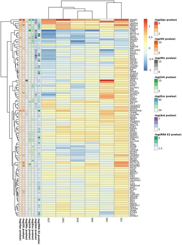
NYAP1, MS4A gene cluster, NDUFAF6, OARD1, significant signals (p

Sex stratified meta-analysis (Supplementary Tables 7, 8        mitochondrial genes, most of them involved in the
and Supplementary Figure 6) identified genome-wide             oxidative phosphorylation pathway. However, several
significant signals for BIN1 and APOE as well as               genes from this pathway were differentially expressed
suggestive signals for PICALM, MYLK, SOX5 and                  in all strata but with opposite expression profiles, such
SCEL in the female population. By contrast in males,           as the electron transport chain genes ATP5F1, UQCRB
only suggestive signals for BIN1, APOE, ZCCHC2, the            or NDUFB3 upregulated in APOE2 cases but
ABI3BP/IMPG2 locus, ESRRB and the 19q13.4                      downregulated in APOE4 cases when compared to
leukocyte receptor cluster were identified. Stratified         controls of the same haplotype. APOE3 genes were
analysis by sex and APOE (Supplementary Tables 9–14            mainly cytoplasmatic genes involved in RNA
and Supplementary Figure 7), yielded genome-wide               metabolism.
significant signals for APOE in the APOE4 stratum in
both sexes and for a 400kb 13q31.3 intergenic region           Cortex APOE-stratified DE included the MAYO,
containing the Ubiquitin Specific Peptidase 7 (Herpes          ROSMAP, MSBB, GSE15222 and GSE48350 studies.
Virus-Associated) (USP7) pseudogene (RP11-464I4.1)             Meta-analysis of cortex datasets resulted in 518, 7714
for APOE3 males. Among APOE4 males, we found                   and 1717 statistically significant genes (FDR
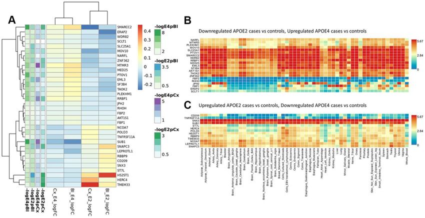
Table 1. GWAS analysis: gene level data (MAGMA results) by APOE stratum.
Rank E2        HUGO          NSNPS      NPARAM          ZSTAT      PJoint   PSNPwise (mean)   PSNPwise (top)
1             RNF152          609         65             4.62    1.90E-06    4.59E-07          4.63E-04
2             DUOX2           150         26             3.97    3.53E-05    4.72E-06          5.12E-03
3             METRN           140         18             3.88    5.29E-05    2.41E-05          9.43E-04
4             WDR24           197         18             3.83    6.31E-05    2.71E-05          1.21E-03
5             FBXL16          196         18             3.84    6.03E-05    2.85E-05          1.11E-03
6            FAM173A          120         17             3.83    6.29E-05    2.99E-05          1.14E-03
7              JMJD8          193         18             3.79    7.59E-05    3.34E-05          1.32E-03
8             CCDC78          115         17             3.81    6.99E-05    3.68E-05          1.14E-03
9            DUOXA2           145         23             3.63    1.41E-04    3.68E-05          6.05E-03
10            HAGHL           107         15             3.83    6.43E-05    3.88E-05          9.08E-04
11            NARFL           105         15             3.83    6.51E-05    4.00E-05          8.92E-04
12             STUB1          193         18             3.72    1.01E-04    4.61E-05          1.56E-03
13            ABCB4           344         35             3.89    5.11E-05    5.27E-05          5.59E-03
14             APOC1          209         32             3.42    3.16E-04    5.40E-05          4.13E-02
15              IRGC          300         45             4.55    2.72E-06    5.52E-05          3.36E-05
Rank E3        HUGO          NSNPS      NPARAM          ZSTAT      PJoint   PSNPwise (mean)   PSNPwise (top)
1               BIN1          633         70             7.02    1.15E-12    8.93E-07          3.51E-12
2               FBN1          533         41             4.71    1.27E-06    4.82E-06          2.76E-04
3              WNT3           239         38             4.69    1.35E-06    6.85E-06          1.26E-04
4            CLEC4M           424         68             4.07    2.35E-05    1.35E-05          1.46E-03
5                NSF          144         22             4.76    9.48E-07    2.88E-05          3.07E-05
6              CASS4          349         50             5.00    2.80E-07    3.43E-05          4.58E-05
7                APP          998         99             3.96    3.75E-05    3.91E-05          3.43E-03
8              GPR27          306         47             3.21    6.62E-04    4.63E-05          6.29E-02
9             CRHR1          1261         23             4.07    2.39E-05    5.46E-05          2.55E-03
10             CD209          482         71             3.80    7.36E-05    6.93E-05          1.55E-03
11            SPPL2C          556         19             4.03    2.83E-05    7.77E-05          1.68E-03
12           KANSL1           978         20             4.01    2.98E-05    7.79E-05          1.44E-03
13           LRRC37A          115         6              4.01    3.09E-05    7.97E-05          4.41E-04
14               STH          460         17             3.94    4.00E-05    8.35E-05          2.88E-03
15             EIF4E3         516         83             3.01    1.29E-03    9.34E-05          1.41E-01
Rank E4        HUGO          NSNPS      NPARAM          ZSTAT      PJoint   PSNPwise (mean)   PSNPwise (top)
1            TOMM40           293         44            14.90    1.59E-50    1.50E-26          1.00E-50
2              APOC1          247         38            15.05    1.71E-51    3.20E-26          1.00E-50
3              APOE           270         40            14.82    5.36E-50    2.83E-24          1.00E-50
4              PVRL2          361         51            14.31    8.88E-47    1.17E-23          1.00E-50
5              APOC4          241         29            11.30    6.96E-30    1.60E-14          2.52E-33
6              APOC2          232         27            13.20    4.31E-40    8.54E-13          1.00E-50
7            CLPTM1           267         34            12.78    1.06E-37    3.43E-11          5.65E-46
8                CLU          351         46             7.60    1.47E-14    5.85E-10          4.89E-13
9            SCARA3           426         51             7.08    7.06E-13    7.15E-07          3.53E-13
10            PICALM          555         43             5.54    1.49E-08    8.39E-07          9.65E-08
11            AKAP2           601         85             4.12    1.86E-05    2.27E-06          5.58E-03
12         PALM2-AKAP2       1542        159             3.61    1.52E-04    3.72E-05          1.46E-02
13               IL6          414         48             3.72    1.01E-04    4.36E-05          7.17E-03
14             EPHX2          467         56             4.23    1.15E-05    5.26E-05          2.17E-04
15              BIN1          635         70             5.70    5.95E-09    5.72E-05          1.11E-08

www.aging-us.com                                 9281                                               AGING
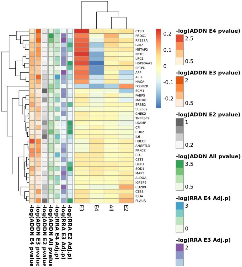
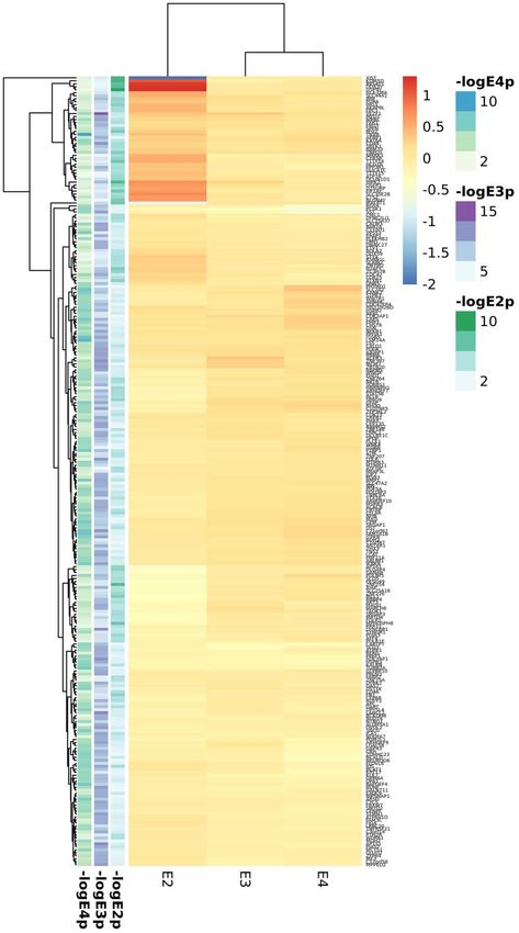
Figure 2. Top candidates from DE analysis in blood datasets.

www.aging-us.com                            9282                                  AGING
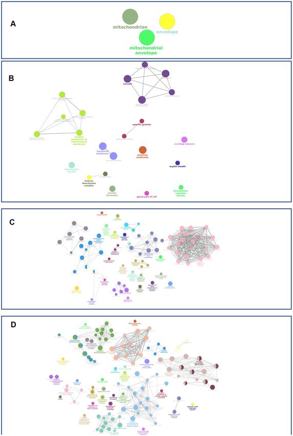
Figure 3. Top candidates from DE analysis in cortex datasets.

www.aging-us.com                            9283                                   AGING
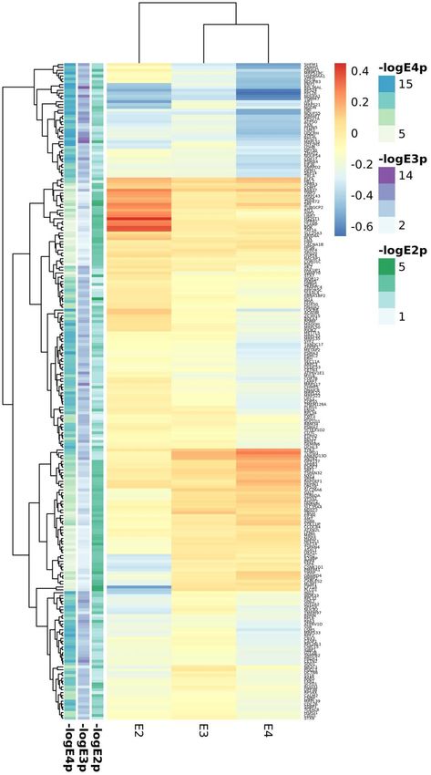
mitochondrial proteins (ATP5F1, ATP6V1D, MRPL51              SRGAP1, TOMM40, TSPAN14, UBE2F, WWOX,
and UQCRH). The list of APOE2 specific genes is the          ZNF264). Enrichment analysis also identified both
largest one (241 genes) and include mitochondrial            common and exclusive pathways. Common pathways
transporters such as SLC25A3 and SLC25A4. APOE3              for AD irrespective of the APOE haplotype were related
specific signatures included MAPT and APP genes,             to adhesion, neuronal development, differentiation, and
along with other neuronal genes such as the cholinergic      lipoprotein metabolism; diverse signals related to
receptors CHRNA10 and CHRNA2. APOE4 specific                 neuronal death are also present in all three strata.
genes included known AD genes involved in vesicle            Again, we observed larger overlap between APOE3 and
transport such as CLU and SORL1; SORL1 has been              APOE4 pathways (glial cell differentiation and
shown to regulate IL6 levels, also identified among          activation,    immunological,     lipid    metabolism,
APOE4 specific signatures. We found however a large          cardiovascular system development and heart function)
overlap among significant genes for the APOE3 and            than for APOE2 and APOE3 (which includes
APOE4 strata (n=51) including BIN1, MS4A4A,                  axonogenesis) or APOE2 and APOE4 (mainly
MS4A6A, PICALM and SLC24A4 AD genes and a good               phospholipid and lipoprotein metabolism due to APOE,
number of ribosomal and electron transport                   APOC1 and APOC2 genes). (Supplementary Tables 34–
mitochondrial proteins (ATP5I, ATP5O, ATP6V1E1,              36 and Supplementary Figure 9). We observed that
COX17, MRPL27, MRPL33, MRPL35, MRPS17,                       APOE2 exclusive pathways include chromatin
MRPS21, UQCRB). APOE2 and APOE3 shared eleven                regulation and telomere maintenance related processes.
genes (ACADM, AK2, ENSA, GPR132, GPR137,                     The APOE3 strata showed the largest number of
HSP90AA1, MCL1, NXF1, TAF1C, UBA7, ZC3H15)                   significant enrichments, but most of them showed a
whereas the overlap between E2 and E4 was the                similar annotation in APOE2, or, more frequently, in
smallest with eight proteins (KTN1, NDST2, POLR1D,           APOE4 strata, with the exception of antigen processing
RHOT2, STK17B, TOMM40, TSPAN32, UCHL3). At                   and presentation, IFNG signalling, astrocyte
the pathway level, we observed a lower overlap               development and activation and myelin sheath. In
between APOE strata, with only three shared                  APOE4 macrophage activation, fructose metabolism,
mitochondrial GO categories among the three                  vitamin D mediated inflammation, inositol phosphate
haplotypes, and little or none overlap between APOE2         metabolism and cholesterol efflux were the most
and APOE3 or APOE2 and APOE4 (Supplementary                  relevant pathways. Clathrin vesicles, amyloid biology,
Tables 31–33 and Supplementary Figure 8). In contrast,       inflammatory and immune response and glial cell
there was considerable overlap between APOE3 and             development and differentiation appear as the most
APOE4 which includes mitochondria biology, secretory         relevant categories shared by APOE3 and APOE4 strata.
vesicles and antigen processing and presentation
functions.                                                   To identify relevant candidate blood biomarkers
                                                             tracking brain changes in AD pathology we compared
In cortex, we found 376, 399 and 366 significant genes       blood and cortex analyses (Figure 4). We identified 68
(FDR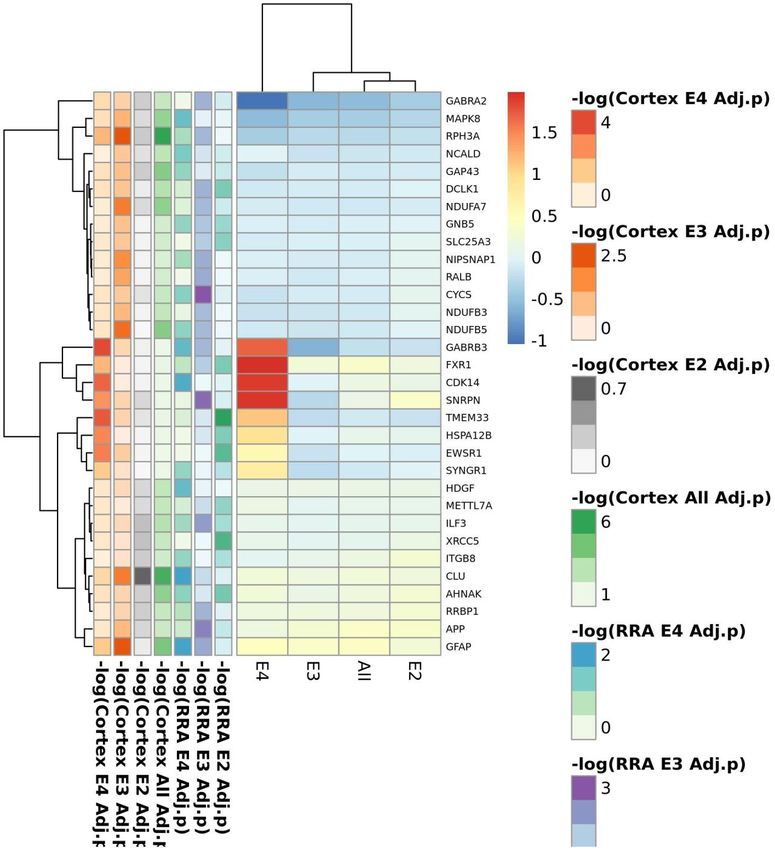
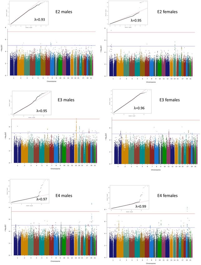
Figure 4. Summary of candidate genes (A–D) and pathways (E–H) from APOE2 (B, F), APOE3 (C, G) and APOE4 (D, H) common candidates
from Blood and Cortex RRA analyses.

www.aging-us.com                                           9285                                                        AGING
blood and cortex (upregulated or downregulated in AD                    expressed genes in the unstratified analysis, 8 genes in the
cases vs controls) and showing opposite profiles in                     APOE4 stratum and 9 in the APOE3 stratum. We could
APOE2 and APOE4, which included 34 genes with                           confirm APOE allele-specific effects identified in the
overrepresentation of the gluconeogenesis and fructose                  RRA analysis for the immune related proteins AIF1,
metabolic pathways (FBP1, FBP2, SLC25A1) (Figure                        METAP2, NCK1, PRDX1, PRKCZ, RPS27A in the
5A). When compared with average expression in                           APOE3 stratum, and FCGR2B and SEZ6L2 (involved in
normal brains, FBP1, FBP2, RHOH, JPH2, ERAp2 and                        SNC development) in the APOE4 stratum. Overall,
SCLT1 were upregulated in APOE4 cases when they are                     among these 38 RRA candidates, we identified a cluster
usually expressed at low levels, whereas, SNX3 and                      of 11 overexpressed proteins in AD cases when compared
SUB1, were downregulated in APOE4 cases when they                       to controls in the APOE3 stratum, but downregulated
are expressed at very high levels in the normal brain                   APOE4 AD cases including AIF1, APP, GDI2,
according to GTEx (Figure 5B).                                          HSP90AA1, METAP2, NACA, NCK1, PRDX1,
                                                                        RPS27A, SFTPD and UFC1 (Supplementary Figure 11);
Validation on proteomic datasets                                        immunological functions associated to these proteins
                                                                        include leukocyte activation (APP, PRDX1, GDI2), Toll-
We aimed at investigating if any of our candidate genes                 like receptors (TLRs) cascade (APP, RPS27A, SFTPD) or
were detected and differentially expressed at the                       phagocytosis (NCK1, HSP90AA1, SFTPD, AIF1) in line
proteomic level using blood proteomics data from the                    with our RRA findings.
ADDN study (931 proteins) and cortex proteomics from
four independent datasets (BANNER, BLSA, MAYO                           In cortex, 234 out of 1,039 RRA candidates were
and MSBB, 2,658 proteins).                                              present in the proteomics DE meta-analysis, 100 of
                                                                        them showing evidences of association (p
RRA candidates involved in neurogenesis (DPYSL4, and microglia populations, and an increase in
EHD1, GABRB3, MAPK8, UNC13A), or more expression in the oligodendrocyte subpopulation
specifically, in glial cell differentiation (CLU, GAP43, (Figure 6). The seven genes in common in all the RRA
GFAP, GSN). Among APOE3 candidates, we analyses, were downregulated in all cell types except
confirmed candidates involved in neurotransmission for APOE and APOC1 in microglia, and CD44 in
such as RPH3A PTK2B, ALDH5A1, GABRA2 and astrocytes (Figure 6). In the stratified analysis, these 7
APP (the later upregulated in all strata) and genes from genes were predominantly downregulated in AD
the electron transport chain (ALDH5A1, NDUFA7, APOE3 carriers and upregulated among APOE4 AD
NDUFB3). Confirmed APOE2 candidates included the cases when compared to controls, particularly APOC1,
choline transporter SLC44A1, involved in myelin DST and CD44 (Supplementary Figure 9). By stratum,
production, and the myelin basic protein MBP; MAPT APOE2 RRA cortex candidates were mostly
was upregulated in all strata but particularly in the upregulated in all cell types (Supplementary Figure 10),
APOE2 stratum. We also confirmed the role of CDC42 and in particular FXR1 and DNAJB1, the latter only
and DST in all the strata, but we did not observe downregulated in microglia. APOE3 RRA candidates
association of CD44 and PGM2L1 with AD in this showed the largest differences between cases and
analysis. controls in microglia cells, where APOC1, ALDOA,
RPLP0 and DYNLRB1 were strongly upregulated
Cell-type-specific expression profiles: cortex whereas ARL17B was downregulated in AD cases
snRNAseq Supplementary Figure 11). Almost all APOE4 candidate
genes were downregulated in both excitatory and
Since the enrichment analysis showed an over- inhibitory neurons and upregulated in the glial lineage,
representation of neuronal development related particularly TMEM163 and CPM in microglia and
pathways in all strata, and of cells from the glial lineage GFAP, PLCE1, CLU, CALN1, DLG2 and PDE5A in
in the APOE3 and APOE4 strata, we investigated which astrocytes Supplementary Figure 12); we also observed
cerebral cell types our cortex RRA candidates were a strong downregulation of the Serine/Threonine Kinase
mainly expressed in, and which cell types showed 17b (STK17B), involved in apoptosis and autophagy, in
largest differences between cases and controls using microglial cells of APOE4 cases.
snRNAseq from the ROSMAP study (Figure 6). We
dropped pericytes and endothelial cells from the DISCUSSION
differential expression analysis because of the low
number of cells (≈100 cells,Figure 6. Cortex snRNAseq data from the ROSMAP study. (A) Cell clustering labelled by reported cell type; (B) APOE expression across cell types; (C) violin plot for APOE expression by cell type; (D) UMAP plot for average expression of cortex RRA APOE2 candidates by total gene expression; (E) UMAP plot for average expression of cortex RRA APOE3 candidates by total gene expression; (F) UMAP plot for average expression of cortex RRA APOE4 candidates by total gene expression; (G) UMAP plot for average expression of blood/cortex biomarkers by total gene expression; (H) expression by cell type of the seven genes in common in all the RRA analyses; (I) APOE expression by cell type and case status. www.aging-us.com 9288 AGING
of the expression profiles) for selecting candidate genes       in AD and even MCI subjects, showing a significant
in expression datasets.                                         increase of oxidative stress markers, such as lipid
                                                                peroxidation and protein oxidation products [30–32].
At the genome level, we were able to detect genome-             We did not observe mitochondrial signatures at the
wide significant signals for ABCA7, BIN1 and PICALM             whole cortex level, mostly enriched in activated genes
in the APOE3 stratum and for APOE, BIN1, CLU and                from neuronal, apoptosis, vesicle mediated transport
PICALM in the APOE4 stratum. We identified a novel              and adhesion related pathways, maybe because
candidate region for APOE4 carriers on 4p15.1                   mitochondrial dysfunction has been reported to be
(33.6Mb-34.3Mb), which, according to the GWAS                   limited to certain hippocampal and temporal cortex
catalogue (https://www.ebi.ac.uk/gwas/) has not been            neurons [33, 34].
previously associated with AD, but with schizophrenia,
total cholesterol change in response to fenofibrate in          Integration of genome data with expression data at
statin-treated type 2 diabetes, and PCSK9 levels, a             blood and cortex levels through the RRA algorithm,
protease that binds to lipoprotein receptors promoting          showed a larger overlap of genes and functions in
their degradation; a homozygous deletion overlapping            APOE3 and APOE4 carriers than in APOE2 carriers,
this region has been described for the offspring of a           which appears as a more distinct entity. In fact, we
consanguineous marriage between first cousins, with             identified signatures for chromatin remodeling and
cognitive impairment and autistic-like behavior [27].           regulation in this stratum at both brain and plasma
Sex-stratified analysis identified genome wide                  levels, not observed in the other two strata. Common
significant signals for APOE and BIN1 only in females;          features of the disease to all three strata are related to
this result is in agreement with the recent report from         lipid metabolism due to APOE (except for the APOE3
Fan et al., who described a genome-wide significant             carriers), APOC1 and APOC2. A recent report has
association for BIN1 only in females [28]. Further              suggested that APOC1 gene, located in the APOE locus,
stratification of male and female populations by APOE           is an independent risk factor for AD, and that genetic
haplotype identified a genome-wide significant                  variability in the region is associated with chromatin
intergenic region on 13q31.3 among APOE3 males.                 regulation [35].
This region has been associated with TREM2 levels,
circulating Interleukin-1-receptor antagonist levels and        AD cases in APOE3 and APOE4 share signaling
triglyceride change in response to fenofibrate in statin-       pathways and functional categories previously reported
treated type 2 diabetes. This region harbors a USP7             by other groups such as amyloid-beta formation
pseudogene        (RP11-464I4.1)      associated      with      (APOE, BIN1, CLU, PICALM) mitochondrial
herpesvirus. Interestingly, a potential role of herpes          physiology (including ATP5H, NDUFS5, MRP
simplex virus infection in AD has recently been object          proteins, SNCA, SOD1, SSBP1, SUCLG1 or UQCRH),
of intense debate [29]. Despite the number of GWAS              vesicle mediated transport (including APOC1, APP,
datasets collected, our study is still underpowered for         BIN1, C1QTNF5, CASS4, CDC42, LDLR, MAPT,
detecting genuine APOE strata-specific signals with             PICALM or PTK2B), actin organization (ACTN1,
low effect sizes, but resulting gene-level statistics were      ACTR2, AIF1, ANTXR1, CALD1, CAPZ1, CD2AP,
instrumental to select those DE signals that better             DST, ITGB5, MACF1, MAPT, PALLD, RHOC …) or
correlate with the disease at genetic level. This helps         immunological functions (CCL5, CD209, CD44, CR1,
maximize high probable loci involved in the                     IL6, LILRA5 or MS4A2 among others), but with
fundamental       pathways     involved     in     disease      specific gene signature (for example IL6 in the APOE4
pathogenesis.                                                   stratum or CD209 in the APOE3 stratum). IL6 plays a
                                                                critical role in inflammation as well as in
The genome-wide expression analysis was performed at            neuroprotection through two different mechanisms.
two levels: blood and brain cortex. In blood,                   Anti-inflammatory effects are mediated by the classical
mitochondrial ribosomal genes and as well as those              signaling pathways, which involves the binding of IL6
encoding proteins of the respiratory chain appeared             to the membrane bound IL6 receptor (IL6R), whereas
downregulated in cases irrespectively of the APOE               proinflammatory effects are mediated by soluble IL6R
haplotype, but more pronounced among APOE4                      forms. Classical signaling occurs in microglia whereas
carriers. Mitochondria are crucial players of energy            trans-signaling is predominant in most neuronal types,
metabolism but are also the main source of Reactive             astrocytes and oligodendrocytes [36]. Cross-talk
Oxygen Species (ROS). Mitochondrial dysfunction has             between TREM2, CD33 and IL6 (among other ILs)
been proposed as the primary process triggering all the         regulating phagocytic capacity, a hallmark of AD
cascade of events that lead to sporadic late-onset AD.          among APOE4 carriers according to our results, has
Although this hypothesis has not been confirmed,                been reported in microglia cells [37]. Interestingly, IL6
diverse mitochondrial functions were observed altered           is degraded by SORL1, encoded by another well-

www.aging-us.com                                         9289                                                    AGING
known AD gene [38]. CD209 is mainly expressed on a process counterbalanced by cholesterol efflux, a
the surface of dendritic cells, specialized antigen- mechanism identified as an APOE4 specific feature in
presenting cells, where regulates DC adhesion, our study. A key protein in this process seems to be
migration and triggering of immune response [39]. In AIF1, a pro-inflammatory molecule expressed primarily
conclusion, our results suggest that APOE-allele in the monocyte/macrophage lineage, which was shown
specific immunological checkpoints may exist in AD. to be downregulated in APOE4 cases and upregulated in
plasma samples of APOE3 cases in this study. A1F1
Although we have identified signatures of the nervous was originally cloned from a rat heart allograft under
system development in all strata, they represent a chronic rejection, and it is involved in several
largest proportion of relevant pathways in the APOE3 inflammatory conditions including atherosclerosis.
stratum. In this stratum, enrichment analysis of RRA Crossbreeding experiments A1F1 and APOE transgenic
cortex candidates showed an over-representation of mice have shown an interaction between these genes
genes involved in cardiac development and function leading to atherosclerotic vasculopathy though
(DLG1, JPH2 or MEF2C among others), supporting a modulation of the incorporation of degenerated LDL by
cardiovascular etiology of dementia in this stratum. In macrophages [47, 48].
line with this finding, we have recently reported a link
between cardiac function and AD, that is mediated, at In brain, the resident macrophages, microglia cells, are
least in part, by CFLAR and caspase dependent the specialized phagocytic cells acting through a
mechanisms [40]. In fact, CFLAR and CASP8 are both complement dependent mechanism coupled to ATP
RRA cortex candidates in this stratum. Another production. The analysis of single cell cortex data
example of the nervous-cardiac connection is GFAP, points to a pivotal role of the glial lineage in the
which participates in the control of heart rate and development of AD in accordance with RRA results
vascular resistance through the sympathetic nervous and current knowledge. Beyond astrocytes and
system (SNS), which controls heart rate and vascular microglia, the main cell types in which APOE is
resistance. We have observed an upregulation of GFAP expressed, oligodendrocytes and oligodendrocyte
protein in cortex of all AD cases irrespective of the precursors (OPCs) also play a role; interestingly, it has
APOE carrier status. Macrophage activation and Fc been suggested that astrocytes and oligodendrocytes
gamma receptor mediated phagocytosis appeared as the could also participate in phagocytosis in the brain [49].
most exclusive pathways in the APOE4 stratum. But the main role of oligodendrocytes is the
Phagocytosis (i.e. the engulfment and digestion of production of myelin in the central nervous system, a
cellular debris) is critical for the degradation of cholesterol dependent mechanism; oligodendrocytes
infectious agents and senescent cells, playing a key role are continuously generated in the healthy adult brain,
in tissue remodeling, immune response, and being the formation of new myelinating
inflammation. Several Fc receptors (FcRs, FCGR2B, oligodendrocytes during adult life an important
FCRLA and FCRLB) and downstream effectors receptor mechanism for neuroplasticity [50]. Astrocytes were
such as CDC42, RHOH, RHOQ and RHOT2, GTPases shown to facilitate all steps of myelination, promoting
that regulate actin cytoskeleton, have been identified as OPC proliferation through PDGF and FGF2, or
APOE4 RRA candidates. While FcRs are constitutively inhibiting the differentiation of OPCs into myelin-
active for phagocytosis, the complement receptor (CR)- forming cells through the CD44 receptor. Furthermore,
mediated phagocytosis is activated in presence of CD44 is a top candidate from cortex RRA analysis
additional stimuli. An additional difference between upregulated in astrocytic cells of AD cases of all
FcR- and CR-mediated phagocytosis is that he former APOE strata, particularly in APOE4, while
have a higher capacity for triggering the release of downregulated in the other cell types including OPCs,
inflammatory mediators [41]. In fact, an enhanced illustrating the complexity of AD related mechanism at
release of inflammatory molecules such as IL-6, an the cellular level. Myeloid basic protein encoding gene
APOE4 RRA candidate, IL1β or TNFα has been (MBP) is one of the top RRA candidates from the
observed in blood among APOE4 carriers [42, 43] and APOE2 stratum, also reinforcing the relevance of
in blood and brain humanized APOE4 mice models myelination in AD in agreement with recent research
[44–46] has been observed. In this study, we found that in the field [51, 52]. In fact, evidence from multiple
CR-related mechanisms were more relevant in APOE2 sclerosis- lesions suggests that Fc receptors and
and APOE3 carriers, with CR1 and ATP5F1 as RRA complement have relevant roles in myelin
candidates in both strata. phagocytosis, while in-vitro blockade of Fc or CRs
reduced myelin phagocytosis [53].
Macrophages are also involved in the development of
atherosclerotic plaques through the intracellular In summary, through the integration of multi-OMICS
accumulation of lipids and the formation of foam cells, datasets we have identified both common and APOE
www.aging-us.com 9290 AGINGspecific signatures of AD. The ADAPTED consortium Quality control (QC) and imputation
has generated isogenic hiPSC derived macrophages, A standard QC was applied to all datasets, including
neurons, astrocytes, and microglia carrying the different removal of individuals with more than 3% missing
APOE haplotypes to further explore presented findings genotypes, with excess autosomal heterozygosity (>0.35
in human samples, in a cell-type specific manner. This or more than 3 standard deviations (SD) from population
will support the further elucidation of APOE dependent mean), those showing a discrepancy between genotypic
pathways that drive the AD risk and potentially support and reported sex, as well as individuals of non-European
developing a therapy for AD patients. ancestry based on SMARTPCA principal component
(PC) analyses (exclusion of subjects more than 6 SDs
MATERIALS AND METHODS away from the population mean) [54]. Duplicated and
related individuals were identified and removed by means
Table 2 summarizes the datasets and number of of IBS estimates (IBS>0.1875) both within and across
individuals by APOE stratum included at each analysis studies. At the genotype level, we removed SNPs with
stage (total number of processed samples: 50,737). A missing genotype rate > 5%, not in Hardy-Weinberg
flow chart of the analyses performed in this report is equilibrium (HWE) (pTable 2. Study datasets.
ApoE2 ApoE3 ApoE4
Controls Cases Controls Cases Controls Cases
GWAS Stage I
ADGC 191 113 810 1070 329 2063
ADDN 22 10 126 104 50 140
ADNI 40 10 161 153 67 303
GNADA 92 35 487 252 164 467
NXC 78 21 510 150 128 150
MAYO 147 24 657 233 286 478
NIA 68 9 353 94 374 297
ROSMAP 51 62 153 353 23 200
TGEN 71 25 281 261 89 420
Total Stage I 760 309 3538 2670 1510 4518
GWAS Stage II
FACE 330 123 2220 1314 639 1115
GERAD 780 145 3572 1090 1574 1634
Total Stage II 1110 268 5792 2404 2213 2749
GWAS Stage III
ARIC 1001 144 4479 688 1666 567
CHS 243 44 1058 240 302 133
FHS 474 27 2346 180 687 88
RS 667 102 2770 548 1001 435
Total Stage III 2385 317 10653 1656 3656 1223
GWAS Stage IV
EADI 819 124 4317 993 1188 1135
Total Stage IV 819 124 4317 993 1188 1135
TOTAL GWAS 5074 1018 24300 7723 8567 9625
Blood GWES
ADDN 14 11 92 79 37 107
ADNI 27 3 118 71 48 127
TOTAL Blood GWES 41 14 210 150 85 234
Cortex GWES
MAYO (TCX) 27 5 112 71 43 118
ROSMAP (DLPFC) 28 26 85 147 13 92
MSBB FP 2 1 10 16 3 18
MSBB OVC 1 3 8 16 4 7
MSBB DLPF 2 0 9 12 5 16
MSBB PCG 0 3 5 14 0 14
MSBB PFC 1 1 9 15 1 17
GSE15222 26 3 114 48 37 108
GSE48350 SFG 9 1 23 8 16 10
GSE48350 EC 8 1 16 5 15 8
GSE48350 PG 10 1 21 8 13 14
TOTAL Cortex GWES 114 45 412 360 150 422
Blood Proteomics
ADDN 12 10 46 76 28 110
TOTAL Blood Proteomics 12 10 46 76 28 110
Brain proteomics
BANNER 6 6 29 35 6 57
BLSA 5 3 7 10 1 7
www.aging-us.com 9292 AGINGMAYO 3 4 23 37 4 43
MSBB 6 10 15 88 7 44
TOTAL Cortex Proteomics 20 23 74 170 18 151
Brain snRNAseq
ROSMAP 4 2 7 7 2 9
Total snRNAseq 4 2 7 7 2 9
TOTAL 5265 1112 25049 8486 8850 10551
method implemented in METAL [57] or PLINK disequilibrium (LD) between markers [60]. All SNPs
software programs. SNPs with MAF >1% that were with MAF above 5% were used in these analyses,
available in at least 60% of the datasets at each stage setting a distance threshold of 50kb. At each stratum,
were included in the meta-analysis. Genomic inflation genes were ranked according to the global p mean
lambda (λ) was calculated using the GenABEL package value.
[58]. Manhattans and QQ plots were generated with the
qqman R package [59]. Genome-wide expression analysis (GWES) and
meta-analysis
Gene-level analysis
Gene level analysis was performed using MAGMA Study cohorts
software, which compute gene-wise statistics taking Whole blood expression profiles for meta-analysis were
into account physical distance and linkage obtained from ADNI and AddNeuroMed studies
Figure 7. Integrative analysis workflow.
www.aging-us.com 9293 AGING(N=734). The cortex gene-expression meta-analysis been described to outperform other methods in terms of
included Mount Sinai Brain Bank (MSBB) dataset power detection, biological association, stability and
(frontal pole, occipital visual cortex, dorsolateral robustness [66]. All the analyses were performed with
prefrontal cortex, precentral gyrus, prefrontal cortex), the metaDE R tool. Heatmap graphs were generated
ROSMAP (dorsolateral prefrontal cortex) and MAYO with the Pheatmap R package.
(temporal cortex) studies and GSE15222 [61] and
GSE48350 [62] (entorhinal cortex, superior frontal Integrative analysis
cortex, post-central gyrus) datasets from the GEO
repository (N=1,503). In order to obtain per-gene single estimates GWAS and
GWES data were combined using the Robust Rank
QC Aggregation (RRA) method [67]. The algorithm,
For these analyses, we used background corrected and integrated in the RobustRankAggreg R package, uses a
normalized intensity values from expression probabilistic model for aggregation that is robust to
microarrays distributed by the dataset providers, except noise and also facilitates the calculation of significance
for GSE48350. For this GEO dataset, raw. CEL files we probabilities for all the elements in the final ranking.
downloaded and processed using the Robust Multi-array Two independent runs of the RRA algorithm were
Average (RMA) algorithm integrated in the affy R performed. In all of them we combined stage I+II+III
package for background correction and normalization GWAS meta-analysis plus blood or cortex GWES
[63]. Diagnostic plots included Residuals vs Fitted, metanalyses (Figure 7). Final gene ranks for blood and
Residual vs Leverage, Scale Location, PCA and QQ cortex were generated according to ascending order of
plots; outlier values identified in these analyses were the exact p values generated by the RRA algorithm.
disregarded. For those datasets provided in different
experimental batches, the ComBat function from the sva Proteomic data analysis
R package [64] was used to minimize batch effects. A
multivariate regression model was fitted to adjust Proteomic data from blood (ADDN study) and brain
intensity values for covariates, including pH, post- (BANNER, BLSA, MAYO and MSBB studies) were
mortem interval (PMI), RNA integrity numbers (RIN), collected. Histograms and boxplots were generated to
age of death, sex, race and use of lipid lowering assess the distribution of normalized intensity protein
medication when available. expression values distributed by data providers.
Differential protein expression analyses by study and
Differential expression analysis APOE stratum were performed using limma, with PMI,
As for GWAS data, differential expression (DE) age, sex and, when available, lipid lowering medication
analysis between cases and controls was performed as covariates. Meta-analysis of the diverse brain
independently in the three APOE subgroups using R datasets was performed as described for GWES
package limma [65] by dataset and brain region when datasets.
available. Limma results were adjusted for multiple
testing using the Benjamin and Hochberg’s (BH) Single nuclei RNAseq (snRNAseq) data analysis
method. Volcano plots and heatmaps were produced to
assess these results. Probes were annotated to gene Additionally, we explored snRNAseq cortex data from
symbols using appropriate specific libraries, keeping the the ROSMAP study [19]. Count matrix provided by
most differentially expressed mRNA isoform for those ROSMAP study was processed using Seurat package
genes showing alternative splicing. [68]. After QC (filtering out cells that have unique
feature counts over 2,500 or less than 200 and cells with
Differential expression meta-analysis (meta-GWES) >5% mitochondrial counts), data were normalized and
Independent APOE stratified meta-analyses were scaled. Prior to clustering the cells, we applied the
performed for combining DE results from the different Uniform Manifold Approximation and Projection
datasets into single ranked gene lists for both blood and (UMAP) dimensional reduction technique. Finally, a
cortex. For cortex, only genes present in at least a 70% differential expression analysis between AD cases and
of the datasets were considered for meta-analysis. controls was performed by each cell type using the
Individual logFCs were combined using the Random edgeR package [69].
Effect Model (REM). Given that the analysis included
data from different brain regions, genes were ranked Enrichment analysis
according to the Fisher statistics to avoid making
assumptions about the directionality of the effect, aimed Enrichment analysis of RRA results was performed
at identifying candidate markers differentially expressed using four different tools: WebGestaltR [70, 71],
in the “majority” of studies, where Fisher methods has FUMA [72] and gPROFILER [73], for genes passing
www.aging-us.com 9294 AGINGthe multiple testing correction threshold (p=0.05), and Hilkka Soininen (Kuopio), Magda Tsolaki GSEA [74] for full gene ranked lists. The databases (Thessaloniki), and Bruno Vellas (Toulouse), imaging being interrogated include GO, KEGG, WikiPathways, leads are Andy Simmons (London), Lars-Olad Wahlund and Reactome. Only pathways and GO categories (Stockholm) and Christian Spenger (Zurich) and selected by at least two enrichment tools with adjusted bioinformatics leads are Richard Dobson (London) and p
The Banner Sun Health Research Institute (BANNER)             necessarily represent the official views of the National
data were provided by Dr. Levey from Emory                    Institutes of Health.
University. A portion of these data were generated from
samples collected through the Sun Health Research             The EADI study has been developed and supported by
Institute Brain and Body Donation Program of Sun              the LABEX (laboratory of excellence program
City, Arizona. The Brain and Body Donation Program            investment for the future) DISTALZ grant
is supported by the National Institute of Neurological        (Development of Innovative Strategies for a
Disorders and Stroke (U24 NS072026 National Brain             Transdisciplinary approach to ALZheimer’s disease)
and Tissue Resource for Parkinson’s Disease and               including funding from MEL (Metropole européenne de
Related Disorders), the National Institute on Aging (P30      Lille), ERDF (European Regional Development Fund)
AG19610 Arizona Alzheimer’s Disease Core Center),             and Conseil Régional Nord Pas de Calais. This work
the Arizona Department of Health Services (contract           was supported by INSERM, the National Foundation
211002, Arizona Alzheimer’s Research Center), the             for Alzheimer’s disease and related disorders, the
Arizona Biomedical Research Commission (contracts             Institut Pasteur de Lille and the Centre National de
4001, 0011, 05-901 and 1001 to the Arizona Parkinson's        Recherche en Génomique Humaine, CEA, the JPND
Disease Consortium) and the Michael J. Fox Foundation         PERADES, the Laboratory of Excellence GENMED
for Parkinson's Research. This study was downloaded           (Medical Genomics) grant no. ANR-10-LABX-0013
from Synapse (10.7303/syn7170616).                            managed by the National Research Agency (ANR) part
                                                              of the Investment for the Future program, and the FP7
The Baltimore Longitudinal Study on Aging (BLSA)              AgedBrainSysBio. The Three-City Study was
study data were generated from postmortem brain tissue        performed as part of collaboration between the Institut
collected through The National Institute on Aging’s           National de la Santé et de la Recherche Médicale
Baltimore Longitudinal Study of Aging and provided by         (Inserm), the Victor Segalen Bordeaux II University and
Dr. Levey from Emory University. This study was               Sanofi-Synthélabo. The Fondation pour la Recherche
downloaded from Synapse (10.7303/syn3606086).                 Médicale funded the preparation and initiation of the
                                                              study. The 3C Study was also funded by the Caisse
Infrastructure for the Cohorts for Heart and Aging            Nationale Maladie des Travailleurs Salariés, Direction
Research in Genomic Epidemiology (CHARGE)                     Générale de la Santé, MGEN, Institut de la Longévité,
Consortium is supported in part by the National Heart,        Agence Française de Sécurité Sanitaire des Produits de
Lung, and Blood Institute grant HL105756 and for the          Santé, the Aquitaine and Bourgogne Regional Councils,
neuroCHARGE phenotype working group through the               Agence Nationale de la Recherche, ANR supported the
National Institute on Aging grants R01 AG033193, RF1          COGINUT and COVADIS projects. Fondation de
AG059421, U01 AG049505 and AG052409.                          France and the joint French Ministry of
                                                              Research/INSERM “Cohortes et collections de données
The Cardiovascular Health Study (CHS) research was            biologiques” programme. Lille Génopôle received an
supported by NHLBI contracts HHSN268201200036C,               unconditional grant from Eisai. The Three-city
HHSN268200800007C,              HHSN268201800001C,            biological bank was developed and maintained by the
N01HC55222,           N01HC85079,        N01HC85080,          laboratory for genomic analysis LAG-BRC - Institut
N01HC85081,           N01HC85082,        N01HC85083,          Pasteur de Lille. This work was further supported by the
N01HC85086, N01HC15103, HHSN268200960009C;                    CoSTREAM project (http://www.costream.eu/) and
and NHLBI grants U01HL080295, R01HL087652,                    funding from the European Union's Horizon 2020
R01HL105756, R01HL103612, R01HL120393, and                    research and innovation program under grant agreement
U01HL130114 with additional contribution from the             667375. Pascual Sanchez Juan was supported by grants
National Institute of Neurological Disorders and Stroke       from, IDIVAL, Instituto de Salud Carlos III (Fondo de
(NINDS). Additional support was provided through              Investigacion Sanitario, PI08/0139, PI12/02288,
R01AG023629, and R01AG033193 from the National                PI16/01652, JPND (DEMTEST PI11/03028) and the
Institute on Aging (NIA). A full list of principal CHS        CIBERNED program. Belgium samples: Research at
investigators and institutions can be found at CHS-           the Antwerp site is funded in part by the Belgian
NHLBI.org. The provision of genotyping data was               Science Policy Office Interuniversity Attraction Poles
supported in part by the National Center for Advancing        program, the Belgian Alzheimer Research Foundation,
Translational Sciences, CTSI grant UL1TR001881, and           the Flemish government-initiated Flanders Impulse
the National Institute of Diabetes and Digestive and          Program on Networks for Dementia Research (VIND)
Kidney Disease Diabetes Research Center (DRC) grant           and the Methusalem excellence program, the Research
DK063491 to the Southern California Diabetes                  Foundation Flanders (FWO), and the University of
Endocrinology Research Center. The content is solely          Antwerp Research Fund, Belgium. The Antwerp site
the responsibility of the authors and does not                authors thank the personnel of the VIB Neuromics

www.aging-us.com                                       9296                                                  AGING
Support Facility, the Biobank of the Institute Born- Peter H. St. George-Hyslop. University of Toronto,
Bunge and neurology departments at the contributing Center for Research in Neurodegenerative Diseases,
hospitals. The authors acknowledge the members of the Toronto, Ontario, Howard Feldman. Vancouver
BELNEU consortium for their contributions to the Hospital and Health Sciences Centre, Vancouver,
clinical and pathological characterization of Belgium British Columbia, Anthony Guzman. Clinical Trial
patients and the personnel of the Diagnostic Service Unit, SCO Health Service, Ottawa, Ontario, Michael
Facility for the genetic testing. Finish sample collection: Borrie. Parkwood Hospital, London, Ontario, Andrew
Financial support for this project was provided by Kertesz. St. Joseph's Hospital, London, Ontario,
Academy of Finland (grant number 307866), Sigrid Richard Delisle. Clinique de Neurologie, Trois-
Jusélius Foundation and the Strategic Neuroscience Riviéres, Quebec. The study was funded by
Funding of the University of Eastern Finland Swedish GlaxoSmithKline, Inc.. Translational Medicine and
sample collection: Financially supported in part by the Genetics, Clinical Imaging Center, R&D Alliances, and
Swedish Brain Power network, the Marianne and Worldwide Epidemiology. The dataset used for
Marcus Wallenberg Foundation, the Swedish Research analyses described in this manuscript were obtained
Council (521-2010- 3134, 2015-02926), the King from NIH dbGaP repository (phs000219.v1.p1).
Gustaf V and Queen Victoria’s Foundation of
Freemasons, the Regional Agreement on Medical Data used in the preparation of this article was obtained
Training and Clinical Research (ALF) between from the Genetic and Environmental Risk for
Stockholm County Council and the Karolinska Alzheimer’s disease (GERAD1) Consortium (Harold et
Institutet, the Swedish Brain Foundation and the al. 2009). Cardiff University was supported by the
Swedish Alzheimer Foundation”. Wellcome Trust, Medical Research Council (MRC),
Alzheimer’s Research UK (ARUK) and the Welsh
This work has been partly supported by the National Assembly Government. Cambridge University and
Heart, Lung and Blood Institute's Framingham Heart Kings College London acknowledge support from the
Study (Contract No. N01-HC-25195 and No. MRC. ARUK supported sample collections at the South
HHSN268201500001I) and its contract with West Dementia Bank and the Universities of
Affymetrix, Inc. for genotyping services (Contract No. Nottingham, Manchester and Belfast. The Belfast group
N02-HL-6-4278). A portion of this research utilized the acknowledges support from the Alzheimer's Society,
Linux Cluster for Genetic Analysis (LinGA-II) funded Ulster Garden Villages, N.Ireland R&D Office and the
by the Robert Dawson Evans Endowment of the Royal College of Physicians/Dunhill Medical Trust.
Department of Medicine at Boston University School of The MRC and Mercer’s Institute for Research on
Medicine and Boston Medical Center. This study was Ageing supported the Trinity College group. The South
also supported by grants from the National Institute of West Dementia Brain Bank acknowledges support from
Aging (R01s AG033040, AG033193, AG054076, Bristol Research into Alzheimer's and Care of the
AG049607, AG008122, AG016495; and U01- Elderly. The Charles Wolfson Charitable Trust
AG049505) and the National Institute of Neurological supported the OPTIMA group. Washington University
Disorders and Stroke (R01-NS017950). We would like was funded by NIH grants, Barnes Jewish Foundation
to thank the dedication of the Framingham Study and the Charles and Joanne Knight Alzheimer's
participants, as well as the Framingham Study team, Research Initiative. Patient recruitment for the MRC
especially investigators and staff from the Neurology Prion Unit/UCL Department of Neurodegenerative
group, for their contributions to data collection. Dr. Disease collection was supported by the UCLH/UCL
DeCarli is supported by the Alzheimer’s Disease Center Biomedical Centre and NIHR Queen Square Dementia
(P30 AG 010129). The views expressed in this Biomedical Research Unit. LASER-AD was funded by
manuscript are those of the authors and do not Lundbeck SA. The Bonn group was supported by the
necessarily represent the views of the National Heart, German Federal Ministry of Education and Research
Lung, and Blood Institute; the National Institutes of (BMBF), Competence Network Dementia and
Health; or the U.S. Department of Health and Human Competence Network Degenerative Dementia, and by
Services. the Alfried Krupp von Bohlen und Halbach-Stiftung.
The GERAD1 Consortium also used samples
The genotypic and associated phenotypic data used in ascertained by the NIMH AD Genetics Initiative.
the study, Multi-Site Collaborative Study for Genotype-
Phenotype Associations in Alzheimer’s Disease The Genome Research @ Fundació ACE project
(GenADA) study was contributed by Luis Fornazzari. (GR@ACE) is supported by Grifols SA, Fundación
Memory Clinic, St. Michaels Hospital, Toronto, Serge bancaria ‘La Caixa’, Fundació ACE, and CIBERNED.
Gauthier. McGill Centre for Studies in Aging, A.R. receive support from the European Union/EFPIA
Alzheimer's Disease Research Unit, Verdun, Quebec, Innovative Medicines Initiative Joint undertaking
www.aging-us.com 9297 AGINGADAPTED and MOPEAD projects (grant numbers has received funding from HHSN268200782096C.
115975 and 115985, respectively) and by national "NIH contract High throughput genotyping for studying
grants PI19/01301, PI16/01861, PI17/01474 and the genetic contributions to human disease". National
PI19/01240. Acción Estratégica en Salud is integrated Institutes of Health, Bethesda, MD, USA. The study
into the Spanish National R + D + I Plan and funded by was downloaded from dbGap (phs000168p2v2).
ISCIII (Instituto de Salud Carlos III)–Subdirección
General de Evaluación and the Fondo Europeo de The ROS/MAP study data were provided by the Rush
Desarrollo Regional (FEDER–‘Una manera de hacer Alzheimer’s Disease Center, Rush University Medical
Europa’). Some control samples and data from patients Center, Chicago. Data collection was supported through
included in this study were provided in part by the funding by NIA grants P30AG10161 (ROS),
National DNA Bank Carlos III R01AG15819 (ROSMAP; genomics and RNAseq),
(http://www.bancoadn.org/, University of Salamanca, R01AG17917 (MAP), R01AG30146, R01AG36042
Spain) and Hospital Universitario Virgen de Valme (5hC methylation, ATACseq), RC2AG036547
(Sevilla, Spain); they were processed following (H3K9Ac), R01AG36836 (RNAseq), R01AG48015
standard operating procedures with the appropriate (monocyte RNAseq) RF1AG57473 (single nucleus
approval of the Ethical and Scientific Committee. RNAseq), U01AG32984 (genomic and whole exome
sequencing), U01AG46152 (ROSMAP AMP-AD,
The Mayo Clinic Alzheimer's Disease Genetic Studies, targeted proteomics), U01AG46161(TMT proteomics),
led by Dr. Nilüfer Ertekin-Taner and Dr. Steven G. U01AG61356 (whole genome sequencing, targeted
Younkin, Mayo Clinic, Jacksonville, FL using samples proteomics, ROSMAP AMP-AD), the Illinois
from the Mayo Clinic Study of Aging, the Mayo Clinic Department of Public Health (ROSMAP), and the
Alzheimer's Disease Research Center, and the Mayo Translational Genomics Research Institute (genomic).
Clinic Brain Bank. Data collection was supported Additional phenotypic data can be requested at
through funding by NIA grants P50 AG016574, R01 https://www.radc.rush.edu/. This dataset was
AG032990, U01 AG046139, R01 AG018023, U01 downloaded from Synapse (doi:10.7303/syn3219045).
AG006576, U01 AG006786, R01 AG025711, R01
AG017216, R01 AG003949, NINDS grant R01 The Rotterdam Study (RS) is funded by Erasmus
NS080820, CurePSP Foundation, and support from Medical Center and Erasmus University, Rotterdam,
Mayo Foundation. This dataset was downloaded from Netherlands Organization for the Health Research and
Synapse (doi:10.7303/syn5550404). Development (ZonMw), the Research Institute for
Diseases in the Elderly (RIDE), the Ministry of
The Mount Sinai Brain Bank (MSBB) study data were Education, Culture and Science, the Ministry for
provided by Dr. Levey from Emory University based on Health, Welfare and Sports, the European
postmortem brain tissue collected through the Mount Commission (DG XII), and the Municipality of
Sinai VA Medical Center Brain Bank provided by Dr. Rotterdam. The authors are grateful to the study
Eric Schadt from Mount Sinai School of Medicine. This participants, the staff from the Rotterdam Study and
dataset was downloaded from Synapse the participating general practitioners and
(10.7303/syn3159438). pharmacists. The generation and management of
GWAS genotype data for the Rotterdam Study (RS I,
The Neocodex-Murcia study was funded by the RS II, RS III) were executed by the Human
Fundación Alzheimur (Murcia), the Ministerio de Genotyping Facility of the Genetic Laboratory of the
Educación y Ciencia (Gobierno de España), Department of Internal Medicine, Erasmus MC,
Corporación Tecnológica de Andalucía and Agencia Rotterdam, The Netherlands. The GWAS datasets are
IDEA (Consejería de Innovación, Junta de Andalucía). supported by the Netherlands Organisation of
The Diabetes Research Laboratory, Biomedical Scientific Research NWO Investments (nr.
Research Foundation. University Hospital Clínico San 175.010.2005.011, 911-03-012), the Genetic
Carlos has been supported by CIBER de Diabetes y Laboratory of the Department of Internal Medicine,
Enfermedades Metabólicas Asociadas (CIBERDEM); Erasmus MC, the Research Institute for Diseases in
CIBERDEM is an ISCIII Project. the Elderly (014-93-015; RIDE2), the Netherlands
Genomics Initiative (NGI)/Netherlands Organisation
The National Institute on Aging (NIA) - Late Onset for Scientific Research (NWO) Netherlands
Alzheimer's Disease Family Study data were provided Consortium for Healthy Aging (NCHA), project nr.
by Richard Mayeux, MD, MSc. Columbia University, 050-060-810. We thank Pascal Arp, Mila Jhamai,
New York, NY, USA andTatiana Foroud, PhD. Marijn Verkerk, Lizbeth Herrera and Marjolein
National Cell Repository for Alzheimer's Disease and Peters, and Carolina Medina-Gomez, for their help in
Indiana University, Indianapolis, IN, USA. This study creating the GWAS database, and Karol Estrada,
www.aging-us.com 9298 AGINGYou can also read